Abstract
During early embryogenesis, before the conceptus forms the placenta, maternal nutrients as well as signaling molecules must reach the embryo proper through a tightly sealed epithelial tissue, the visceral endoderm (VE). The VE serves as a signaling center for embryogenesis, where exocytic and endocytic processes integrate signal production, perception and termination. However, the endocytic process in this important tissue has not been well characterized. We show that endocytic delivery to the lysosomes occurs via RAB7-dependent microautophagy. This process is essential for early mammalian development.
Keywords: autophagy, apical vacuole, microautophagy, RAB7, early mouse development
A single fertilized egg increases in cell number through multiple rounds of cell division, with the cells eventually acquiring their own characteristics. They interact with one another to organize various tissues of distinct architecture. This early embryogenesis proceeds rapidly after the embryo acquires the critical number of cells. The cell-cell communication is regulated by secretory signaling molecules, whose production and perception are highly dependent on exocytosis and endocytosis. Efficient termination of the signals is important to execute the patterning programs within a relatively short duration. The endocytic pathway shuts off the signaling activity by sequestering and degrading the cytosolic signaling components. In addition to the signaling function, the endocytosis also plays a role in nutrition. During early embryogenesis, maternal nutrients must reach the embryo proper through a tightly sealed VE. The transepithelial transport depends most likely on the endocytic pathway.
Early electron microscopy observations showed that the VE actively internalizes the extracellular components into the cytoplasm. These studies also showed the presence of unusually large vacuoles at the apical side of the nucleus. This large compartment, called the apical vacuole, shares several characteristics with lysosomes: it is positive for lysosome-associated membrane proteins (LAMPs) and lysosomal proteinases, and accumulates endogenous and exogenous endocytic markers. Furthermore, its assembly requires the function of RAB7, VPS41/Vam2, and LAMTOR2/p14, which are involved in membrane trafficking in the late stages of the endocytic pathway. These observations highly support the view that the apical vacuole is a specialized form of lysosome in the VE of mouse embryos.
We have been interested in the endocytic pathway in the VE. We combined fluorescent labeling of endocytic compartments with embryo culture techniques to visualize the endocytic dynamics in pre-gastrulation mouse embryos. The elaborated embryo culture technique, which contributed to the recent understanding of regulatory mechanisms in developmental programming, makes embryonic tissues an interesting system for cell biological studies.
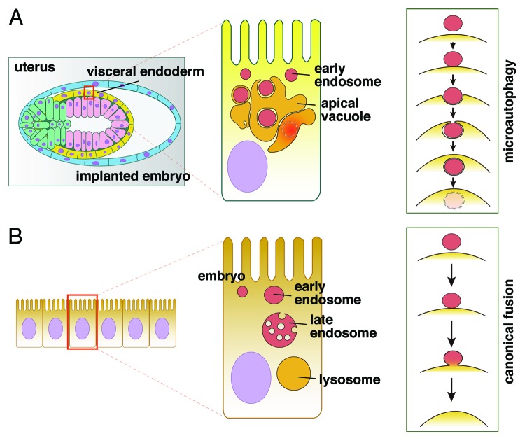
During our studies, we noted that delivery of endosomes to the apical vacuole may occur via a process different from the normal membrane fusion between endosomal and vacuolar membranes (Fig. 1). In the canonical fusion scheme, the limiting membranes of endosomes and apical vacuoles fuse and form a continuous membrane; however, this was unlikely given the following observations: (1) numerous spherical bodies are observed in the apical vacuoles; (2) endocytically labeled endosomes appear inside the apical vacuoles as spherical bodies; (3) RAB7, a key cytosolic regulator of the late stages of the endocytic pathway, often appears inside LAMP2-positive compartments; (4) the morphology of the spherical bodies inside the apical vacuoles well corresponds with those of the endosomes located between the apical vacuoles and the apical plasma membrane; (5) double membranes encircle these spherical bodies inside the apical vacuoles, presumably one originating from the limiting membrane of apical vacuoles and the other from the endosome itself; and (6) an irreversible inhibitor of lysosomal acid lipase, tetrahydrolipstatin (orlistat), blocks the release of endosomal contents into the apical vacuoles.
Figure 1. (A) A schematic model of microautophagy in the visceral endoderm (VE, shown in yellow) of an implanted mouse embryo at the egg-cylinder stage. The VE cells contain large apical vacuoles (AVs), which share several characteristics with lysosomes. Delivery of early endosomes to the AVs occurs via microautophagy, a process in which the endosomes are internalized into the lysosomal lumen as internal vesicles with double membranes. The release of endosomal contents into the AVs requires lysosomal lipase activity. (B) The canonical scheme of delivery to lysosomes and lysosomal fusion. The limiting membranes of the endosomes and lysosomes fuse and form a continuous membrane, allowing for the exchange of contents between them.
These lines of evidence suggested that the delivery of endosomes to the apical vacuoles occurs via the engulfment of whole endosomes by the apical vacuoles. These membrane dynamics are known as microautophagy, a process by which organelles and/or a portion of the cytosol are pinched off by invaginations of the limiting membranes of lysosomes, and then delivered to the lysosomal lumen as small internal vesicles with a single membrane derived from the lysosome/vacuole and, when the substrate is an organelle, a second membrane corresponding to the limiting membrane of the organelle. In yeast cells, peroxisomes can be engulfed directly by vacuoles, a process known as micropexophagy, in response to changes in the carbon source in the medium. The nucleus also undergoes a type of microautophagy referred to as micronucleophagy, or piecemeal microautophagy of the nucleus. However, in mammalian cells, microautophagy has been less frequently reported, and its physiological relevance remains unknown.
The small GTP-binding protein RAB7 is required for the assembly of the large apical vacuoles in VE cells. RAB7-deficient mouse embryos lack the large vacuoles, but accumulate smaller vesicles in the VE. The fragmented vesicles morphologically resemble the endosomes in wild-type embryos, and pulse-chase experiments showed that the endosomes are defective in merging with each other. RAB7 regulates membrane recognition and fusion during the endosome/lysosome interaction, and, obviously, its function is not restricted to the VE. It is also required for proper endocytic function in various cell types, and thus it is not specifically involved in microautophagy. Furthermore, we also showed that the function of VPS41, which is part of the fusion machinery in late endocytic compartments, is essential for the assembly of the large apical vacuoles and the microautophagic delivery of endosomes to the apical vacuoles. Both VPS41 and RAB7 are not specific to microautophagy, but also participate in endocytosis in various cells.
Microautophagy delivers both endosomal and lysosomal membranes to the digestive lumen; thus, these membrane dynamics may participate in the maintenance of membrane ratios at the cell surface and on intracellular organelles in cells with very active endocytosis. Our studies showed that defective microautophagy due to the loss of either RAB7 or VPS41 function impairs gastrulation, the key developmental process by which animals establish the three germ layers, ectoderm, endoderm, and mesoderm.
Disclosure of Potential Conflicts of Interest
No potential conflicts of interest were disclosed.
Footnotes
Previously published online: www.landesbioscience.com/journals/autophagy/article/22585


