Abstract
The poly-γ-d-glutamic acid capsule of Bacillus anthracis is a barrier to infection by B. anthracis-specific bacteriophages. Capsule expression was found to completely inhibit lytic infection by γ phage, an observation supported by the demonstration that this phage does not elaborate a hydrolase that would facilitate penetration through the protective capsule outer layer.
TEXT
Inhalation of endospores of Bacillus anthracis causes lethal infection due to the elaboration by the vegetative form of a protein exotoxin complex and a poly-γ-d-glutamic acid (PDGA) capsule; these virulence factors are plasmid associated, pXO1 harboring the toxin structural genes and pXO2 carrying the capsule biosynthetic operon (1, 2). The threat posed by the malicious dissemination of aerosolized B. anthracis spores is a major and growing concern; countermeasures include vaccination, quarantine, and chemotherapy, and a limited number of antibiotics have been identified for prophylaxis and treatment (3). B. anthracis resistant to these (4, 5) and other (6) antibiotics can be readily selected in the laboratory, and barriers to the engineering of multidrug-resistant strains are low. Thus, alternative therapeutic modalities that will not be compromised by the acquisition of antibiotic resistance genes have attracted attention and include proposals for the therapeutic use of bacteriophage (7, 8) and enzyme-mediated degradation of the PDGA capsule (9). Phages infecting encapsulated bacteria generally carry capsule depolymerases that facilitate interaction with their cell surface receptor (10), and such phage enzymes have enabled the evaluation of capsule degradation as an alternative therapeutic modality (11). Although the genomes of lytic phages infecting Bacillus subtilis carry genes that encode depolymerases that remove the capsular barrier to phage absorption (12), expression of the B. anthracis capsule appears to be restricted to environments high in atmospheric CO2 (1). High CO2 conditions pertain during infection but not necessarily in soil, and B. anthracis-specific phages, as defined by Abshire and coworkers (13), are mainly derived from soil.
Indeed, phage binding is most readily demonstrated using the B. anthracis Sterne strain that lacks the PDGA capsule (14). We have therefore investigated the capacity of B. anthracis-specific phages to lyse B. anthracis grown under conditions that modulate capsule expression with a view to determining penetration of the capsule barrier.
Gamma phage has been frequently employed to differentiate B. anthracis from closely related members of the Bacillus genus (15). The provenance of γ phage can be traced to phage W, an inducible prophage isolated from an atypical Bacillus cereus strain in 1951 by McCloy (16). Phage W lysed all members of a large collection (171 isolates) of B. anthracis isolates and 2 of 54 B. cereus strains; it infected only nonencapsulated forms of B. anthracis, limiting its utility as a diagnostic tool (17). Brown and Cherry later isolated a variant form of phage W, which they termed γ phage, and reported it to be highly specific for B. anthracis and capable of lysing both encapsulated and nonencapsulated phenotypes (18). The mechanism enabling γ phage to penetrate the protective capsular layer is unknown, and it is unclear whether the capsule interferes with phage absorption by perturbation of phage-host cell interactions.
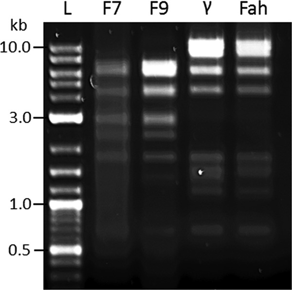
We examined the lytic capacity of four phages. Gamma phage was obtained from the Felix d'Herelle Reference Centre for Bacterial Viruses, Université Laval, Canada, and Fah phage, a γ phage variant widely used in the former Soviet Union for B. anthracis identification (19), from the George Eliava Institute, Tblisi, Georgia. Phages F7 and F9 were isolated from samples of bovine feces in Puławy, Poland. F7 and F9 host specificity was established by their capacity to lyse a panel of 57 Bacillus isolates, including four strains of B. anthracis (Table 1). Phages were propagated in B. anthracis Sterne or B. cereus hosts. Transmission electron microscopy of phage stained with 1% (vol/vol) uranyl acetate determined that F7 and F9 belonged to the Siphoviridae; both possessed long noncontractile tails (∼100 nm) and icosahedral heads (data not shown). Genomic variation was analyzed by restriction digestion of phage DNA; F7 and F9 were clearly distinct from γ and Fah (Fig. 1). The capacity of these four phages to form plaques and zones of lysis on B. anthracis strains Vollum (pXO1+ pXO2+; ASC 6), Ames (pXO1+ pXO2+; ASC 68) and Sterne (pXO1+ pXO2−; ASC 1) was determined. B. anthracis strains were streaked onto blood agar, and isolated colonies were used to prepare turbid bacterial suspensions (optical density at 600 nm [OD600] of ∼1) in brain heart infusion broth (Oxoid, United Kingdom). Blood agar plates were seeded with 100 μl of the bacterial suspension and allowed to air dry. Phage suspension (2 × 10¹⁰ PFU/ml; 20 μl) was dropped onto the seeded area (in duplicate), and the plates were incubated at 37°C for ∼16 h. These conditions yielded lawns of nonencapsulated bacteria. For induction of capsule expression, nutrient broth-yeast extract (NBY) agar (20) containing 0.7% (wt/vol) NaHCO3 and 10% (vol/vol) horse serum was employed for both preparation of the bacterial inoculum and for phage spot tests. The plates were incubated in an atmosphere containing 5% CO2. Capsule expression was confirmed by mucoid lawn morphology.
Table 1.
Susceptibility of Bacillus strains to lysis by phages F7 and F9
| Speciesa | Strain | Susceptibilityb of strain to lysis by phage: |
|||
|---|---|---|---|---|---|
| F7 | F9 | γ | Fah | ||
| Bacillus anthracis | 1584 | + | + | + | + |
| 211 | + | + | + | + | |
| SL 1809 | + | + | + | + | |
| Sterne 34F2 | + | + | + | + | |
| Bacillus cereus | ATCC 14579 | − | − | − | − |
| ATCC 13472 | + | + | − | − | |
| ATCC 19637 | − | − | − | − | |
| ATCC 23261 | − | − | − | − | |
| ATCC 10876 | + | + | − | − | |
| ATCC 16959 | − | − | − | − | |
| ATCC 17285 | − | − | − | − | |
| UW 85 | − | − | − | − | |
| F 171202 | − | − | − | − | |
| Bacillus subtilis | ATCC 6633 | − | − | − | − |
| Bacillus thuringiensis | ATCC 10792 | − | − | − | − |
| ATCC 33679 | + | + | − | − | |
| ATCC 35646 | − | − | − | − | |
| T07-153 | − | − | − | − | |
| T07-128 | − | − | − | − | |
| T07-019 | − | − | − | − | |
| 37 | − | − | − | − | |
| Bacillus sp. strain Ba 813 | 6 (I/2) | − | − | − | − |
| 15 (11614/2) | − | − | − | − | |
An additional 34 strains of non-phage-susceptible Bacillus spp., including representatives of B. megaterium and B. thuringiensis, were also examined.
+, susceptible to phage; −; not susceptible to phage.
Fig 1.

Restriction digests of phages F7, F9, γ, and Fah. Genetic variation was assessed by digestion of genomic DNA using restriction enzymes NdeI and XhoI. Phages F7 and F9 were found to be genetically distinct from γ and its variant, Fah. Lane L contains molecular size standards (in kilobases).
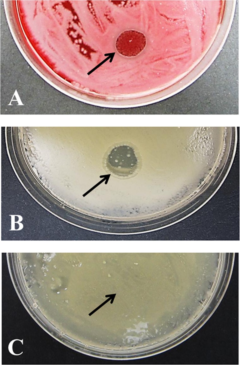
Under conditions which did not induce capsule expression, challenge with high-titer suspensions of all four phages produced large zones of lysis or growth inhibition on lawns of B. anthracis strains Vollum, Ames, and Sterne (Table 2). After culture and incubation under conditions conducive to capsule expression, all phages, including γ phage, failed to produce zones of lysis/inhibition on lawns of the pXO2+ strains Vollum and Ames. However, all four phages elicited zones of lysis/inhibition on the noncapsular pXO2− Sterne stain under both culture conditions. The results for γ phage are shown in Fig. 2.
Table 2.
Effect of capsule expression on phage-mediated lysis of B. anthracis
| B. anthracis strain | Culture medium | Zone of lysis or growth inhibitiona mediated by phage: |
|||
|---|---|---|---|---|---|
| γ | Fah | F7 | F9 | ||
| Ames | Blood agar | +++ | +++ | ++ | +++ |
| NBY agar + CO2 | − | − | − | − | |
| Vollum | Blood agar | +++ | ++ | ++ | +++ |
| NBY agar + CO2 | − | − | − | − | |
| Sterneb | Blood agar | +++ | +++ | +++ | +++ |
| NBY agar + CO2 | +++ | ++ | +++ | +++ | |
+++, zone of lysis or inhibition 1 cm in diameter; ++, zone of lysis or inhibition 1 cm in diameter with some regrowth; −, no zone of lysis or inhibition.
B. anthracis Sterne lacks capsule genes.
Fig 2.

B. anthracis capsule expression inhibits γ phage infection. (A and C) Phage preparations (20 μl) were spotted onto lawns of B. anthracis Ames (pXO1+ pXO2+) seeded on blood agar (no capsule expression) (A) or NBY agar with supplements inducing capsule expression (C). (B) B. anthracis Sterne (pXO1+ pXO2−) was seeded on NBY agar with supplements to ensure that lack of γ phage infection was not influenced by the composition of the solid growth medium. The area where phage was applied is indicated by an arrow in each panel.
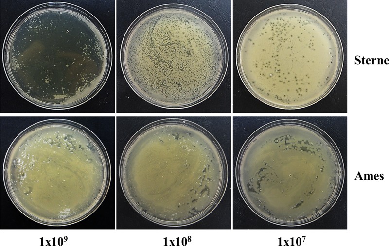
As these data contradict the observation of Brown and Cherry that γ phage lyses both encapsulated and nonencapsulated forms (18), we determined the efficiency of plating (EOP) of γ phage by titration on encapsulated and nonencapsulated phenotypes of B. anthracis Ames relative to the Sterne strain. Bacterial lawns were allowed to air dry on blood agar and supplemented NBY agar, and 100 μl of γ phage preparation was serially diluted 10-fold in SM buffer (100 mM NaCl, 8 mM MgSO4 · 7H2O, 50 mM Tris HCl) distributed evenly across the agar prior to incubation at 37°C for ∼16 h (5% CO2 atmosphere to enhance capsule expression on NBY). Dilutions of high-titer (1010 PFU/ml) preparations of Sterne- and B. cereus-propagated γ phage plated less efficiently on acapsular Ames and Sterne strains (∼104) than on the propagation strain B. cereus ATCC 4342; they were, however, unable to form plaques on capsule-induced Ames at any dilution employed (EOP of 0.0; Fig. 3). These observations strongly imply that the B. anthracis capsule hinders access of γ phage to its cell wall-anchored GamR receptor at the bacterial surface and that in contrast to the conclusions drawn from the study of Brown and Cherry (18), this phage, in similar fashion to other anthrax phages, is unable to infect encapsulated vegetative cells.
Fig 3.
Capacity of γ phage to form plaques on B. anthracis strains Sterne (pXO1+ pXO2−) and Ames (pXO1+ pXO2+). Tenfold dilutions of γ phage preparations (1010 PFU/ml; 100 μl) were spread on NBY agar with supplements that had been seeded with B. anthracis Sterne or Ames. Phage titers represent the number of phage particles applied to each plate. Capsule expression by the Ames strain prevented phage infection and plaque formation at all dilutions over the PFU range 109 to 105.
Brown and Cherry examined the capacity of γ phage to lyse B. anthracis by spotting ∼108 phage particles onto heart infusion agar plates containing 10% mule serum seeded with overnight bacterial cultures grown in heart infusion broth. The plates were incubated at 37°C in an atmosphere of 60 to 80% CO2. Published images (18) show more-extensive zones of clearing or inhibition formed by γ phage than by W phage; bacterial seeding was less dense in the region of the γ phage zone, and the bacteria do not appear mucoid. No attempt was made to examine the capacity of these two phages to form plaques using diluted phage suspensions. It had been previously established (21) that B. anthracis isolates failed to optimize capsule production when bicarbonate was omitted from the growth medium; this was not taken into account by Brown and Cherry. We observed that in order to obtain consistent phage susceptibility data, it was necessary to prepare the B. anthracis inoculum under conditions that permitted capsule expression. Failure to do so resulted in a partial zone of lysis/inhibition much reduced in size (data not shown); thus, it would appear that seeding plates with acapsular B. anthracis and incubating under conditions permitting capsule synthesis provides a window of opportunity for phage adsorption and initiation of the phage lytic cycle. Once capsule is fully expressed, phage adsorption no longer takes place. We suggest that the apparent susceptibility to γ phage of encapsulated B. anthracis reported by Brown and Cherry was due to a failure to ensure that test plates were seeded with capsule-replete bacteria and to optimize conditions favoring capsule expression.
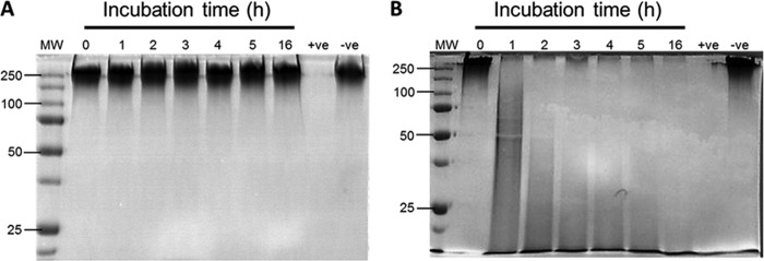
Phages infecting encapsulated bacterial hosts possess capsule hydrolases that remove this outermost protective layer to facilitate docking with surface-associated receptors (10). Such enzymes exist in both phage-bound and soluble form (22) and strip away capsular material at the cell surface, allowing unhindered phage access. To determine whether γ phage possesses such a potentially valuable enzyme, 109 PFU/ml was incubated (37°C) for 16 h with 8 μg PDGA. The B. subtilis phage ΦNIT1 encodes γ-PGA depolymerase PghP (12) and served as a positive control. We found no evidence that γ phage hydrolyzed the PDGA polymer to produce low-molecular-weight fragments; in contrast, ΦNIT1 elicited a time-dependent fragmentation of PDGA (Fig. 4). The absence of a phage-associated capsule depolymerase supports our contention that γ phage does not have the capacity to infect encapsulated phenotypes of B. anthracis.
Fig 4.

Phage-mediated hydrolysis of poly-γ-d-glutamic acid (PDGA). (A and B) Aliquots (1 × 10⁹ PFU) of γ phage (A) or ΦNIT1 phage (B) were incubated with 8 μg PDGA for 16 h at 37°C. Degradation of capsular material was determined by SDS-PAGE. Lanes: MW, molecular weight markers (in thousands); +ve, positive control (contains 35 μg CapD hydrolase [24] with PDGA); -ve, negative control (contains PDGA and no phage). PDGA was obtained from high-Mn-grown cultures of Bacillus licheniformis.
A comparison of the genome sequence of γ phage isolate d'Herelle with the genome of its temperate progenitor Wβ indicated that missense mutations in the tail fiber locus orf14 resulted in a more basic protein that could allow γ phage to more readily penetrate the negatively charged capsule of B. anthracis (23). Sequencing of orf14 from the γ phage stock used in the current study revealed 100% sequence homology with other sequenced γ phage isolates deposited in the NCBI database (accession numbers DQ222853 and DQ222855); none possess the missense mutations in the tail fiber locus orf14 described for the variant d'Herelle. The potential of such tail fiber mutations in the facilitation of capsule penetration has yet to be determined.
In summary, our results show that, in contrast to previous reports, B. anthracis-specific phages, in particular γ phage, are unable to infect encapsulated B. anthracis cells. Additionally, γ phage does not elaborate a PDGA hydrolase that would enable access of the phage to its underlying bacterial surface receptor, and as a consequence, such B. anthracis-specific phages are unlikely to have utility as therapeutic agents.
ACKNOWLEDGMENTS
D.N. was supported by a Medical Research Council Capacity Building Studentship award to P.W.T. and is currently a Fellow of the Maplethorpe Trust.
We thank Agnès Fouet, Institut Cochin, Paris, France, for the gift of CapD and the late Yoshifumi Itoh, National Food Research Institute Tsukuba for ΦNIT1 phage.
Footnotes
Published ahead of print 2 November 2012
REFERENCES
- 1. Koehler TM. 2002. Bacillus anthracis genetics and virulence gene regulation. Curr. Top. Microbiol. Immunol. 271:143–164 [DOI] [PubMed] [Google Scholar]
- 2. Mock M, Fouet A. 2001. Anthrax. Annu. Rev. Microbiol. 55:647–671 [DOI] [PubMed] [Google Scholar]
- 3. Franz DR, Jahrling PB, Friedlander AM, McClain DJ, Hoover DL, Bryne WR, Pavlin JA, Christopher GW, Eitzen EM. 1997. Clinical recognition and management of patients exposed to biological warfare agents. JAMA 278:399–411 [DOI] [PubMed] [Google Scholar]
- 4. Brook I, Elliott TB, Pryor HI, Sautter TE, Gnade BT, Thakar JH, Knudson GB. 2001. In vitro resistance of Bacillus anthracis Sterne to doxycycline, macrolides and quinolones. Int. J. Antimicrob. Agents 18:559–562 [DOI] [PubMed] [Google Scholar]
- 5. Price LB, Vogler A, Pearson T, Busch JD, Schupp JM, Keim P. 2003. In vitro selection and characterization of Bacillus anthracis mutants with high-level resistance to ciprofloxacin. Antimicrob. Agents Chemother. 47:2362–2365 [DOI] [PMC free article] [PubMed] [Google Scholar]
- 6. Kim HS, Choi EC, Kim BK. 1993. A macrolide-lincosamide-streptogramin B resistance determinant from Bacillus anthracis 590: cloning and expression of ermJ. J. Gen. Microbiol. 139:601–607 [DOI] [PubMed] [Google Scholar]
- 7. Inal JM. 2003. Phage therapy: a reappraisal of bacteriophages as antibiotics. Arch. Immunol. Ther. Exp. 51:237–244 [PubMed] [Google Scholar]
- 8. Walter MH. 2003. Efficacy and durability of bacteriophages used against spores. J. Environ. Health 66:9–15 [PubMed] [Google Scholar]
- 9. Scorpio A, Chabot DJ, Day WA, O'Brien DK, Vietri NJ, Itoh Y, Mohamadzadeh M, Friedlander AM. 2007. Poly-gamma-glutamate capsule-degrading enzyme treatment enhances phagocytosis and killing of encapsulated Bacillus anthracis. Antimicrob. Agents Chemother. 51:215–222 [DOI] [PMC free article] [PubMed] [Google Scholar]
- 10. Sutherland IW, Hughes KA, Skillman LC, Tait K. 2004. The interaction of phage and biofilms. FEMS Microbiol. Lett. 232:1–6 [DOI] [PubMed] [Google Scholar]
- 11. Mushtaq N, Redpath MB, Luzio JP, Taylor PW. 2004. Prevention and cure of systemic Escherichia coli K1 infection by modification of the bacterial phenotype. Antimicrob. Agents Chemother. 48:1503–1508 [DOI] [PMC free article] [PubMed] [Google Scholar]
- 12. Kimura K, Itoh Y. 2003. Characterization of poly-gamma-glutamate hydrolase encoded by a bacteriophage genome: possible role in phage infection of Bacillus subtilis encapsulated with poly-gamma-glutamate. Appl. Environ. Microbiol. 69:2491–2497 [DOI] [PMC free article] [PubMed] [Google Scholar]
- 13. Abshire TG, Brown JE, Ezzell JW. 2005. Production and validation of the use of gamma phage for identification of Bacillus anthracis. J. Clin. Microbiol. 43:4780–4788 [DOI] [PMC free article] [PubMed] [Google Scholar]
- 14. Davison S, Couture-Tosi E, Candela T, Mock M, Fouet A. 2005. Identification of the Bacillus anthracis (gamma) phage receptor. J. Bacteriol. 187:6742–6749 [DOI] [PMC free article] [PubMed] [Google Scholar]
- 15. Brown ER, Moody MD, Treece EL, Smith CW. 1958. Differential diagnosis of Bacillus cereus, Bacillus anthracis, and Bacillus cereus var. mycoides. J. Bacteriol. 75:499–509 [DOI] [PMC free article] [PubMed] [Google Scholar]
- 16. McCloy EW. 1951. Studies on a lysogenic Bacillus strain. I. A bacteriophage specific for Bacillus anthracis. J. Hyg. (Lond.) 49:114–125 [DOI] [PMC free article] [PubMed] [Google Scholar]
- 17. McCloy EW. 1951. Unusual behaviour of a lysogenic Bacillus strain. J. Gen. Microbiol. 5:xiv–xv [PubMed] [Google Scholar]
- 18. Brown ER, Cherry WB. 1955. Specific identification of Bacillus anthracis by means of a variant bacteriophage. J. Infect. Dis. 96:34–39 [DOI] [PubMed] [Google Scholar]
- 19. Minakhin L, Semenova E, Kiu J, Vasilov A, Severinova E, Gabisonia T, Inman R, Mushegian A, Severinov K. 2005. Genome sequence and gene expression of Bacillus anthracis bacteriophage Fah. J. Mol. Biol. 354:1–15 [DOI] [PubMed] [Google Scholar]
- 20. Vidaver AK. 1967. Synthetic and complex media for the rapid detection of fluorescence of phytopathogenic pseudomonads: effect of the carbon source. Appl. Microbiol. 15:1523–1524 [DOI] [PMC free article] [PubMed] [Google Scholar]
- 21. Thorne CB, Gómez CG, Housewright RD. 1952. Synthesis of glutamic acid and glutamyl polypeptide by Bacillus anthracis. II. The effect of carbon dioxide on peptide production on solid media. J. Bacteriol. 63:363–368 [DOI] [PMC free article] [PubMed] [Google Scholar]
- 22. Tomlinson S, Taylor PW. 1985. Neuraminidase associated with coliphage E that specifically depolymerizes the Escherichia coli K1 capsular polysaccharide. J. Virol. 55:374–378 [DOI] [PMC free article] [PubMed] [Google Scholar]
- 23. Schuch R, Fischetti VA. 2006. Detailed genomic analysis of the Wβ and γ phages infecting Bacillus anthracis: implications for evolution of environmental fitness and antibiotic resistance. J. Bacteriol. 188:3037–3051 [DOI] [PMC free article] [PubMed] [Google Scholar]
- 24. Candela T, Fouet A. 2005. Bacillus anthracis CapD, belonging to the γ-glutamyltranspeptidase family, is required for the covalent anchoring of capsule to peptidoglycan. Mol. Microbiol. 57:717–726 [DOI] [PubMed] [Google Scholar]


