Abstract
The pathogenesis of intussusception caused by enterohemorrhagic Escherichia coli (E. coli) O157 infection is unknown. In our case, colonoscopy was useful for confirming O157 infection. The intussusception was caused by focally damaged edematous mucosa in the cecum. This case helped in elucidating the pathogenesis of the disease.
Key words: colonoscopy, intussusception, O157.
Introduction
Intussusception is reportedly a complication of hemorrhagic enteritis caused by enterohemorrhagic Escherichia coli (E. coli) O157.1,2 However, the pathogenesis of this condition is unknown. We present the case of a 12-year-old girl who developed intussusception due to O157 infection. She was diagnosed with colonoscopy, which also helped in elucidating the pathogenesis of the disease.
Case Report
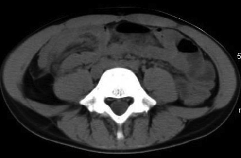
A 12-year-old girl was admitted to Saitama Children's Medical Center (Japan) for repositioning of the intussusception. She had been well until 2 days before admission when she developed abdominal pain and diarrhea. On the day of admission she went to a clinic because of diarrhea accompanied by abdominal pain. Physical examination revealed tenderness in the right lower quadrant of the abdomen. Appendicitis was suspected, and contrast abdominal computed tomography was performed at the clinic, which revealed ensheathing of one portion of the colon into another. She was diagnosed with colonic intussusception (Figure 1), and was admitted to our hospital for repositioning of the intussusception. On admission, the patient was alert and communicative. Her temperature was 36.6°C, blood pressure was 108/52 mmHg, and pulse was 88 beats per minute. On palpation, her abdomen was soft and tender, with the most severe tenderness in the right lower quadrant. There was no abdominal rebound or guarding.
Figure 1.

Abdominal computed tomography showing the ensheathing of one portion of the colon into another.
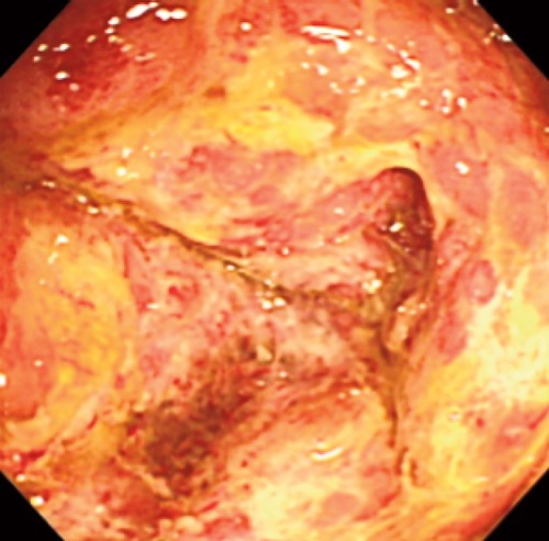
Blood tests revealed a hemoglobin level of 13.4 g/dL, white blood cell count of 10,300/µL, blood urea nitrogen level of 9 mg/dL, creatinine level of 0.42 mg/dL, and C-reactive protein level of 0.23 mg/dL. Results of urine analysis were normal, but her stool tested positive for fecal occult blood. It later became clear that her stool culture was negative for recognized pathogens, including O157, Salmonella, Shigella, Campylobacter, and Yersinia. An ultrasound-guided saline enema was performed for the intussusception by a pediatric radiologist; repositioning of the intussusception was immediately confirmed. The patient was transferred to the Division of General Pediatrics because of persistent intermittent abdominal pain and the appearance of grossly bloody stool after repositioning. A abdominal ultrasound showed no findings of the target shaped lesion or the multilayered lesion and we excluded a relapse of the intussusception. On the forth day, a colonoscopy under vein anesthesia was undertaken without bowel preparation for investigating the cause of intermittent abdominal pain and bloody stool because the stool culture was negative for pathogens including O157. The procedure revealed significant mucous membrane swelling with pus formation in the cecum. Examination of the terminal ileum revealed normal mucosa (Figure 2). Enterohemorrhagic E. coli O157, which produces verotoxins (VT1 and VT2), was later cultured by fecal aspiration. Recovery was uneventful with fasting and saline infusion. She improved without antibiotics and was discharged on the eighth postoperative day without developing hemolytic-uremic syndrome, and there has been no recurrence of intussusception till date.
Figure 2.

Colonoscopic image indicating significant mucous membrane swelling with pus in the cecum.
Discussion and Conclusions
The 0157 E. coli strain belongs to a group of bacteria known as enterohemorrhagic E. coli, and is a common cause of foodborne illness.3 Infection with this strain frequently leads to hemorrhagic diarrhea, intense abdominal pain, and occasionally, hemolytic-uremic syndrome, intussusception, and intestinal perforation. Lopez reported the first case of intussusception due to E. coli O157 infection.1 In mid-July, 1996, a massive outbreak of hemorrhagic colitis (HC) occurred among elementary school children in Sakai City, Japan. A total of 425 children with HC were treated at the local hospital. Hashizume et al. reported that 8 (1.9%) of these 425 patients developed intussusception.4 Intussusception is presumably caused by edematous changes in the intestinal mucosa accompanied by intense inflammation, enlargement of intestinal membrane lymph nodes, and changes in the intestinal tract, including enteric ischemia-related changes. However, the precise causes remain unknown. In our case, the intussusception was presumably caused by focally damaged edematous mucosa in the cecum because there was no evidence of other leading points, such as lymph node enlargement. Colonoscopy was useful for confirming O157 infection because the O157 strain was cultured by fecal aspirate rather than by repeated fecal cultures. Although it is more difficult to perform colonoscopy in children than in adults, we consider this procedure to be a useful diagnostic tool in cases that deviate from the conventional mean age usually reported for the onset of intussusception.
References
- 1.Lopez EL, Devoto S, Woloj M, et al. Intussusception associated with Escherichia coli O157:H7. Pediatr Infect Dis J. 1989;8:471–3. [PubMed] [Google Scholar]
- 2.Siegler RL. Spectrum of extrarenal involvement in postdiarrheal hemolyticuremic syndrome. J Pediatr. 1994;125:511–8. doi: 10.1016/s0022-3476(94)70001-x. [DOI] [PubMed] [Google Scholar]
- 3.Karch H, Tarr PI, Bielaszewska M. Enterohaemorrhagic Escherichia coli in human medicine. Int J Med Microbiol. 2005;295:405–18. doi: 10.1016/j.ijmm.2005.06.009. [DOI] [PubMed] [Google Scholar]
- 4.Fukushima H, Hashizume T, Morita Y, et al. Clinical experiences in Sakai City Hospital during the massive outbreak of enterohemorrhagic Escherichia coli O157 infections in Sakai City, 1996. Pediatr Int. 1999;41:213–7. doi: 10.1046/j.1442-200x.1999.4121041.x. [DOI] [PubMed] [Google Scholar]


