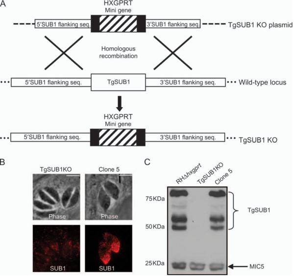
Figure 3. Generation of a TgSUB1 KO parasite.
A) A diagram of the strategy used to disrupt the TgSUB1 gene by homologous recombination in the RH background is shown. The TgSUB1KO plasmid is seen on the top line showing 2.9 kb of 5′ and 3′ DNA flanking the coding region of TgSUB1. Dashed lines indicate the rest of the targeting plasmid sequence. TgSUB1 is replaced by the HXGPRT cassette, which includes the HXGPRT gene surrounded by the DHFR promoter and terminator sequences. Dots signify the rest of the genomic locus. B and C) IFA and western blot analysis of TgSUB1KO and clone 5 parasites (TgSUB1 was revealed with AE653 antibody). MIC5 is used as internal control for western blot loading. TgSUB1 immunofluorescence signal depicts the conventional apical microneme labeling. Scale bar: 5 μm.

