Abstract
Metastasis from breast cancer to other parts of the body is very common, but the spread of the tumor to pituitary gland, especially to infandibulum, is a rare presentation. At the time of pituitary metastasis, a majority of the patients have clinical and radiological evidence of the disease. It seems that the posterior area of the gland is the most common site of metastasis, probably due to highly rich blood supply through the hypophyseal artery.
The present report introduces a case of a 55-years-old woman presented with diabetes insipidus resulting from metastasis of the tumor to pituitary infandibulum, which is a rare site for metastasis, without significant complaint resulting from metastasis to other part of the body, or other primary diseases. Further evaluation revealed that in spite of previous reports, which metastasis usually happens in end stage of cancer, the patients had primary breast cancer. In subsequent evaluations of the case, hypofunction of adenohypophysis was also detected.
Key Words: Breast cancer, pituitary gland, diabetes insipidus, infundibular metastasis
Introduction
Breast cancer is one of the most common cancers with multiple metastases to different sites of the body. It can spread to pituitary gland, particularly to posterior part, possibly via the direct blood supply by the arterial system. Diabetes insipidus (DI) is the most common presentation of posterior pituitary involvement, but headache, visual symptoms, endocrine abnormality are also present. Metastasis to the anterior part is usually seen concurrent with posterior part involvement. In spite of the posterior and anterior lobe involvements, infundibular metastasis, especially in early stage of cancer, is a rare event.1
In most of the patients, the signs and symptoms of metastasis to other parts of the body such as lymph nodes, lung or bone are the first manifestations, and metastasis to pituitary gland occurs later and is usually detected incidentally.1 At the time of diagnosis of the pituitary metastasis, many patients have widespread evidence of the disease; however, pituitary dysfunction, as the first presentation of primary breast cancer and infundibular involvement, occurs rarely.2
Case Presentation
A 55-year-old woman with polyuria and polydypsia, who had been diagnosed as having DI since one year earlier. The patients was referred to the Department of Neurology, Motahhari Clinic complaining of an intractable frontal headache. The pain was radiating to the vertex, and was accompanied by nausea and occasional vomiting.
Since seven months prior to referral to the Clinic, she had anorexia, general weakness, sleepiness, and a central type of hypothyroidism in addition to DI. Therefore, she had started receiving thyroid hormone replacement therapy. After receiving replacement therapy, the patient's general condition had improved; however, she was still suffering from moderate constant headache. Therefore, she was referred to a neurologist (one of the authors) for neurological evaluations.
General examination by the neurologist revealed nothing remarkable, except for asymmetric breasts with left breast being smaller than the right one. Physical examination of the breasts showed a non mobile, 3×4 cm mass with irregular border in the left breast. Retraction of the left nipple was apparent, but exam of the axillary area was negative for lymph nodes or other abnormalities.
All of the examinations related to cranial nerves including visual acuity and visual field and fundoscopy, motor system (bulk, tone and power), sensory system, deep tendon reflex, planter reflex, cerebellar signs and gait were normal.
Hematocrit, platelet counts, blood urea nitrogen, serum creatinine, fasting blood sugar, stool examination for occult blood/parasite, liver function test, lumbar puncture for protein-sugar-cell count and cytology, erythrocyte sedimentation rate, C-reactive protein were all within relevant normal ranges. Confrontation perimetry for visual field evaluation was normal as well. She did not permit repeated lumbar puncture for excessive cytological study.
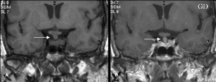
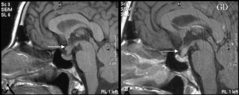
In brain Magnetic Resonance Imaging (MRI) a thickening of pituitary infundibulum with moderate enhancement after the injection of MR contrast was seen. However, the pituitary gland was normal (figure 1: A and B, and Figure 2: C and D).
Figure 1.
Coronal view of T1 weighted magnetic resonance images of the pituitary region before (A) and after (B) the injection of gadolinium. White arrows ahow enlarged pituitary infandibulum and moderate diffuse enhancement. Pituitary gland and optic chiasma were normal.
Figure 2.
Sagittal views of T1 weighted magnetic resonance images of the pituitary region before (A) and after (B) the injection of gadolinium. White arrows show enlargement and diffuse enhancement of pituitary infandibulum.
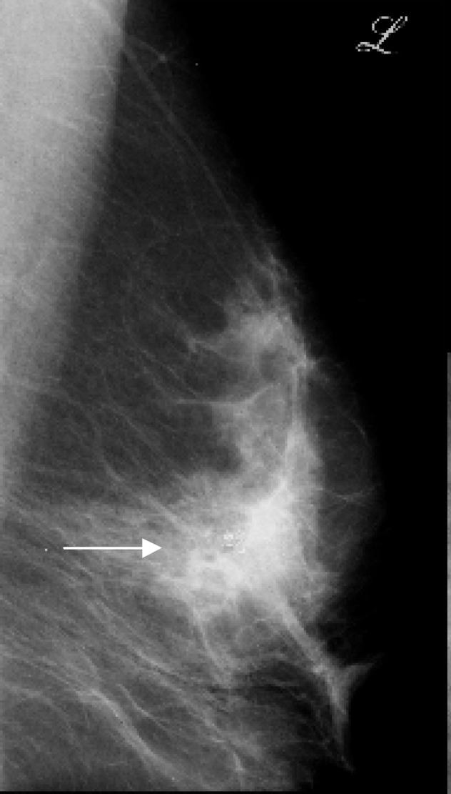
Mammography of breasts, revealed a punctuated dens mass with multiple micro calcification in subareolar region of the left breast (figure 3).
Figure 3.
Mediolateral view of left mammogram shows a punctuated mass with multiple micro calcifications (white arrows) in subareolar region on the left breast, which show nipple retraction
Subsequent evaluation using fine needle aspiration (FNA) revealed few small groups of ductal epithelial cells with mild anisonucleosis, some hyperchromatic nuclei and irregular nuclear borders. The FNA and smear was low cellular and suspected for malignancy. For investigation sites of metastases, total body scan was recommended for the patient. The scan showed two sites of metastases in skull and vertebral body. She was finally diagnosed as primary breast cancer with multiple metastases, and was referred to an oncologist for chemotherapy and radiotherapy.
Discussion
In most of the studies on metastatic involvement of the pituitary gland, breast and lung cancers were the most primary tumors comprising approximately two-thirds of cases, but metastasis from lymphoma, leukemia, melanoma, kidney, colon, and prostatic cancer were also reported.1
A review of the literature suggests that when pituitary gland is involved in a malignancy, posterior lobe is the most common affected site. The spread of malignancy to pituitary might have occurred through direct blood supply by arterial system. Therefore, hematogenous spreads of malignant cells disseminate easier to posterior part of hypophysis than to the anterior lobe, which is supplied by hypophyseal portal system.3,4 However, compared to metastasis to posterior and anterior lobes, metastasis to infundibulum is a rare incident. The present case presented first with signs and symptoms of DI such as polydypsia and polyuria, which implied hypophyseal involvement. This finding is similar to those of other studies demonstrating the presence of DI upon metastasis spreads to hypophyseal gland.1,5,6 However, headache, visual symptoms due to chiasmal compression, endocrine abnormality and extraoccular nerves palsy due to cavernous sinus invasion have also been reported.1,3 The patient gradually deteriorated to a hypothyroid state, indicating adenohypophyseal hypofuction. Concomitant hypofunction of both anterior and posterior hypophysis was described in the literature as a rare event.5
Pituitary MRI of the present case revealed an enlargement of stalk with a moderate diffuse enhancement after injection of the contrast, and a normal appearance of pituitary gland and optic chiasma. The location of stalk involvement in imaging provided a higher possibility of differentiation between malignant and benign (e.g. adenoma) diseases, because malignant metastasis to pituitary gland may mimic benign tumors in clinical and radiological manifestations. Junea and colleagues,7 reported three cases with hypophyseal tumor. One of them had carcinoma of lung, the other had breast cancer and the third one had sellar plasmacytoma. In physical exams, these cases shared several important features of pituitary adenoma such as progressive visual loss and extraocular nerves palsy.7 Kovacs and colleagues reported two cases with pituitary metastasis, which had widespread metastasis to several organs with disseminated carcinomatous processes. They concluded that pituitary metastasis usually happens in the terminal stage of cancer, when widespread metastasis is already present.8 However, the present case first presented the signs and symptoms of hypophyseal metastasis and other sites of metastasis such as skull and vertebral bodies were revealed after further evaluations.
Treatment of malignant pituitary involvement consists of transcranial and trasnsphenoidal surgeries, chemotherapy, and radiotherapy. The regression of metastasis is achieved after each of these treatments. However, for the relief of DI, desmopressin (1-deamino-8-D-arginine vasopressin, DDAVP) is usually needed. Mark and colleagues showed that gamma knife surgery could eliminate the symptoms of DI with minimum radiation to optic apparatus and an effective radiation to the tumor margins.9
This report shows that in addition to adenohypophysis and neurohypophysis, infundibulum may be involved in the metastasis of a malignant tumor to pituitary gland resulting in both anterior and posterior lobe-related manifestations. The involvement of infundibulum can occur in the early stage of the disease or even as a first presentation of cancer, as is the case for the patient in this report.
Conflict of Interest: None declared
References
- 1.Kurkjian C, Armor JF, Kamble R, et al. Symptomatic metastases to pituitary infundibulum resulting from primary breast cancer. Int J Clin Oncol. 2005;10:191–4. doi: 10.1007/s10147-004-0458-5. [DOI] [PubMed] [Google Scholar]
- 2.Chaudhuri R, Twelves C, COX TCS, Bingham JB. MRI in diabetes insipidus due to metastatic breast carcinoma. Clin Radiol. 1992;46:184–8. doi: 10.1016/s0009-9260(05)80442-8. [DOI] [PubMed] [Google Scholar]
- 3.Snell RS. Clinical neuroanatomy. 7th ed. Philadelphia, PA: Williams & Wilkins; 2010. pp. 388–9. [Google Scholar]
- 4.Sturm I, Kirschke S, Krahl D, Dörken B. Panhypopituitareism in a patient with breast cancer. Onkologie. 2004;27:280–2. doi: 10.1159/000080370. [DOI] [PubMed] [Google Scholar]
- 5.Fassett DR. Couldwell WT. Metastases to the pituitary gland. Neurosurg Focus;16:E8. [PubMed] [Google Scholar]
- 6.Junea P, Schoene WC, Black P. Malignant tumors in the pituitary gland. Arch Neurol. 1992;49:555–8. doi: 10.1001/archneur.1992.00530290147025. [DOI] [PubMed] [Google Scholar]
- 7.Kovacs K. Metastatic cancer of the pituitary gland. Oncology. 1973;27:533–42. doi: 10.1159/000224763. [DOI] [PubMed] [Google Scholar]
- 8.Mark P, Paul D, Paul C, Michael J. Resolution of diabetes insipidus following gamma knife surgery for a solitary metastasis to the piyuitary stalk. Case report. J Neurosurg. 2004;101:1053–6. doi: 10.3171/jns.2004.101.6.1053. [DOI] [PubMed] [Google Scholar]
- 9.Rajput R, Bhansali A, Dutta P, Gupta SK, Radotra BD, Bhadada S. Pituitary metastasis masquerading as non-functioning pituitaryadenoma in a woman with adenocarcinoma lung. Pituitary. 2006;9:155–7. doi: 10.1007/s11102-006-8326-0. [DOI] [PubMed] [Google Scholar]


