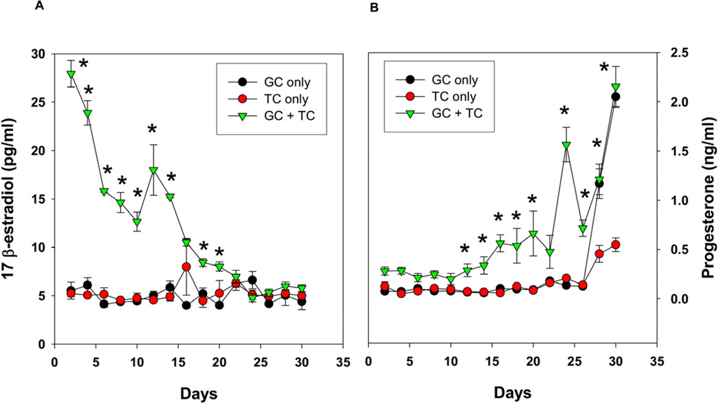
Fig 3.
(A) 17 β-estradiol production as a function of time by co-cultured granulosa cells with theca cells in the presence of FSH and LH in a 2D co-culture system. (B) Progesterone production by co-cultured granulosa cells with theca cells in response to FSH and LH in 2D system. The results indicate the loss of estrogen production with time and the increase in progesterone, indicating luteal differentiation. Each data point represents mean ± SEM of 6 values (3 wells/group assayed in duplicates). * denotes statistical significance at P <0.05 compared to granulosa and/or theca cells alone. The figures represent data from one of two separate experiments.

