Abstract
Background and Objective
Generalized argyria is a blue-gray hyperpigmentation of the skin resulting from ingestion or application of silver compounds, such as silver colloid. Case reports have noted improvement after Q-Switched Neodymium--Yttrium Aluminum Garnet laser (1064nm QS Nd:YAG) laser treatment to small surface areas. No reports have objectively monitored laser treatment of generalized argyria over large areas of skin, nor have long-term outcomes been evaluated.
Study Design/Materials and Methods
An incremental treatment plan was developed for a subject suffering from argyria. A quantitative near infrared spectroscopic measurement technique was employed to non-invasively analyze tissue-pigment characteristics pre- and post-laser treatment. Post-treatment measurements were collected at weeks 1, 2, 3, and 4, and again at 1 year.
Results
Immediate apparent removal of pigment was observed with 1 Q-switched 1064 nm Nd:YAG laser treatment (3-6 mm spot; 0.8-2 J/cm2) per area. Entire face, neck, upper chest and arms were treated over multiple sessions. Treatments were very painful and general anesthesia was utilized in order to treat large areas. Near-infrared spectroscopy was used to characterize and quantify the concentration of silver particles in the dermis based on the absorption features of the silver particles as well as the optical scattering effects they impart. We were able to estimate that there was, on average, 0.042 mg/mL concentration of silver prior to treatment and that these levels went below the minimum detectable limit of the instrument post-treatment. There was no recurrence of discoloration over the 1-year study period.
Conclusion
QS 1064 nm laser treatment of argyria is a viable method to restore normal skin pigmentation with no evidence of recurrence over study period. Quantitative spectroscopic measurements, 1) confirmed dyspigmentation was due to silver, 2) validated our clinical assessment of no recurrence up to one year post-treatment and 3) indicated no collateral tissue damage with treatments.
Keywords: Argyria, (Nd:YAG), quantitative spectroscopy, NIR spectroscopy, tissue optics
Introduction
Argyria is an uncommon but well-defined pigmentary disorder caused by the cutaneous accumulation of silver. This is usually achieved by oral ingestion of silver-containing compounds, usually in homeopathic remedies, but has also been reported to occur via localized mucocutaneous application of creams, dressings, or eye drops containing non-ionized silver.[1-4] Silver particles are deposited around adnexal structures in the dermis, classically in the basement membrane of eccrine coils, but they can also be found on the dermal side of the dermal-epidermal junction and around nerves, capillary walls, arrector pilori muscles, and collagen and elastin fibers.[5, 6]
Argyria manifests as blue-gray macular dyspigmentation, often enhanced on sun-exposed skin.[4, 7, 8] Patients are typically distressed by the prominent appearance, which has been likened to the coloring of a corpse and clinically can be mistaken for cyanosis or methemoglobinemia.[9]
The grey-blue appearance of the skin tone in argyria is the result of pigmented particles in skin and light scattering effects that produce the resultant grey-blue coloration. Silver colloid in the absence of tissue actually presents itself as brown in color, but since it is buried within the dermis, there is a volume of tissue above it that scatters light. This scattering volume preferentially allows shorter visible wavelengths of light (blue) to be diffusely reflected and exit the tissue, while allowing longer wavelengths of light (red) to reach the dermal tissue and be preferentially absorbed. This effectively “shifts” the skin coloration towards a bluish hue. This “Tyndall effect” is the same phenomenon that results in the clinical appearance of blue nevi, even though the pigmentation is due to melanin, or bluish discoloration reported with superficially injected hyaluronic acid.[10, 11]
Optical spectroscopy can be a useful tool for characterizing bulk turbid media, such as biological tissue[12-20]. Optical properties derived from these techniques can be used to report intrinsic and distinct physiological and chemical properties of the biological sample. The absorption coefficient can be used to quantify concentrations of endogenous chromophores such as oxy- and deoxy- hemoglobin, water, and melanin as well as exogenous chromophores (e.g. silver colloid) in tissue. Similarly, reduced scattering coefficient has been shown to be sensitive to structural aspects of cells, cellular components as well as extracellular materials.
A novel spectroscopic technique based on quantitative modeling of light transport in tissue has been developed at the Beckman Laser Institute as a method to quantify tissue optical properties, targeting breast and skin.[12-14] This approach has been developed to interrogate tissue optical properties in the NIR regime (650-1000nm), which is the wavelength region in which photons can penetrate tissue up to several cm. In the context of targeting the optical properties specific to skin, a specialized fiber probe, has been designed to measure optical properties in superficial volumes, such as the epidermal and dermal layers within skin.[14]
In terms of this technique, argyria presents two sources of optical contrast that can be used to characterize and monitor the presence of silver colloid in skin: Absorption and Scattering. Wavelength dependent absorption can be used to identify and quantify the concentration of silver present in tissue and scattering can be used to describe the nature of light transport in tissue and to characterize the structural properties of subcellular objects the interrogating light encounters. As demonstrated in histopathologic sections of hyperpigmented skin in argyria, silver particles accumulate in the extra-cellular space within the dermis.[4, 5] Optical scattering properties can help describe the size and distribution of these particles in contrast to normal tissue constituents that scatter light.
Two case reports evaluated the 1,064 nm Q-Switched (QS) Neodynium-Doped Yttrium Aluminum Garnet laser (1064nm QS Nd:YAG) for localized treatment of argyria.[21, 22] While both these reports exhibited successful clinical pigment clearing, follow-up periods were short (4 weeks and 3 months) and treatment sites were focal (forehead and face). Very recently, another case study has also been reported [23]. In this study, they provided treatment of the face and clinical evaluation based on visual assessment of the treatment efficacy after 1 year.
We report a case of a 52-year-old female with generalized argyria who was successfully treated with the 1064nm QS Nd:YAG. A quantitative spectroscopic measurement technique was employed to non-invasively analyze tissue-pigment characteristics pre- and post-laser treatment. The goals of this preliminary study are to (a) characterize the spectral signature of silver colloid in the skin in terms of absorption and scattering properties detected in vivo, and (b) use some or all of these identified features to quantify the amount of silver present in skin post treatment as a function of time to verify whether this treatment method is effective in permanently removing the clinical appearance of the silver particles.
Materials and Methods
A 52-year-old female (Fitzpatrick skin type II) presented with a 10-year history of confluent blue-gray discoloration of the skin. The patient’s medical history was notable for oral ingestion of a concentrated colloidal silver solution (Angstrom Minerals, 300 mg/L) as a homeopathic remedy for a duration of 9-10 months in 2000. She subsequently developed gradual cutaneous blue-gray hyperpigmentation of the entire body, with enhancement in sun-exposed areas. She denied any history of medications or products commonly implicated in cutaneous hyperpigmentation, such as minocycline or gold-containing remedies. The patient had undergone a series of homeopathic remedies including detoxification treatments without success.[7, 24] She had not had prior laser therapy.
After the initial consultation, a test-spot was performed on the nape of the neck using the 1064nm QS Nd:YAG laser (RevLite®, ConBio). Immediate and dramatic lightening of the skin to a creamy-pink hue was observed after each pulse at 2 joules/cm2 and a 4mm spot size. The improvement in this area was maintained at 1 week, 2 months, 1 year, and 18 months. Over time, other body sites including the entire face, neck, upper chest and arms were also treated successfully, with fluences ranging from 0.8-2 joules/cm2 (4-6 mm spot). A larger spot size in conjunction with a lower energy was used in later treatments to cover more area while achieving the same results. Each area was only treated one time. The patient developed significant transient swelling in treated areas, but there was minimal to no scabbing and no bruising or other tissue effects. Dexamethasone was administered to minimize swelling when large areas were treated.
Each treatment session was complicated by the complaint of severe pain associated with each laser pulse. Initially, the patient could only tolerate less than 5 pulses per treatment session on the posterior neck and forehead despite the use of topical anesthetics or locally injected 1% lidocaine. Concomitant intramuscular meperidine (Demerol) did not provide adequate pain relief. Ultimately, the patient underwent outpatient general anesthesia for 1-hour treatment sessions.
The targeted treatment sites were measured using the spectroscopic technique immediately prior to treatment, and repeat measurements were obtained of both the treated and adjacent untreated areas on a weekly basis for 4 weeks and then at 1 year.
Results
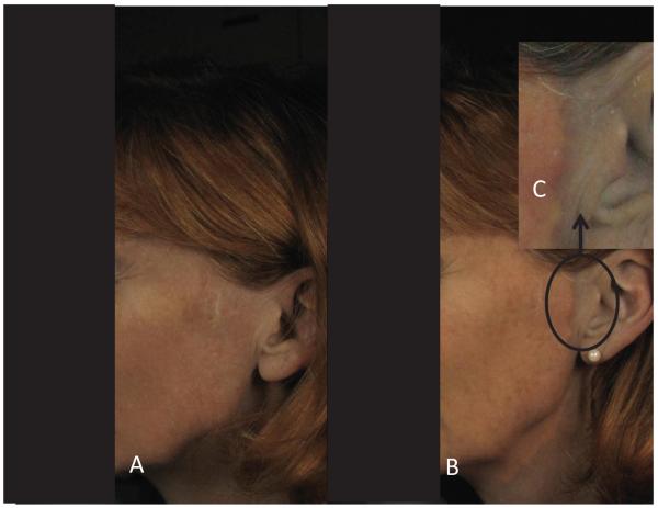
Clear changes in skin appearance were observed immediately after treatment (Fig 1). Photographs, however, only provide a qualitative assessment of the efficacy of this treatment modality and cannot quantify the changing spectral characteristics of the silver particles in the tissue and whether or not it is being affected by the laser. Using the quantitative spectroscopic instrument indicated above, the optical properties of the silver particles were determined and used as a quantitative measure to assess the amount of silver present (Absorption), and to provide deeper insight to the size and organization of the particles in the tissue (Scattering).
Figure 1.
Clinical photography of Argyria patient (A) pre- and (B) post- full-face treatment, the call-out image, (C), demarks the edge of the treatment area to better illustrate the apparent pigmentation contrast between treated and untreated skin tissue.
Absorption
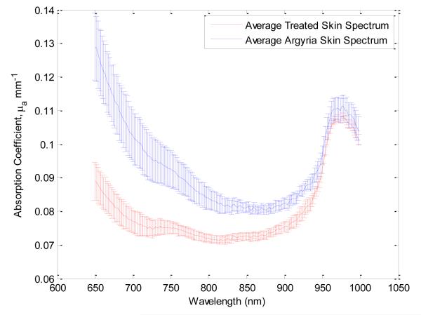
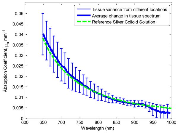
Figure 2 shows the significantly different absorption spectra from treated and untreated skin across multiple areas of the body and over multiple time points. The increased absorption (blue trace) in the untreated skin was hypothesized to represent the presence of silver in skin. To test the hypothesis that this was indeed due to the presence of silver, reference measurements of silver colloid (Sigma Aldrich, 85131-10g, purity: 65-75%) in solution (H2O) were taken in a spectrophotometer (Shimadzu, UV-1800) at concentrations of 0.085, 0.25 and 0.50 mg/ml. Not only does this approach provide a spectral absorption reference for the silver colloid present within the skin tissue, it also provides a quantitative basis from which we can non-invasively estimate the concentration of silver within the skin. The absorption spectra of treated skin (clinically clear of argyria) additively combined with the measured reference absorption spectra of isolated silver colloid is equal to the absorption spectra of untreated, clinically argyric skin (Fig 3). This indicates that our quantitative spectroscopic method can 1) isolate the absorption characteristics of exogenous, dermally-deposited silver colloid from the baseline absorption features in skin due to endogenous cutaneous chromophores (melanin, hemoglobin species, lipids and water) and 2) estimate that for this particular subject there was an average concentration of 0.042 mg/ml of silver present within the tissue.
Figure 2.
The average quantitative absorption spectra (across all locations and measurement time points) pre- (blue) and post- (red) treatment.
Figure 3.
Averaged site-specific difference spectra in absorption between pre- and post-treatment tissue. The green line represents the independently measured absorption signature of silver colloid in solution. Here the difference spectra match the spectral profile of the silver colloid solution reference, indicating that the change in tissue due to treatment is the targeted removal of silver.
Scattering
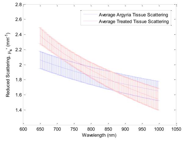
There was also a differentiation of tissue scattering properties observed between treated and untreated tissues. Figure 4 shows the distributions of wavelength-dependent scattering spectra for all treated and untreated tissues. It has been shown that the reduced scattering coefficient, μs,’ can be modeled by a simple power law relation.[25]
Figure 4.
The average quantitative reduced scattering spectra (across all locations and measurement time points) pre- (blue) and post- (red) treatment.
Scattering in the near infrared can be described by two parameters: A, b. These parameter values are summarized across all tissue locations in Table 1, for both treated and untreated skin. The parameter, b, is related to the average size of the scatterers present in the tissue. The differences in b value between treated and untreated skin as measured using our quantitative spectroscopy approach supports the assertion that the presence of silver manifests itself as a distribution of particles which is significantly larger than typical endogenous scatterers in tissue (collagen fibrils, nuclei, etc.) for this wavelength regime. The shift from b values of 0.5 in the treated skin, which is typical for normal Caucasian skin[13], to values that approach 1.0 indicate the presence of scatterers that are an order of magnitude larger than the typical average endogenous scatter (~0.2 microns).
Table 1.
Average parameters used to describe the reduced scattering spectra for treated and untreated tissue. The P-values are also reported to demonstrate the statistical significance of differentiation between the individual distributions of these parameters between tissue cohorts
| Untreated | Treated | P-value | |
|---|---|---|---|
| <ln(A)> | 3.93 | 7.21 | 7.93E-05 |
| <-b> | −0.494 | −0.979 | 1.1E-04 |
The other component that defines scattering in the equation above is the parameter “A”. “A” is typically related to the density of scatterers.[25] This parameter is more specifically correlated with the number of scattering events the photons encounter when interrogating the volume of tissue. It is our working hypothesis, based on Mie theory models [26], that the silver accumulated in the dermis contributes to the measured reduced scattering coefficient. However, this assumption does not necessarily imply that the “A” value will increase relative to skin tissue volumes without argyria. Scattering of near infrared light, like that used by our spectroscopic method, in normal tissue typically originates from sub-cellular components (e.g. mitochondria, lysosomes, vesicles, collagen) and is highly forward directed. This is largely because the index of refraction mismatch between these endogenous scattering objects and the surrounding environment is small. On the other hand, silver particles exhibit markedly different optical index of refraction properties than the surrounding environment. Mie theory predicts that silver colloid particles are approximately 3 orders of magnitude greater scatterers in the backward direction than endogenous scatterers[27]. Hence, scattering objects like colloidal silver will limit the penetration depth of detected photons in a reflectance geometry such as that used to make spectroscopic measurements. This manifests as a reduction in the number of scattering events that photons encounter (compared to normal tissue) prior to detection, thus lowering the value of “A” for regions containing silver compared to silver free regions.
As summarized in Table 1, there is a clear differentiation in distribution of A values between treated and untreated tissues, irrespective of any intrinsic location-specific tissue scattering. There is a statistically significant reduction in the A parameter in untreated skin tissue relative to the treated skin. There was no temporal correlation with changes to the A or b parameters as a function of time from treatment, suggesting that this parameter change is due to the apparent removal of silver (treatment) and not related to any other physiological, long-term response to the laser therapy (as in tissue restructuring or wound healing). Additionally, as the spectroscopic instrument is capable of determining the concentrations of water and hemoglobin species[28], there were no significant changes in these values that would indicate edema (increase in water) or of vascular damage or bruising (increases in deoxy- or methemoglobin) within the affected tissue volumes.
Monitoring of treated skin over time
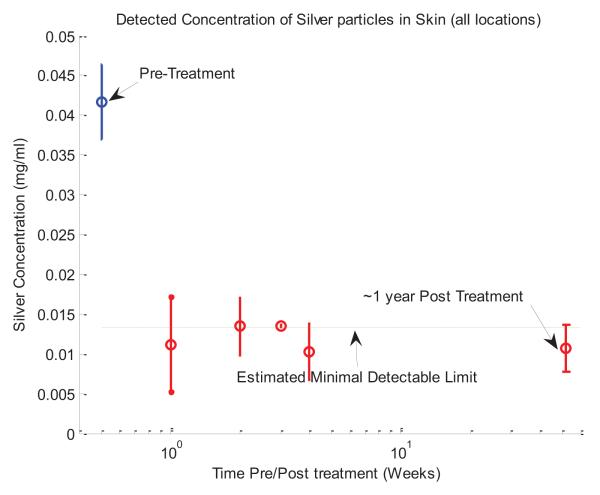
Using the quantitative absorption data collected over the one year time frame, we can determine the amount of silver concentration present in the skin relative to the pre-treatment value. For this given measurement and instrumentation geometry, this particular spectroscopic approach is capable of detecting concentrations of silver down to ~0.013 mg/mL. This minimal detectable signal limit arises due to the fact that this instrument’s probe geometry and subsequent data processing are based on analytical methods of solving light transport based on the Diffusion Approximation[15]. Also, the spectral features of silver are quite broad and featureless in the near infrared regime. The spectral decomposition of the measured tissue components into the characteristic basis set (i.e. silver, hemogobins, lipids, water and melanin absorption) are not perfectly orthogonal and hence there is some amount of cross-talk error present in any decomposition.
Figure 5 shows the detected amount of silver present as a function of time. Day zero represents the concentration detected in regions of tissue prior to treatment. The change in detected silver concentration after treatment is statistically significant from the pre-treatment levels (p-value: 2.6×10⁻⁸). Whereas there are some degrees of variance present in the subsequent, post-treatment measurements, they remain within the detection limit of the spectroscopic instrument and exhibit no significant trends toward the re-accumulation of silver in the skin. Additionally, the resulting treated skin tissue exhibits no significant difference or spectral features from that of normal skin of the same skin type,[13] further indicating that there are no collateral changes or side-effects to the skin or skin appearance that are induced by this treatment method.
Figure 5.
The detected concentration of silver in the skin as a function of time based on the determination through the decomposition of the absorption spectra (averaged over all skin locations)
Discussion
We successfully achieved immediate apparent removal of pigment with 1 Q-switched 1064 nm Nd:YAG laser treatment per area. Improvement was noted clinically and confirmed with spectroscopic analysis. Entire face, neck, upper chest and arms were treated over multiple sessions and improvement was maintained over the 1 year study period.
Argyria is believed to occur in the setting of a cumulative elemental silver dose of as little as 3.8gm [29], although an older but criticized study from 1935 calculated that 1.84gm of actual silver could induce clinical argyria [30]. The Environmental Protection Agency’s reference dose for daily oral silver exposure is 5 μg/kg/day, and the typical diet yields an average of 27-88 μg of silver/day [5, 31]. According to this data, approximately 0.49g – 1.61g of silver may be ingested by the age of 50 for the typical adult. Although this does not reflect the total retained silver dosage, which may be more relevant for developing argyria, as silver can also be excreted via the gastrointestinal and urinary tracts. As with other deposition diseases, it is the excess material that is deposited and becomes pathogenic. It has been estimated that only ~10% of ingested silver is absorbed, with only 2-4% actually being retained within tissues [7]. Moreover, several authors have reported high levels of urinary, blood, or fecal silver several months to years after supposed cessation of silver intake in patients, possibly reflecting a slow leaching of excess tissue silver[32-34]. Despite this supposition, there has been no data to indicate that cutaneous silver deposition is reduced with time, and formation of silver sulfide and silver selenide are thought to result in a more permanent form of silver that is unlikely to leach from the skin due to its tight adherence to tissue proteins. As such, argyria has been considered by some sources to be a permanent dyspigmentation, with no effective treatments.[7, 35] Recent data[21-23], including our own findings, suggest that the QS 1064 nm Nd:YAG laser can be clinically effective for removing dyspigmentation.
Laser treatment of cutaneous argyria has been reported twice in the literature, both times using the 1064nm QS Nd:YAG laser.[21, 22] However, long-term clinical follow-up was not provided. Our results indicate not only long-term clinical improvement in the treatment of argyria with the QS laser, but also shows that the character of the cutaneous deposits was permanently altered, corresponding with the change in clinical phenotype.. This finding is particularly exciting as some current toxicology and dermatologic literature describe argyria as a permanent dyspigmentation whose main treatment modality is secondary prevention with sunscreens and discontinuation of silver ingestion. Chelation and hypopigmenting creams are ineffective.[36]
Pain was a significant hindrance in the treatment of large areas. Multiple analgesic modalities were employed but ultimately failed to control this patient’s pain adequately, necessitating general anesthesia to allow for more widespread treatment. Etiology of severe pain is unclear. We speculate that destruction of perineural silver granules, one of the sites of silver deposition in argyria, is the primary cause of the exaggerated pain response. Further research would be required to confirm this.
We have previously reported on the treatment of chrysiasis (cutaneous hyperpigmentation due to gold in the skin) with a pulsed dye laser.[37] In this case, spectroscopic measurements showed much less change in the absorption of the treated skin after laser treatment. Further, multiple treatments were required, pigment removal was not complete, and recurrence was noted with time. It remains unclear why the same treatment approach produces such different clinical results in argyria versus chrysiasis. Further study will be required of light-based interactions for in both of these conditions.
While we show in this paper that quantitative near infrared absorption spectroscopy is an effective method to both determine the concentration of silver present in tissue and also to monitor its presence over time, concomitant analysis of the scatter properties has the potential to provide more detailed information regarding the pathology and subsequent dyspigmentation of skin due to cutaneous silver deposition. From this study, there is a clear indication of: 1) exogenous micron-scale particles present in the skin prior to treatment, 2) limitation of light penetration into the tissue from these particles, and 3) a match between the optical properties of these tissue-bound particles and those of colloidal silver solution. Further study is warranted, to gain a better understanding of the scattering properties of these particles. By refining our understanding of the specific scattering properties of tissue-deposited silver, more accurate estimations of particle size, distribution, and probable responses to laser treatment can be determined for argyria and other pigment deposition disorders such as drug-induced hyperpigmentation, melasma, and chrysiasis.
Conclusion
QS 1064 nm laser treatment of argyria is a viable method to restore normal skin pigmentation with no evidence of recurrence over study period. Quantitative spectroscopic measurements 1) confirmed dyspigmentation was due to silver, 2) validated our clinical assessment of no recurrence up to one year post-treatment and 3) indicated no collateral tissue damage with treatments.
Acknowledgments
We acknowledge various sources of support including the Beckman Foundation and NIH. The project described was supported by award number: P41EB015890 from the National Institute of Biomedical Imaging and Bioengineering. The content is solely the responsibility of the authors and does not necessarily represent the official views of the National Institute of Biomedical Imaging and Bioengineering or the National Institutes of Health.
Funding: The project described was supported by award number: P41EB015890 from the National Institute of Biomedical Imaging and Bioengineering. The content is solely the responsibility of the authors and does not necessarily represent the official views of the National Institute of Biomedical Imaging and Bioengineering or the National Institutes of Health.
References
- 1.Schwieger-Briel A, Kiritsi D, Schumann H, Meiss F, Technau K, Bruckner-Tuderman L. Grey spots in a patient with dystrophic epidermolysis bullosa. Br J Dermatol. 163(5):1124–6. doi: 10.1111/j.1365-2133.2010.09900.x. [DOI] [PubMed] [Google Scholar]
- 2.Karcioglu ZA, Caldwell DR. Corneal argyrosis: Histologic, ultrastructural and microanalytic study. Can J Ophthalmol. 1985;20(7):257–60. [PubMed] [Google Scholar]
- 3.Wang XQ, Chang HE, Francis R, Olszowy H, Liu PY, Kempf M, Cuttle L, Kravchuk O, Phillips GE, Kimble RM. Silver deposits in cutaneous burn scar tissue is a common phenomenon following application of a silver dressing. J Cutan Pathol. 2009;36(7):788–92. doi: 10.1111/j.1600-0560.2008.01141.x. [DOI] [PubMed] [Google Scholar]
- 4.Chan MW. Disorders of hyperpigmentation. In: Bolognia JL, Jorizzo JL, Rapini RP, editors. Dermatology. Elsevier; 2008. p. 943. [Google Scholar]
- 5.Wadhera A, Fung M. Systemic argyria associated with ingestion of colloidal silver. Dermatol Online J. 2005;11(1):12. [PubMed] [Google Scholar]
- 6.Hristov AC, High WA, Golitz LE. Localized cutaneous argyria. J Am Acad Dermatol. 65(3):660–1. doi: 10.1016/j.jaad.2010.05.016. [DOI] [PubMed] [Google Scholar]
- 7.Drake PL, Hazelwood KJ. Exposure-related health effects of silver and silver compounds: A review. Ann Occup Hyg. 2005;49(7):575–85. doi: 10.1093/annhyg/mei019. [DOI] [PubMed] [Google Scholar]
- 8.Shelley WB, Shelley ED, Burmeister V. Argyria: The intradermal “Photograph,” A manifestation of passive photosensitivity. J Am Acad Dermatol. 1987;16(1 Pt 2):211–7. doi: 10.1016/s0190-9622(87)80065-8. [DOI] [PubMed] [Google Scholar]
- 9.Glenn JB, Walker AN. Argyria in an elderly man. The Internet Journal of Dermatology. 2002;1(2) [Google Scholar]
- 10.Tièche M. über benigne melanome (“chromatophorome”) der haut—“blaue naevi”. Virchows Archiv. 1906;186(2):212–229. [Google Scholar]
- 11.Hirsch RJ, Narurkar V, Carruthers J. Management of injected hyaluronic acid induced tyndall effects. Lasers Surg Med. 2006;38(3):202–4. doi: 10.1002/lsm.20283. [DOI] [PubMed] [Google Scholar]
- 12.Cerussi A, Shah N, Hsiang D, Durkin A, Butler J, Tromberg BJ. In vivo absorption, scattering, and physiologic properties of 58 malignant breast tumors determined by broadband diffuse optical spectroscopy. J Biomed Opt. 2006;11(4):044005. doi: 10.1117/1.2337546. [DOI] [PubMed] [Google Scholar]
- 13.Tseng SH, Grant A, Durkin AJ. In vivo determination of skin near-infrared optical properties using diffuse optical spectroscopy. J Biomed Opt. 2008;13(1):014016. doi: 10.1117/1.2829772. [DOI] [PMC free article] [PubMed] [Google Scholar]
- 14.Tseng SH, Hayakawa C, Tromberg BJ, Spanier J, Durkin AJ. Quantitative spectroscopy of superficial turbid media. Opt Lett. 2005;30(23):3165–7. doi: 10.1364/ol.30.003165. [DOI] [PubMed] [Google Scholar]
- 15.Tseng SH, Hayakawa CK, Spanier J, Durkin AJ. Determination of optical properties of superficial volumes of layered tissue phantoms. IEEE Trans Biomed Eng. 2008;55(1):335–9. doi: 10.1109/TBME.2007.910685. [DOI] [PMC free article] [PubMed] [Google Scholar]
- 16.Bevilacqua F, Berger AJ, Cerussi AE, Jakubowski D, Tromberg BJ. Broadband absorption spectroscopy in turbid media by combined frequency-domain and steady-state methods. Appl Opt. 2000;39(34):6498–507. doi: 10.1364/ao.39.006498. [DOI] [PubMed] [Google Scholar]
- 17.Cerussi AE, Jakubowski D, Shah N, Bevilacqua F, Lanning R, Berger AJ, Hsiang D, Butler J, Holcombe RF, Tromberg BJ. Spectroscopy enhances the information content of optical mammography. J Biomed Opt. 2002;7(1):60–71. doi: 10.1117/1.1427050. [DOI] [PubMed] [Google Scholar]
- 18.Lee J, Kim JG, Mahon S, Tromberg BJ, Mukai D, Kreuter K, Saltzman D, Patino R, Goldberg R, Brenner M. Broadband diffuse optical spectroscopy assessment of hemorrhage- and hemoglobin-based blood substitute resuscitation. J Biomed Opt. 2009;14(4):044027. doi: 10.1117/1.3200932. [DOI] [PMC free article] [PubMed] [Google Scholar]
- 19.Shah N, Cerussi A, Eker C, Espinoza J, Butler J, Fishkin J, Hornung R, Tromberg B. Noninvasive functional optical spectroscopy of human breast tissue. Proc Natl Acad Sci U S A. 2001;98(8):4420–5. doi: 10.1073/pnas.071511098. [DOI] [PMC free article] [PubMed] [Google Scholar]
- 20.Tromberg BJ, Shah N, Lanning R, Cerussi A, Espinoza J, Pham T, Svaasand L, Butler J. Noninvasive in vivo characterization of breast tumors using photon migration spectroscopy. Neoplasia. 2000;2(1-2):26–40. doi: 10.1038/sj.neo.7900082. [DOI] [PMC free article] [PubMed] [Google Scholar]
- 21.Han TY, Chang HS, Lee HK, Son SJ. Successful treatment of argyria using a low-fluence qswitched 1064-nm nd:Yag laser. Int J Dermatol. 50(6):751–3. doi: 10.1111/j.1365-4632.2010.04796.x. [DOI] [PubMed] [Google Scholar]
- 22.Rhee DY, Chang SE, Lee MW, Choi JH, Moon KC, Koh JK. Treatment of argyria after colloidal silver ingestion using q-switched 1,064-nm nd:Yag laser. Dermatol Surg. 2008;34(10):1427–30. doi: 10.1111/j.1524-4725.2008.34302.x. [DOI] [PubMed] [Google Scholar]
- 23.Hovenic W, Golda N. Treatment of argyria using the quality-switched 1,064-nm neodymiumdoped yttrium aluminum garnet laser: Efficacy and persistence of results at 1-year follow-up. Dermatol Surg. doi: 10.1111/j.1524-4725.2012.02536.x. [DOI] [PubMed] [Google Scholar]
- 24. [cited 2012 5/24/12];Cure for argyria thread… Silver toxicity. Online User Forum. Patient Testimonials]. Available from: http://www.eytonsearth.org/forum/about7.html.
- 25.Mourant JR, Freyer JP, Hielscher AH, Eick AA, Shen D, Johnson TM. Mechanisms of light scattering from biological cells relevant to noninvasive optical-tissue diagnostics. Appl Opt. 1998;37(16):3586–93. doi: 10.1364/ao.37.003586. [DOI] [PubMed] [Google Scholar]
- 26.Bohren CF, Huffman DR. References, in Absorption and scattering of light by small particles. Wiley-VCH Verlag GmbH; 2007. pp. 499–519. [Google Scholar]
- 27.Edward DP. List of contributors for volume iii, in Handbook of optical constants of solids. Academic Press; Burlington: 1997. pp. xiii–xv. [Google Scholar]
- 28.Tseng SH, Bargo P, Durkin A, Kollias N. Chromophore concentrations, absorption and scattering properties of human skin in-vivo. Opt Express. 2009;17(17):14599–617. doi: 10.1364/oe.17.014599. [DOI] [PMC free article] [PubMed] [Google Scholar]
- 29.Fung MC, Bowen DL. Silver products for medical indications: Risk-benefit assessment. J Toxicol Clin Toxicol. 1996;34(1):119–26. doi: 10.3109/15563659609020246. [DOI] [PubMed] [Google Scholar]
- 30.Gaul LE, Staud AH. Clinical spectroscopy - seventy cases of generalized argyrosis following organic and colloidal silver medication, including a biospectrometric analysis of ten cases. Journal of the American Medical Association. 1935;104:1387–1390. [Google Scholar]
- 31.Hamilton EI, Minski MJ. Abundance of the chemical elements in man’s diet and possible relations with environmental factors. Science of The Total Environment. 1973;1(4):375–394. [Google Scholar]
- 32.DiVincenzo GD, Giordano CJ, Schriever LS. Biologic monitoring of workers exposed to silver. Int Arch Occup Environ Health. 1985;56(3):207–15. doi: 10.1007/BF00396598. [DOI] [PubMed] [Google Scholar]
- 33.Furchner JE, Drake GA. Lack of effect of pilocarpine nitrate on the excretion of strontium-85 by mice. La-3848-ms. LA Rep. 1968:89. [PubMed] [Google Scholar]
- 34.Scott KG, Hamilton JG. The metabolism of silver in the rat with radiosilver used as an indicator. Univ’ Calif Pubs Pharmacol. 2:241–62. Other Information:Orig Receipt Date: 31-DEC-50, 1950: p. Medium: X; Size: [PubMed] [Google Scholar]
- 35.Thompson R, Elliott V, Mondry A. BMJ Case Rep. Argyria: Permanent skin discoloration following protracted colloid silver ingestion. 2009;2009 doi: 10.1136/bcr.08.2008.0606. [DOI] [PMC free article] [PubMed] [Google Scholar]
- 36.Kwon HB, Lee JH, Lee SH, Lee AY, Choi JS, Ahn YS. A case of argyria following colloidal silver ingestion. Ann Dermatol. 2009;21(3):308–10. doi: 10.5021/ad.2009.21.3.308. [DOI] [PMC free article] [PubMed] [Google Scholar]
- 37.Wu JJ, Papajohn NG, Murase JE, Verkruysse W, Kelly KM. Generalized chrysiasis improved with pulsed dye laser. Dermatol Surg. 2009;35(3):538–42. doi: 10.1111/j.1524-4725.2009.01092.x. [DOI] [PMC free article] [PubMed] [Google Scholar]


