Abstract
Rdc2 is the first flavin-dependent halogenase identified from fungi. Based on the reported structure of the bacterial halogenase CmlS, we have built a homology model for Rdc2. The model suggests an open substrate binding site that is capable of binding the natural substrate, monocillin II, and possibly other molecules such as 4-hydroxyisoquinoline (1) and 6-hydroxyisoquinoline (2). In vitro and in vivo halogenation experiments confirmed that 1 and 2 can be halogenated at the position ortho to the hydroxyl group, leading to the synthesis of the chlorinated isoquinolines 1a and 2a, respectively, which further expands the spectrum of identified substrates of Rdc2. This work revealed that Rdc2 is a useful biocatalyst for the synthesis of various halogenated compounds.
Keywords: Fungal halogenase, Flavin-dependent, Homology model, Isoquinolines, Substrate specificity
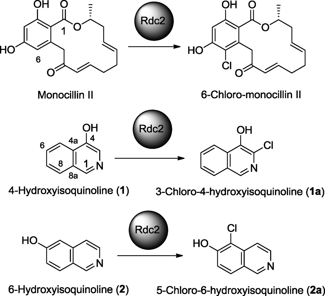
Halogenated molecules are an important group of natural products because of their significant biological activities. Up to now, more than 4,500 halogenated compounds have been isolated from nature,1, 2 such as rebeccamycin, chloramphenicol and vancomycin. Recently, biological halogenation has drawn more and more attention as a highly selective and efficient substitute for classical organic chemistry approaches.3, 4 Consequently, discovery of novel halogenases with broad substrate specificity from natural product biosynthetic pathways is critical. Our group has recently discovered a promising halogenase Rdc2 that is involved in radicicol biosynthesis in Pochonia chlamydosporia.5 This enzyme specifically chlorinates at C-6 of monocillins such as monocillin II, a biosynthetic precursor of radicicol, as shown in Fig. 1. We have found that this enzyme can also work on other similar macrolactones such as dihydroresorcylide. This enzyme can catalyze both mono- and dichlorination. Additionally, it also accepts bromide as a halogen donor to conduct both mono- and dibromination.5 Taken these unique promising properties together, this enzyme has shown great potential as a powerful halogenating biocatalyst. Thus, we decided to further study this enzyme to understand its structure and test its applicability to other types of substrates. In this work, we report a homology model of Rdc2 for substrate docking and chlorination of two isoquinolines by this enzyme.
Figure 1.
Chlorination of 1 and 2 by Rdc2.
Several structures of bacterial flavin-dependent halogenases have been reported, such as PrnA (AAB97504),6 RebH (CAC93722)7 and CmlS (AAK08979).8 Both PrnA and RebH are tryptophan 7-halogenase, involved in the biosynthesis of pyrrolnitrin and rebeccaymcin, respectively, while CmlS is characterized from the biosynthetic pathway of chloramphenicol.
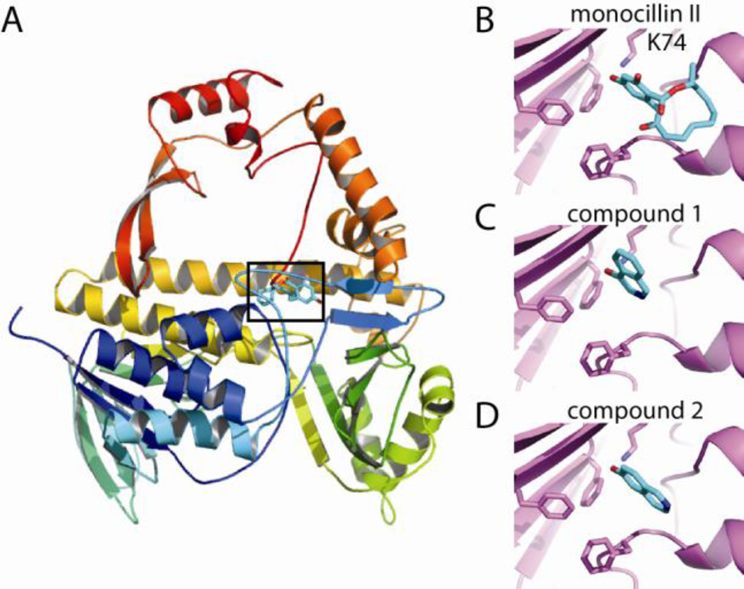
We constructed a homology model of Rdc2 using the LOMETS server.9 The highest scored solution was based on the crystal structure of CmlS (pdb ID 3i3l), using MUSTER10 to perform the threading alignment and MODELLER11 to build the final structure. The model contains 505 residues, which corresponds to 94.7% of the Rdc2 sequence. The final model contains an N-terminal flavin monooxygenase domain and a C-terminal “winged-helix” domain (Fig. 2A). Additional models produced by the server vary in the C-terminal domain but show strong agreement in the N-terminal domain.
Figure 2.
Homology model of Rdc2. (A) Overview of model colored from N (blue) to C terminus (red). The box indicates the putative active site. Modeling of monocillin II and two unnatural substrates 1 and 2 into the active site region are indicated in B–D, respectively.
The putative substrate binding site is located in a hydrophobic region of the N-terminal domain adjacent to the strictly conserved K74. The Rdc2 substrate monocillin II was modeled into the structure using a superimposed PrnA structure bound to 7-chlorotryptophan (PDB ID 2AR8) as a guide (Fig. 2B). The model suggests an open binding site that may accommodate a variety of compounds, raising the possibility that Rdc2 may be able to utilize various substrates.
To further expand the substrate spectrum and the application of Rdc2, we tested the ability of Rdc2 to chlorinate isoquinoline substrates. Isoquinoline is a common structural component of many bioactive natural products, such as renieramycin M from Xestospongia sp. and jorunnamycin C from Jorunnafunebris, both having promising anticancer activity with IC50 values in the nanomolar range.12 In this paper, we chose two typical isoquinolines, 4-hydroxyisoquinoline (1) and 6-hydroxyisoquinoline (2), to test whether Rdc2 could chlorinate this important type of molecule. As shown in Fig. 2C and 2D, both compounds can fit into the active site of Rdc2 and thus represent potential substrates for this enzyme.
We then conducted in vitro enzymatic assays to examine whether they can really be chlorinated by Rdc2. Rdc2 and the partner flavin reductase Fre from Escherichia coli were expressed and purified as previously reported.5 As shown in Fig. 3, LC-MS analysis revealed that both reactions13 yielded a product peak with lower polarity. ESI-MS revealed that both 1a and 2a had two [M+H]+ ion peaks with a 3:1 ratio in the positive mode (data not shown), which is a characteristic of monochlorinated molecules. 1 and 2 are isomers with a molecular weight of 145, while the molecular weight of both 1a and 2a is 179, which is 34 Da greater than the substrates, indicating that they are the chlorinated products.
Figure 3.
HPLC analysis of the in vitro chlorination of 1 (left) and 2 (right) by Rdc2.
In order to obtain sufficient amounts of 1a and 2a for structure elucidation to characterize the chlorination positions, we used an in vivo biocatalytic approach. A total of 30 mg of 1 and 2 were separately fed into the isopropyl β-D-1-thiogalactopyranoside (IPTG) induced fermentation broth of E. coli BL21-CodonPlus (DE3)-RIL/pJZ54 to yield 8.5 mg of 1a and 11.4 mg of 2a, respectively.14
The purified products were dissolved in methanol-d4 and the NMR spectra were acquired on a JEOL instrument (300 MHz). The 1H NMR spectrum of 1a showed two doublet and two triplet signals that belong to H-5, H-6, H-7 and H-8, indicating that the halogenation has not occurred on the benzene ring. In contrast, only one singlet was observed for the pyridine ring, suggesting that either C-1 or C-3 is halogenated. The chemical shift of this signal is δ 8.64, which belongs to H-1. Thus, we can deduce that the halogenation occurred at C-3 position, which is ortho to the hydroxyl group. Thus, 1a was identified as 3-chloro-4-hydroxyisoquinoline.
To determine the chlorination position in 2a, both 1H and 1H-1H COSY spectra were recorded. As expected, there were only five proton signals in the 1H NMR spectrum, confirming that one proton has been substituted by Cl. The 1H-1H COSY correlations of H-3 to H-4 as well as H-7 to H-8 clearly revealed that these protons are not chlorinated. Meanwhile, the signal of H-1 can be easily located in the low field at δ 9.53, indicating that C-5 of 2 has been chlorinated, which is also ortho to the hydroxyl group. Therefore, 2a was characterized as 5-chloro-6-hydroxyisoquinoline. The 1H NMR signals of both products are shown in Table 1.
Table 1.
1H NMR data for 1a and 2a (CD3OD, 300 MHz, J in Hz)
| Position | 1a | 2a |
|---|---|---|
| 1 | 8.64 (1H, s) | 9.53 (1H, s) |
| 3 | 8.51 (1H, d, J = 6.9) | |
| 4 | 8.47 (1H, d, J = 6.9) | |
| 5 | 8.26 (1H, d, J = 8.3) | |
| 6 | 7.79 (1H, dd, J = 8.3, 6.9) | |
| 7 | 7.68 (1H, dd, J = 8.3, 6.9) | 7.68 (1H, d, J = 9.0) |
| 8 | 8.06 (1H, d, J = 8.3) | 8.34 (1H, d, J = 9.0) |
Almost all the previously characterized flavin-dependent halogenases are highly substrate specific, such as those reported tryptophan halogenases.15–18 In order to develop a useful halogenating biocatalyst, an enzyme with broad substrate specificity is needed. Rdc2 is a halogenase different from those previously reported. It is a late tailoring enzyme in radicicol biosynthesis and can halogenate other macrolactones besides its natural substrates monocillins.5 In this work, we report a homology model of the first identified fungal flavin-dependent halogenase Rdc2, which predicts that this enzyme can accommodate various substrates. We demonstrated that Rdc2 can function on another important group of molecule, isoquinolines. This is the first report of enzymatic synthesis of chlorinated isoquinolines. Structural characterization of 1a and 2a indicated that the chlorine atom has been introduced at the position ortho to the hydroxyl group. A hydroxyl group is a strongly activating substituent. The resonance effect of the hydroxyl group directs the electron toward the ring and may contribute to the chlorination of the ortho position. Further investigation of the Xray structure of Rdc2 will reveal more information about the catalytic mechanism of this enzyme, which will allow us to use the structure model to find more potential substrates. Moreover, this research also demonstrated a convenient and effective way to prepare halogenated molecules with Rdc2. In vitro enzymatic reaction allows a quick screening of the substrates and initial analysis of the products, while in vivo biocatalysis through engineered E. coli provides an economic way to obtain sufficient amounts of halogenated products on a large scale for structure elucidation and bioactivity studies.
Acknowledgments
This work was partly supported by a Utah Science Technology and Research (USTAR) Veterinary Diagnostics and Infectious Disease Seed Project and a National Institutes of Health grant (AI065357). We thank Dr. Chad Testa at Frontier Scientific, Inc. for providing the substrates.
Footnotes
Publisher's Disclaimer: This is a PDF file of an unedited manuscript that has been accepted for publication. As a service to our customers we are providing this early version of the manuscript. The manuscript will undergo copyediting, typesetting, and review of the resulting proof before it is published in its final citable form. Please note that during the production process errors may be discovered which could affect the content, and all legal disclaimers that apply to the journal pertain.
References and notes
- 1.Gribble GW. J. Chem. Educ. 2004;81:1441. [Google Scholar]
- 2.Vaillancourt FH, Yeh E, Vosburg DA, Garneau-Tsodikova S, Walsh CT. Chem. Rev. 2006;106:3364. doi: 10.1021/cr050313i. [DOI] [PubMed] [Google Scholar]
- 3.Wagner C, El Omari M, Konig GM. J. Nat. Prod. 2009;72:540. doi: 10.1021/np800651m. [DOI] [PubMed] [Google Scholar]
- 4.Zeng J, Valiente J, Zhan J. Nat. Prod. Commun. 2011;6:223. [PubMed] [Google Scholar]
- 5.Zeng J, Zhan J. ChemBioChem. 2011;11:2119. doi: 10.1002/cbic.201000439. [DOI] [PubMed] [Google Scholar]
- 6.Dong C, Flecks S, Unversucht S, Haupt C, van Pée K-H, Naismith JH. Science. 2005;309:2216. doi: 10.1126/science.1116510. [DOI] [PMC free article] [PubMed] [Google Scholar]
- 7.Bitto E, Huang Y, Bingman CA, Singh S, Thorson JS, Phillips GN., Jr Proteins: Struct. Funct. Bioinf. 2007;70:289. doi: 10.1002/prot.21627. [DOI] [PubMed] [Google Scholar]
- 8.Podzelinska K, Latimer R, Bhattacharya A, Vining LC, Zechel DL, Jia Z. J. Mol. Biol. 2010;397:316. doi: 10.1016/j.jmb.2010.01.020. [DOI] [PubMed] [Google Scholar]
- 9.Wu S, Zhang Y. Nucleic Acids Res. 2007;35:3375. doi: 10.1093/nar/gkm251. [DOI] [PMC free article] [PubMed] [Google Scholar]
- 10.Wu S, Zhang Y. Proteins: Struct. Funct. Bioinf. 2008;72:547. doi: 10.1002/prot.21945. [DOI] [PMC free article] [PubMed] [Google Scholar]
- 11.Sali A, Blundell TL. J. Mol. Biol. 1993;234:779. doi: 10.1006/jmbi.1993.1626. [DOI] [PubMed] [Google Scholar]
- 12.Charupant K, Suwanborirux K, Daikuhara N, Yokoya M, Ushijima-Sugano R, Kawai T, Owa T, Saito N. Mar. Drugs. 2009;7:483. doi: 10.3390/md7040483. [DOI] [PMC free article] [PubMed] [Google Scholar]
- 13.A 100-µL halogenation assay consisted of 100 µM FAD, 10 mM NADH, 0.1 mM 1 or 2, 10 mM NaCl, 16 µM Fre, and 16 µM Rdc2 in100 mM phosphate buffer (pH 7.0). The reaction mixtures were placed at 30°C for 5 h and then quenched by addition of 50 µL of methanol. The mixtures were then subjected to analysis on an Agilent 6130 LC-MS instrument using a Zorbax SB-C18 (5 µm, 4.6 × 150 mm). A gradient of acetonitrile/H2O system (10–90% over 25 min) containing 0.1% trifluoroacetic acid (TFA) was programmed for the analysis.
- 14.30 mg of 1 was fed into the IPTG induced fermentation broth of E. coli BL21-CodonPlus (DE3)-RIL/pJZ54 that expresses Rdc2. The culture was maintained at 28°C with shaking at 250 rpm for 36 h. The ethyl acetate extract of the broth was fractionated on a Sephadex LH-20 (20 g) column eluted with methanol to give 14 fractions, 5 mL each. Fractions 3~6 were combined and further separated by reverse-phase HPLC (Eclipse XDB-C18 column, 5 µm, 4.6 × 150 mm) with isocratic elution of 25% acetonitrile in H2O (each containing 0.1% TFA) for 20 min at a flow rate of 1 mL/min to yield 8.5 mg of 1a. Similarly, 30 mg of 2 was also incubated with E. coli BL21-CodonPlus (DE3)-RIL/pJZ54 under the same conditions. The ethyl acetate extract was fractionated on a Diaion HP-20 (30 g) column eluted with a stepwise gradient of isopropanol-water (0:100, 20:80, 40:60, 60:40, 80:20, 100:0, each 250 mL) to give 6 fractions. Further separation of fraction 3 by reverse-phase HPLC with isocratic elution of 10% acetonitrile in H2O (each containing 0.1% TFA) over 20 min at a flow rate of 1 mL/min afforded 11.4 mg of 2a in pure form.
- 15.Glenn WS, Nims E, O'Connor SE. J. Am. Chem. Soc. 2011;133:19346. doi: 10.1021/ja2089348. [DOI] [PubMed] [Google Scholar]
- 16.Jiang W, Heemstra JR, Jr, Forseth RR, Neumann CS, Manaviazar S, Schroeder FC, Hale KJ, Walsh CT. Biochemistry. 2011;50:6063. doi: 10.1021/bi200656k. [DOI] [PMC free article] [PubMed] [Google Scholar]
- 17.van Pée K-H. ChemBioChem. 2011;12:681. doi: 10.1002/cbic.201100016. [DOI] [PubMed] [Google Scholar]
- 18.Zeng J, Zhan J. Biotechnol. Lett. 2011;33:1607. doi: 10.1007/s10529-011-0595-7. [DOI] [PubMed] [Google Scholar]


