Abstract
Introduction:
Colorectal cancer is the third most common tumor which causes high percentage of mortality in the general population. Etiologic factors which cause this disease are various, while diagnostic methods involve very complex protocols from detection of tumor markers to a combination of endoscopic and imaging methods.
Goal:
To determine the number of patients suffering from colon cancer for a period of two years and with endoscopic methods to verify and localize the tumor and its spread. Histopathological determination of the tumor type. Determine the concentration of CEA and CA 19-9 in the serum. Depending on the tumor location asses its progression, severity and extent by radiological imaging methods.
Material and Methods:
The study was prospective and retrospective, performed at the Gastroenterohepatology Clinic of the Clinical Center of Sarajevo University. During the two-year follow-up, 91 patients were hospitalized underwent endoscopy, targeted biopsy and histologically proven adenocarcinoma of the colon in which a pathologist determined grade of the cancers. Samples were eosin stained and underwent pathological histological analyzes. All patients according to tumor localization underwent CT scan and MRI of the rectum and pelvis.
Results:
The most common location of the cancer regardless of sex was in the recto sigmoid colon. Prevalence of colorectal cancer spread to other organs was not related to location. No significant dependence of the localization of the tumor by gender was found (p-value = 0.313). Ca 19-9 had the highest value in localization of tumors in the rectum. There was no statistically significant difference in age between men and women. The largest number of patients has adenocarcinoma grade 2 and the localization at the rectum.
Conclusion:
The combination of laboratory parameters (CEA and CA 19-9) with endoscopic and radiological imaging methods is essential in diagnosis of colorectal cancer and assessment of the process progression. There is a need to impose additional diagnostic parameters to detect the disease at an earlier stage.
Key words: colon cancer, diagnostic protocols.
1. INTRODUCTION
Colon cancer is among the most common cancers and it is at the third place among most common in men, after lung cancer and also in women is on the third place, after breast cancer and gynecological cancers. It is very common in well-developed countries and least common in Asia, except Japan, South America and Africa. Studying the formation of colon cancer led to the conclusion that it is linked to diet and lifestyle. It has been observed that people who eat in a large amount of fatty food more frequently suffer from colon cancer. People, who have irregular constipation, have greater chances of developing colon cancer. In people whose diet is low in fruit and vegetables and with lower intake of vitamin C also are more frequently affected. Investigations of the effect of bile and bile salts have shown that they are carcinogenic. Lately there is an impression that colon cancer is increasing. It must be recognized that there are very advanced diagnostic capabilities, but our research shows that most of our patients can came late with advanced cancer. Patients with a positive family history are likely to get cancer due to mutations in genes suppressor that are not able to recognize the cell is different from the others and that should undergo programmed cell death–apoptosis. In the diagnostic assessment phase of the disease and its operability, we use laboratory tests, determination of serum levels of cancer antigens such as CEA and CA19-9, followed by endoscopy and imaging diagnostic tests that are necessary to use in making the conclusion of the stage of disease and the patient prognosis.
1.1. Endoscopy
Colonoscopy is a macroscopic, optical examination, which includes visualization, localization and targeted biopsy of changes in colonic wall mucosa. By this method we are able to visualize all macroscopic changes, if necessary, stain and performed targeted biopsy with bioptic samples sent for histopathological analysis. The pathologist will give us a histologic diagnosis of lesions according to which we will be reflected the further treatment of the patient. If it is a case of lesion that affects the lumen of the intestine and becomes impassable for the instrument, to have had the insight on the length of infiltrative processes used by other methods such as barium enema, MRI rectum and pelvic CT and CT colonography bowel wall and abdomen.
1.2. CT of the abdomen and bowel wall
This is an imaging method which by transverse tomography screening of layers produces a clear picture of organs or regions that are followed. Computed tomography (CT) is a computer reconstruction of one layer of the body plane. CT images are reconstructed from a large number of X-ray absorption measurements of the beams that passes through the patient. When measuring density of pathological process we measured the density of the middle of the process because the edges can give false absorption ratios. Pathological lesions in relation to the organ in which they are found, as described isodens, hypo-or hyperdense relative to the targeted organ. In staging is essential to determine the thickness of wall malignancy or tumor penetration depth and the environment as well as metastasis in adjacent organs.
1.3. MRI of the rectum and the pelvis
Diagnostic methods which is necessary in rectal cancer staging. With this method we can gain insight into the depth of penetration into the wall or surrounding adipose tissue, lymph nodes and perirectal fascia. It is necessary to assess the surgery.
Carcinoembryonic antigen (CEA)
CEA is an oncofetal tumor marker discovered 1965. In 60 to 70% of cases is specific to colon cancer. Normal range is up to 2.5ng/ml. In smoking population there are somewhat higher concentrations in serum. Elevated concentrations speak in favor of colon cancer. After the surgery it is normalized but in case of recurrences and metastases concentration increases.
1.4. Carbohydrate antigen (CA19-9)
CA19-9 carbohydrate presents in the serum as a high molecular mucin rich with carbohydrates. It possesses clinical significance in pancreatic cancer and cancer of the gallbladder. Antigen is used in the diagnosis of rectal cancer and other parts of the colon, after the determination of CEA in serum and ovarian cancer, after measurement of CA 125. CA 19-9 can sometimes be a false positive and false negative. Higher values are found in patients suffering from liver cirrhosis, chronic hepatitis and diabetes mellitus. 7-10% of the population cannot produce CA 19-9.
2. GOAL
During the period from 2 years to determine the number of patients with colon cancer, with endoscopic methods verify and localize the tumor and its spread.
Target lesion biopsy and histologic confirmation of the clinical diagnosis. Determine the concentration of CEA and CA 19-9 in the serum. Depending on localization changes, CT of the abdomen and bowel wall and MRI of the rectum and pelvis.
3. MATERIAL AND METHODS
The study was prospective and retrospective, performed at the Clinic of Gastroenterohepatology, Clinical Center of Sarajevo University.
During the two-year follow-up, 91 patients were hospitalized underwent endoscopy, targeted biopsy and histologically was proven adenocarcinoma of the colon in which a pathologist determined grade of the cancers. Samples were eosin stained and pathological-histological analyzed.
All patients according to tumor localization underwent CT scan of the abdomen and MRI of the rectum and pelvis. We want to be certain to diagnose metastases, then the depth of penetration into the bowel wall, the surrounding adipose tissue, adjacent lymph nodes and organs. MRI of the pelvis and the rectum enabled us to visualize the involvement of the perirectal fascia as well as the depth of penetration to other organs in the pelvis, towards bones and muscles.
To all patients we determined the concentration of CEA and CA 19-9 in the serum. They were evaluated at the Institute of Biochemistry by 1-877-4 Abbott immunoassay. Architect CA 19-9XR carbonylmetalloimmunoassay (CMIA) for the quantitative determination of 1116-NS 19-9 reactive determinants in human serum or plasma on the ARCHITECT system (1). Immunoassay also determined the concentration of CEA antigen in the serum (2).
The patients underwent targeted biopsy and bioptic samples were sent to the Institute of Pathology, where they were further cut, stained and analyzed with histologic tumor grade determination.
Patients are put in correlation by sex, age and location of the tumor.
4. RESULTS
In Tables 1 and 2 MRI and CT scans show the distribution of tumor spreading (whether metastasis are present or not) according to localization. CT was done for 64 patients and for 27 did not. Although in the two fields is located less than 5 elements, we performed chi-square test for dependence of columns and rows in a contingency table. The analysis does not include the colon transverse and testing the significance of differences was done for 63 patients for whom CT was done. The result of chi-square test is 0, 987078, from which it follows that with 95% probability can be argued that the prevalence of cancer is not dependent on localization.
Table 1.
CT findings according to tumor location
 |
Table 2.
MRI in case of process spreading
 |
MRI was performed in 39 patients, 11 females and 24 males.
Table 3 shows the distribution of patients by sex and tumor location.
Table 3.
Localization of tumors by sex
 |
Table 4 shows that 18% of all female patients had tumor localization in ascendens or transversum, while other tumors were localized in the descending parts, in male patients, the relationship is 9% to 91% while the total ratio is 12% to 88%.
Table 4.
Localization of tumors by sex
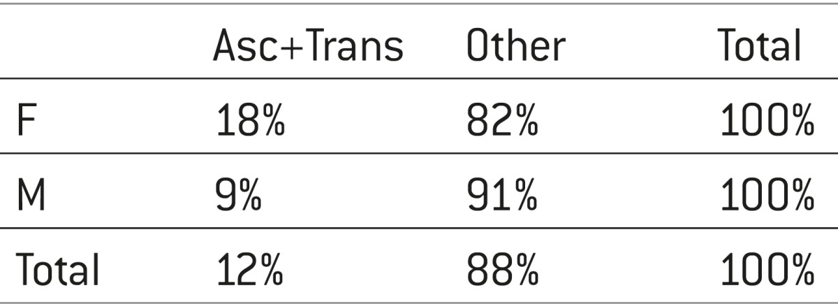 |
Performed is Chi-square test to see whether there is a dependence of the localization on gender, with the localization grouped as ascendens + transversum versus other; test result is with p-value of 0.313 so we cannot accept the hypothesis that there are characteristics of a dependency, although at first glance seemed to be association of localization and sex (see Table 5).
Table 5.
The average age of the patients by sex and tumor location
 |
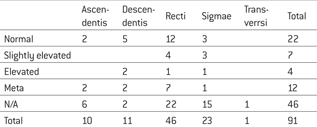
Distribution of patients according to the level of CA19-9 markers and tumor location is presented in Table 6. In the same table are presented laboratory analysis of the CA19-9 which was not performed in 46 patients.
Table 6.
Distribution of patients according to the level of CA19-9 markers and tumor location
 |
The average age of the patients by gender and tumor grade is presented in Table 7.
Table 7.
Tumor grade by sex
 |
From this and Table 8 is evident that the average age of the patients is very similar, the female patients were slightly older than men and this difference was not significant. Patients according to localization and grade are presented in Table 9.
Table 8.
Mean age by gender and tumor grade
 |
Table 9.
Patients according to localization and tumor grade
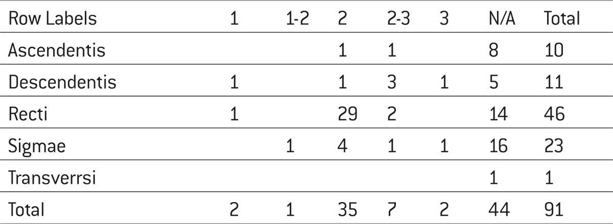 |
Grade was calculated for 47 patients. The table shows that most patients had grade 2 and the localization at the rectum.
5. DISCUSSION
Carcinoembryonic antigen (CEA) and Carbohydrate antigen (Ca 19-9) are well known as the most common tumor markers of colorectal cancer, while their levels are not used only in the preoperative assessment of tumor spread, but also for the monitoring of postoperative relapse. Combined data on the increase in value of preoperative CEA and CA 19-9 levels may be helpful in predicting the prognosis of patients with colorectal cancer (3).
In our sample, we analyzed data on colon adenocarcinoma in 91 patients, of whom 33 women and 58 men for a period of 2 years (2009-2011).
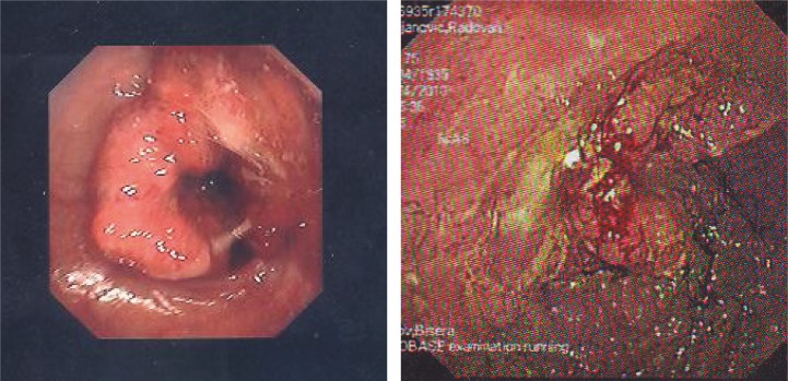
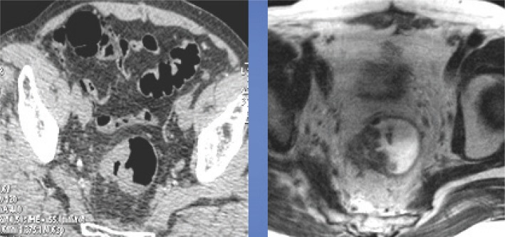
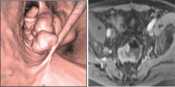
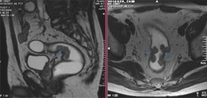
All patients underwent a diagnostic protocol that included: colonoscopy (Figure 1 and 2), targeted biopsy of infiltrative changes, the value of tumor markers (CEA and CA 19-9) and in order to evaluate spread of the process was performed MRI of the rectum in case of rectal tumor location or in the case of abdominal localization CT (Figure 3 and 4).
Figure 1.
Endoscopic view on rectal adenocarcinoma.
Figure 2.
Colonography CT and MRI image of the rectal adenocarcinoma- T3 infiltration of mesorectal fascia.
Figure 3.
Colonography 3d CT and MRI views of the rectal adenocarcinoma at T3 stage with infiltration of mesorectal fascia Colonography 3d CT and MRI views of the rectal adenocarcinoma at T3 stage with infiltration of mesorectal fascia.
Figure 4.
MRI of the rectum, sagittal and axial-adenocarcinoma, Stage T2, N0.
According to the results of previous research was found that 10% of colorectal cancers infiltrate also surrounding organs, the incidence of direct invasion of the pancreatic head and duodenum is less than 1% (4-7).
The results of our study also did not find any significant spread of colon cancer that is associated with tumors location. Therefore, the results of our research are consistent with the results of other published studies.
Analyzing tumor localization in relation to gender, we determined that the most common tumor location, regardless of sex was the recto sigmoid part of the colon. These results are fully in accordance with the results of other studies that have determined same localization of colon cancer. Also, according to the results of our study it did not show a statistically significant dependence of localization of tumors on gender (p-value=0.313).
Determining the value of tumor marker CA 19-9, we concluded that CA 19-9 had the highest value in localization of tumors in the rectum. In the available literature we did not find results with which we could compare our values, while transient elevation of Ca 19-9 in the study of Li YH et al. (2009), was not shown statistically significant in the diagnosis of colorectal cancer (8). Far more important is to follow the dynamics of this tumor marker as disease progression in patients with metastatic colorectal cancer.
Also, there was no statistically significant difference in age between men and women at the time of diagnosis, while the number of male patients was significantly higher the number of women.
On the basis of histopathological analysis it was found that the tumors were usually at grade 2 and at the rectum, for which we also could not find comparable results in the literature. The most common tumor location in our research was the rectum, for which we did not find comparable results in the literature.
Previously published studies have analyzed the causal relationship between the values of tumor markers, tumor location and stage of the tumor but also did not found a significant association (9).
On the basis of histopathological analysis it was found that the tumors were mostly at stage II which is partly in accordance with the results of the study by Omranipour et al (2012) who found that the most common stage of colon cancer was stage II and stage III (10).
6. CONCLUSION
The most common location of cancer regardless of sex was recto sigmoid part of the colon. Spreading of colorectal cancer to other organs does not depend on gender. No significant dependence of the localization of the tumor by gender (p-value=0.313). Ca 19-9 had the highest value in localization of tumors in the rectum. There was no statistically significant difference in age between men and women. The largest number of patients has adenocarcinoma grade 2 and the localization at the rectum.
Conflict of interest
None declared.
REFERENCES
- 1.Ochiai H, Ohishi T, Osumu K, Tokuyama J, Urakami H, Seki S, Shimada A, Matsui A, Isobe Y, Murata Y, Endo T, Ishii Y, Hasegawa H, Matsumoto S, Kitagawa Y. Reevaluation of serum p53 antibody as a marker in colorectal cancer patients. Surg Tuday. 2012;42(2):164–168. doi: 10.1007/s00595-011-0044-1. [DOI] [PubMed] [Google Scholar]
- 2.Tagi T, Matsui T, Kikuchi S, Hoshi S, Ochiai T, Kokuba Y, Kinoshita-Ida Y, Kisumi-Hayashi E, Morimoto K, Imai T, Imoto I, Inazawa J, Otsuji E. Dermokine as a novel biomarker for earlystage coloreectal cancer. J Gastroenterol. 2010;45(12):1201–1211. doi: 10.1007/s00535-010-0279-4. [DOI] [PubMed] [Google Scholar]
- 3.Nozoe T, Rikimaru T, Mori E, Okuyama T, Takahashi T. Increase in both CEA and Ca 19-9 in sera as an independent prognostic indicator in colorectal carcinoma. J Surg Oncol. 2006;94(2):132–137. doi: 10.1002/jso.20577. [DOI] [PubMed] [Google Scholar]
- 4.Berrospi F, Celis J, Ruiz E, Payet E. En block pancreaticoduodenectomy for right colon cancer inading adjacent organs. J Surg Oncol. 2002;79:194–197. doi: 10.1002/jso.10072. [DOI] [PubMed] [Google Scholar]
- 5.Saiura A, Yamamotoo J, Ueno M, Koga R, Seki M, Kokudo N. Long –term survival in patients with localy advanced colon cancer aft er en block pancreaticoduodenectomy and colectomy. Dis. Colon rectum. 2008;51:1545–1551. doi: 10.1007/s10350-008-9318-0. [DOI] [PubMed] [Google Scholar]
- 6.Eldar S, Kemeny MM, Terz JJ. Extended resections for carcinoma of the colon and rectum. Surg Gynecol Obstet. 1985;161:319–322. [PubMed] [Google Scholar]
- 7.Perez RO, Coser RB, Kiss DR, Iwashita RA, Jukemura J, Cunha JE, Habr-Gama A. Combined resection of the duodenum and the pancreat for localy advanced colon cancer. Curr Surg. 2005;62:613–617. doi: 10.1016/j.cursur.2005.03.021. [DOI] [PubMed] [Google Scholar]
- 8.Li YH, An X, Xiang XJ, Wang Zq, et al. Clinical significance of a transient increase in carcinoembrionic antigen and carbohydrate antigen 19-9 in patient with metastatic colorectal cancer receiving chemotherapy. Ai zeng. 2009;28(9):939–944. doi: 10.5732/cjc.009.10001. [DOI] [PubMed] [Google Scholar]
- 9.Davidson BR, Sams VR, Styles J, Dean C, Boulos PB. Comparative study of carcinoembryonic antigen and epithelial membrane antigen expression in normal colon, adenomas and adenocarcinomas of the colon and rectum. Gut. 1989 Sep;30(9):1260–1265. doi: 10.1136/gut.30.9.1260. [DOI] [PMC free article] [PubMed] [Google Scholar]
- 10.Omranipour R, Doroudian R, Mahmoodzadeh H. Anatomical distribution of colorectal carcinoma in Iran: a retrospective 15-yr study to evaluate rightward shift. Asian Pac J Cancer Prev. 2012;13(1):279–282. doi: 10.7314/apjcp.2012.13.1.279. [DOI] [PubMed] [Google Scholar]


