Abstract
The serine protease HtrA is involved in the folding and maturation of secreted proteins, as well as in the degradation of proteins that misfold during secretion. Depletion of HtrA has been shown to affect the sensitivity of many organisms to thermal and environmental stresses, as well as being essential for virulence in many pathogens. In the present study, we compared the behaviors of several different HtrA mutants of the gram-positive pathogen Streptococcus pyogenes (group A streptococcus). Consistent with prior reports, insertional inactivation of htrA, the gene that encodes HtrA, resulted in a mutant that grew poorly at 37°C. However, an identical phenotype was observed when a similar polar insertion was placed immediately downstream of htrA in the streptococcal chromosome, suggesting that the growth defect of the insertion mutant was not a direct result of insertional inactivation of htrA. This conclusion was supported by the observation that a nonpolar deletion mutation of htrA did not produce the growth defect. However, this mutation did affect the production of several secreted virulence factors whose biogenesis requires extensive processing. For the SpeB cysteine protease, the loss of HtrA was associated with a failure to proteolytically process the zymogen to an active protease. For the streptolysin S hemolysin, a dramatic increase in hemolytic activity resulted from the depletion of HtrA. Interestingly, HtrA-deficient mutants were not attenuated in a murine model of subcutaneous infection. These data add to the growing body of information that implies an important role for HtrA in the biogenesis of secreted proteins in gram-positive bacteria.
Proteases in pathogenic bacteria contribute many functions essential to virulence, including the acquisition of nutrients, biogenesis of virulence factors, cleavage of key host proteins for modulation of the host response, and protein quality control (8, 20, 21, 40, 55). The last function becomes particularly important for the survival of the bacterium in stressful environments encountered during infection. In gram-negative bacteria, proteins of the HtrA (DegP) family function as periplasmic serine proteases involved in the degradation of exported proteins that are misfolded or aggregated (for reviews, see references 7 and 46). A significant body of work has also illustrated the important role of HtrA in pathogenesis (10, 14, 29, 34, 48). While it has been suggested that the decreased virulence of HtrA mutants may be a consequence of the accumulation of damaged proteins in the periplasmic space (25), the hypersensitivity to thermal and osmotic stresses that is typical of these mutants may also play a role (51).
Orthologs of HtrA have been found in many gram-positive bacteria, and several have been implicated in virulence (30, 50). However, the specific contribution that HtrA makes to virulence is much less clear. These bacteria lack the outer membrane of the gram-negative bacteria, and as a consequence, lack a periplasmic space. Thus, the folding of secretory proteins does not take place in the confined compartment of the periplasmic space but rather likely occurs at the membrane-cell wall interface following translocation across the cytoplasmic membrane (54, 56). It is known that the peripheral membrane contains some accessory proteins to promote folding, including chaperones and disulfide oxidoreductases (54, 56). However, this peripheral membrane compartment is exposed to the environment and it is not clear that misfolded secreted proteins accumulate at this site, although it is likely that misfolded surface-anchored proteins would accumulate. The fact that the gram-positive orthologs of HtrA are predicted to be peripheral membrane proteins anchored to the membrane by a single transmembrane domain located near their N termini (44, 47) suggests that HtrA may function in protein quality control at this site. In addition, its dual role as a chaperone to promote folding of certain exported proteins in gram-negative bacteria (2, 52) may indicate a more central role in the biogenesis of secreted proteins in gram-positive bacteria. Consistent with this, HtrA has been implicated as the sole extracellular protease responsible for degradation of abnormal exported proteins, for processing secreted proproteins, and for maturation of native proteins in Lactococcus lactis (47).
Secreted proteins play critical roles in the pathogenesis of diseases caused by gram-positive bacteria. For example, culture supernatants of the group A streptococcus (Streptococcus pyogenes) contain at least 16 polypeptides with an identifiable export sequence (33). This bacterium is the causative agent of numerous suppurative infections of the pharynx (e.g., “strep throat”) and soft tissues (impetigo, cellulitis, and necrotizing fasciitis), as well as several systemic diseases that can result from toxigenic (scarlet fever and toxic shock syndrome) or immunopathological (rheumatic fever) processes (11). For the most part, the contribution of any secreted factor to the pathogenesis of any disease caused by S. pyogenes is poorly understood. However, it has been reported that depletion of HtrA was shown to diminish the virulence of S. pyogenes in a mouse model of systemic infection (30), suggesting a possible role for HtrA in the biogenesis of secreted virulence factors.
Two important secreted virulence factors of S. pyogenes are the cysteine protease SpeB and the hemolysin streptolysin S (SLS) (18, 24). Both of these factors require extensive processing for the generation of their biologically active forms via pathways that are not well understood (37, 42). The SpeB protease is secreted across the cytoplasmic membrane and folds into an enzymatically inactive zymogen (37), whose subsequent maturation to a proteolytic active form may require at least six intermediate structures generated by sequential cleavages within the zymogen's prodomain (13). While the protease is autocatalytic under certain conditions (37), efficient activation is an intermolecular event (13). The activation pathway is influenced both by environmental factors (37) and by other streptococcal gene products (9, 38, 39). The SLS hemolysin is a predominantly cell-associated small peptide of ∼30 amino acids that is posttranslationally processed and likely further modified by a poorly understood pathway that is encoded by a cluster of nine genes in the sag (for streptolysin-associated gene) locus (42). The sequence of the toxin precursor is highly enriched in amino acid residues that are the substrates for thio-ether bond modification found in other cyclic peptide toxins, suggesting that the biogenesis of SLS is similar to that of bacteriocins (42). Consistent with this, several of the genes in the sag locus have similarity to genes required for the synthesis of peptide bacteriocins (42). The roles of extracellular processing factors in the biogenesis of the SpeB protease or SLS have not been well defined.
The aim of the present study was to further investigate the role of HtrA in the pathogenesis of S. pyogenes disease. In particular, the contributions of HtrA to the biogenesis of the highly processed SpeB protease and SLS were examined. As previously reported (30), insertion of a polar element into htrA, the gene encoding HtrA, resulted in a mutant that grew very poorly under normal culture conditions. However, this phenotype was not observed for a nonpolar mutation of htrA, suggesting that the growth phenotype was the result of a polar effect on expression of an adjacent gene. Mutants of HtrA did display altered expression of proteolytic and hemolytic activities to imply a role for HtrA in the activation of the SpeB protease and the SLS hemolysin. Finally, examination of nonpolar HtrA mutants in a murine subcutaneous-infection model revealed that they displayed no significant defect in the ability to cause disease in the subcutaneous tissues.
MATERIALS AND METHODS
Strains, media, and culture conditions.
Molecular cloning experiments used Escherichia coli DH5α (Life Technologies), and experiments with S. pyogenes used strain HSC5 (22). Routine culture of S. pyogenes employed Todd-Hewitt medium (BBL) supplemented with 0.2% yeast extract (Difco) (THY medium) in sealed tubes without agitation. Analyses of protease and hemolytic phenotypes were conducted on cultures grown in C medium (38). To produce solid media, Bacto Agar (Difco) was added to THY medium to a final concentration of 1.4%, and unless otherwise indicated, all solid cultures were incubated under anaerobic conditions produced using a commercial gas generator (GasPack, catalog no. 70304; BBL). When appropriate, antibiotics were added to the medium at the following concentrations: kanamycin, 50 μg/ml for E. coli and 500 μg/ml for S. pyogenes; erythromycin 750 μg/ml for E. coli and 1 μg/ml for S. pyogenes.
DNA techniques.
Plasmid DNA was isolated by standard techniques and was used to transform E. coli by the method of Kushner (32) and to transform S. pyogenes by electroporation as previously described (5). Restriction endonucleases, ligases, kinases, and polymerases were used according to the manufacturers' recommendations. Chromosomal DNA was purified from S. pyogenes as previously described (5). Fluorescently labeled dideoxynucleotides (Big Dye terminators; PE Applied Biosystems) were used in DNA-sequencing reactions according to the recommendations of the manufacturer for the confirmation of the DNA sequences generated by PCR.
Growth rate comparison.
The growth rates of various strains described in this study were determined through the change in optical density at 600 nm (OD600) over time. Cultures were initiated from overnight growth in THY media that had been washed once with an equal volume of phosphate-buffered saline (PBS) (pH 7.4). The initial OD600 of the cultures was adjusted to 0.01, and absorbance values were determined at routine time intervals during incubation.
Insertional inactivation of htrA.
For the construction of a polar insertion, a region internal to htrA (open reading frame Spy2Z16 [15]) was amplified using the primers HtrAinternal1 (CACAA CGAAT TCTAC TAAAG CTGTC AAAGC) and HtrAinternal2 (CTGAA TAGCA TCTGC AGAGA CAGTC TCACC). Subsequent insertion of the fragment between the EcoRI and PstI sites of the integrational plasmid pCIV2 using the sites embedded in the primers (underlined) generated pHTR1. For construction of a polar insertion immediately downstream of htrA, a fragment containing the 3′ end of htrA and the adjacent chromosome was amplified using the primers HtrAinternal1 and HtrAafterstop (CATAA AACAG TCCTG CAGAC TGTTTT ATGT). This fragment was then inserted into pCIV2 as described above to generate pHTR2. Integration of pHTR1 and pHTR2 into the HSC5 chromosome via homologous recombination produced strains HTR1 and HTR2, respectively. The former contains a polar insertion in htrA, while the latter contains a polar insertion immediately downstream of an unaltered copy of htrA. The chromosomal structures of these mutants were verified through PCR and sequence analyses.
Construction of an in-frame deletion in htrA.
Primers htrA5720PstI (GGTAG GTCTG CAGAT AATTC TTTTG TC) and htrA7100BamHI (AAGGT ATAAG GATCC AAAGT TCTAT AAGC) were used to amplify a fragment containing the entire htrA open reading frame, which was inserted into pTOPO2.1 (Invitrogen) through a TA cloning procedure. The resulting plasmid (pHTR3) was used as a template in an “inside-out” PCR with the primers htrAIFDdown (GACCT GCTCT TGGAA TACAT ATGGT C) and htrAIFDup (GCTTT GACAG CTTTA GTCAT ATGGG TTGTG). Cleavage of the resulting product with NdeI (the sites are underlined), followed by subsequent religation, resulted in an in-frame deletion of the region of htrA that encodes A153-I299. The deletion allele (htrAΔ153-293) was then inserted between the PstI and BamHI sites of the streptococcal-E. coli shuttle vector pJRS233 using the sites embedded in the original primers (underlined). The resulting plasmid (pHTR10) was then used to replace the wild-type allele of htrA using the method of Ji et al. (28). This method produces a partial duplication with both wild-type and mutant alleles in the chromosome, and the duplication is then resolved to either the wild-type or mutant allele (28). Further analyses were conducted using one isolate that resolved to the mutant allele (HTR10) and a sibling that resolved to the wild-type allele (HTR11). Chromosomal structures were verified by PCR and sequence analysis using primers with the appropriate sequences.
Measurement of protease activity.
Expression of the SpeB protease was analyzed in culture supernatants as follows. Cultures in C medium were initiated using cells from overnight growth in C medium, which were washed in PBS (pH 7.4) to remove any residual protease. The initial OD600 of the cultures was adjusted to 0.01; samples were removed at various time points during incubation at 37°C, and cells were removed by filtration (0.45-μm-pore-size Sterile Acrodisc; Gelman Sciences). The resulting supernatant fluids were diluted in fresh C medium to normalize for any differences in growth between samples based upon the OD600 of the culture at the time of harvest. The presence of the proprotein and processed forms of SpeB was determined through Western blot analysis as described previously (38). The proteolytic activities of supernatants were quantified by the method of Hauser et al. (23), which measures the increase in relative fluorescence generated by the proteolytic cleavage of fluorescein isothiocyanate-casein (Sigma). The activity of uninoculated culture medium was used to derive background values that were typically undetectable under the conditions of this assay. To ensure that all proteolytic activity was specifically the result of SpeB, the cysteine protease-specific inhibitor E-64 (final concentration, 10 mM; Sigma) was routinely added to selected samples. This treatment typically reduced activity by >95%.
Measurement of SLS activity.
The production of SLS-specific hemolytic activity was determined as follows. An overnight culture of the strain under analysis was diluted to an OD600 of 0.01 in C medium and incubated at 37°C. At the times indicated in the text, aliquots were harvested from the culture, washed once in PBS (pH 7.4), and resuspended in PBS to an OD600 of 0.5. The cell-associated SLS activity was then determined by the method of Ofek et al. (45). Hemolytic activity was represented as the reciprocal of the minimum dilution that contained unlysed erythrocytes.
Murine subcutaneous-infection model.
The method of Bunce et al. (4) as modified by others (36, 49) was used to establish an infection of S. pyogenes in the subcutaneous tissues of mice as described in detail elsewhere (3). Mock-infected animals received a subcutaneous injection of saline at a volume equivalent to the volume of the dose of streptococci injected. Ulcer formation was documented by digital photography, and the precise area contained by each ulcer was calculated from the digital record using MetaMorph image analysis software (version 4.6; Universal Imaging Corp.). The difference between the numbers of mice developing an ulcer following subcutaneous challenge with wild-type or mutant bacteria was tested for significance by the chi-square test with Yates' correction (19), and differences in the areas of the resulting ulcers were tested by the Mann-Whitney U test (19). For all test statistics, the null hypothesis was rejected when P was <0.05. The data presented were derived from two independent experiments.
RESULTS
Construction of htrA mutants.
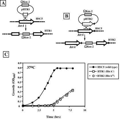
Previous reports have identified htrA (degP) in the genomes of S. pyogenes and Streptococcus mutans, and analyses of mutants constructed by insertional inactivation of htrA have identified key roles for the protein encoded by this gene in surviving thermal and oxidative stresses (12, 30). In addition, HtrA serves to process multiple extracellular proteins in lactococci (47). Thus, it was of interest to examine the role of htrA in the elaboration of secreted virulence factors and in the pathogenesis of S. pyogenes. To accomplish this, an insertion-duplication strategy similar to those used earlier was performed (12, 30), using homologous recombination to target the integration of a circular plasmid (pHTR1) onto which an internal region of htrA had been cloned. Similar to prior reports (12, 30), the HtrA-deficient mutant (HTR1) (Fig. 1A) exhibited normal kinetics of growth at 30°C (data not shown) but had a profound defect for growth at 37°C (Fig. 1C) and failed to demonstrate any observable growth at 40°C, the highest temperature at which the wild-type strain (HSC5) can grow. To control for possible polar effects on expression of any downstream genes, an additional mutant was constructed by insertion-duplication with a plasmid (pHTR2) that results in a functional copy of htrA followed immediately in tandem by a polar insertion (HTR2) (Fig. 1B). However, despite the presence of an intact copy of htrA, the polar control strain grew poorly at 37°C and demonstrated a thermal stress profile identical to that of the HtrA-deficient mutant (Fig. 1C).
FIG. 1.
Both HtrA− and HtrA+ polar mutants are thermally sensitive. (A) Integration of pHTR1 into the HCS5 chromosome by a single recombination event (indicated by an X) with selection for the ΩKm-2 antibiotic resistance determinant insertionally inactivated htrA and produced HTR1. (B) Integration of pHTR2 into the HSC5 chromosome by a single recombination event regenerated htrA and placed the polar ΩKm-2 element immediately downstream of htrA in strain HTR2. (C) Growth of the indicated strains during culture in C medium is presented as the increase in OD600 over time. The data shown are from a single experiment representative of three independent experiments.
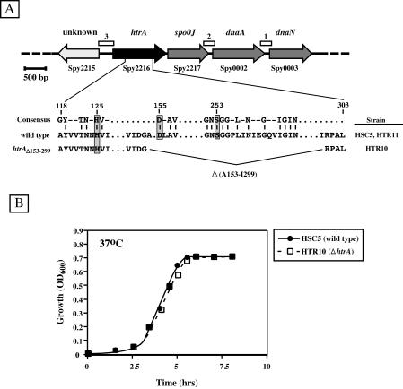
The regulation and organization of the htrA locus are very different in gram-positive bacteria than in gram-negative bacteria. Interestingly, in the streptococci, hrtA is located in the region containing the origin of replication of the chromosome and is immediately upstream of two genes involved in cell division (17) (Fig. 2A). Thus, to reduce the possibility of polar effects, an in-frame deletion mutant in htrA was constructed (HTR10 [Fig. 2A]). In contrast to the polar mutants, examination of the thermal-stress characteristics of the deletion mutant showed no differences from those of a wild-type strain (Fig. 2B). Taken together, these data indicate that polar insertion into or immediately downstream of htrA is detrimental to cell division.
FIG. 2.
An in-frame deletion mutant of htrA is not thermally sensitive. (A) (Top) Organization of open reading frames in the htrA region. Gene assignments (above) and gene numbers (below) are based on the genomic sequence information available for S. pyogenes SF370 (GenBank accession no. AE004092). The boxes labeled 1, 2, and 3 indicate the locations of sequences containing several copies of the consensus binding site for DnaA. (Bottom) Consensus htrA sequence in the active-site region (Consensus, derived from reference 46) and the specific htrA sequences of S. pyogenes HSC5 and HTR11 (wild type). The region removed by the in-frame deletion in HTR10 (htrAΔ152-299) is also shown. The conserved catalytic residues of htrA are boxed and shaded. (B) Growth of the indicated strains during culture in C medium. The data shown are from a single experiment representative of three independent experiments.
Contribution of HtrA to biogenesis of SpeB.
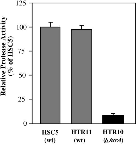
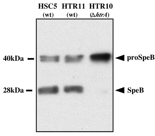
The SpeB cysteine protease is one of the most abundant proteins secreted by S. pyogenes as cultures enter stationary phase (6). The protease is secreted as a zymogen whose maturation to an active protease requires the contribution of multiple accessory gene products, with the result that the propeptide of the enzyme is removed (reviewed in reference 8). In L. lactis, HtrA has been shown to play a role in the processing of the propeptides of several enzymes that are also secreted as zymogens, including nuclease (NucA) and autolysin (AcmA) (47). Thus, it was of interest to evaluate the contribution of HtrA to the biogenesis of SpeB in S. pyogenes. In the examination of the htrA deletion mutant strain (HTR10) for SpeB activity, the mutant strain demonstrated a profound reduction in protease activity. At the time of maximal expression for a wild-type strain (early stationary phase), the mutant exhibited a 12-fold reduction in protease activity compared to the wild-type strain (HSC5) (Fig. 3). A deletion control strain that is a sibling of the deletion mutant but contains wild-type htrA (HSC11) (see Materials and Methods) demonstrated wild-type levels of protease activity (Fig. 3). Analysis of the SpeB polypeptide itself at this time revealed that culture supernatants from wild-type, deletion control, and mutant strains all contained approximately equivalent total amounts of the SpeB protease (Fig. 4). However, while both the wild-type and deletion control strains exhibited the characteristic mixture of zymogen (40 kDa) and processed active protease (28 kDa), the htrA deletion mutant exhibited only the zymogen form, indicating that it was unable to efficiently process the protease proprotein (Fig. 4).
FIG. 3.
Depletion of HtrA is associated with a decrease in SpeB proteolytic activity. The proteolytic activities of the indicated strains were determined using the substrate fluorescein isothiocyanate-casein following growth in C medium. Activity was measured at a time point 1 h after the strains reached the stationary phase of growth. Strain HTR11 is a sibling of the deletion mutant HTR10 but contains a wild-type (wt) allele of htrA. Activity is presented relative to the mean activity produced by wild-type strain HSC5. The data represent the mean and standard error of the mean of at least three independent experiments.
FIG. 4.
Mutation of htrA disrupts the maturation of SpeB. The indicated strains were cultured to early stationary phase in C medium. Cell-free supernatants were prepared and subjected to Western blot analysis using a SpeB-specific antiserum. The molecular mass of the zymogen (proSpeB) and processed (SpeB) forms of the SpeB cysteine protease are indicated. wt, wild type.
HtrA has a negative influence on SLS hemolytic activity.
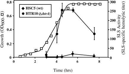
The biogenesis of SLS hemolytic activity is poorly understood, but it likely involves extensive processing of a precursor polypeptide in a manner similar to those of other cyclical bacteriocins (41). Since HtrA has been implicated in the processing of a lactococcal bacteriocin (16), it was of interest to determine the effect of mutation of htrA on the expression of SLS hemolytic activity. The production of SLS is tightly controlled, and the wild-type strain demonstrated the characteristic pattern of expression, with hemolytic activity beginning in late log phase and peaking during early stationary phase, followed by a decline (HSC5) (Fig. 5). Unexpectedly, the htrA deletion mutant demonstrated extremely high levels of SLS activity, with maximal activity >20-fold higher than that of the wild type (Fig. 5, compare HTR10 to HSC5). In addition, in the wild-type strain, peak SLS activity was observed in early stationary phase (Fig. 5) (39), but in the htrA mutant, SLS activity was readily detected toward the end of the logarithmic phase of growth. While the levels of SLS activity began to decrease earlier than in the wild-type strain, the levels remained eightfold higher than the maximal activity of the wild type at a point late in stationary phase. Expression of SLS by the deletion control strain was identical to that of the wild-type strain (data not shown).
FIG. 5.
SLS hemolytic activity is overexpressed in an htrA deletion strain. Expression of SLS-specific hemolytic activity for the various strains during growth in C medium is shown. Because all strains grew equivalently under these conditions, only the growth of wild-type (wt) strain HSC5 is shown. In addition, HTR11, the sibling of the deletion mutant HTR10 with a wild-type allele, produced SLS-specific hemolytic activity identically to the wild type, and for clarity, these data are not shown. The data represent the mean and standard error of the mean of at least three independent experiments.
An HtrA deletion mutant is virulent in a murine model of subcutaneous infection.
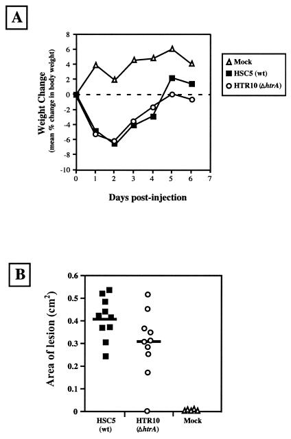
Insertional inactivation of htrA has been associated with reduced ability to grow at 37°C and with reduced virulence of S. pyogenes in a murine model of systemic infection (30). However, because the deletion mutant did not demonstrate a growth defect at 37°C but did exhibit aberrant biogenesis of several virulence factors, it was of interest to evaluate whether the deletion mutant would also show a defect in its ability to cause disease. The murine model of subcutaneous infection was used for this analysis because, similar to most infections caused by S. pyogenes (11), the organism must grow in a local tissue compartment to cause disease and also because the model assesses the ability of the streptococcus to survive and cause disease in the stressful environment produced by the intense host inflammatory response (3, 4, 36, 49). By 18 to 24 h after injection into the subcutaneous tissue of a hairless mouse at a dose of 107 CFU, the wild-type strain used in this study typically produces an ulcer whose margins expand to reach a maximum around day 3 (3). At ∼8 days postinfection, the lesion begins to resolve; it is typically fully healed by day 14, and the animals rarely develop systemic infection (3). Weight loss, time to formation of the ulcer, ulcer size, and time to heal the ulcer are the quantitative parameters typically used to evaluate the severity of disease. When the wild-type and deletion mutant were compared, there was no significant difference in (i) the pattern of weight loss (Fig. 6A), (ii) the time to ulcer formation (data not shown), (iii) ulcer size at the time of maximal ulceration caused by the wild-type strain (day 3 [Fig. 6B]) or at any other time point (data not shown), or (iv) the time to heal the ulcer (data not shown). Thus, deletion of htrA is not associated with a significant reduction in virulence in this model of infection.
FIG. 6.
Depletion of HtrA is not associated with attenuation of virulence in a murine model of subcutaneous infection. Mice were infected with the indicated strains or injected with saline alone (mock), and the course of disease was monitored by change in weight over several days (A) or by comparison of the area of resulting ulcer formation at 3 days postinfection (B). The data for change in weight represent the mean value obtained for infected groups of five mice each. The data for ulcer formation are pooled from two independent experiments, both of which involved infection of groups of five mice for each strain tested. The difference in ulcer formation between the wild type (wt; HSC5) and the htrA deletion mutant (HTR10) was not significant (P < 0.06).
DISCUSSION
In this study, we have shown that the serine protease HtrA of S. pyogenes influences the expression of at least two virulence factors whose biogenesis requires extensive processing. These data contribute to a growing body of evidence that suggests that HtrA plays a central role in protein secretion in gram-positive bacteria.
Interestingly, while insertional mutagenesis of htrA in S. pyogenes has been associated with a reduced capacity to cause disease in an animal model of systemic infection (30), the in-frame deletion mutation analyzed in the present study indicated that deletion of htrA did not result in attenuation in the murine model of subcutaneous infection. One possible explanation for this difference is the observation that the deletion mutant did not demonstrate the growth defect at 37°C that was exhibited in the mutants derived by insertional inactivation of htrA. In addition, the growth defect of the insertion mutant did not appear to be due to the loss of htrA itself but to a polar effect. Immediately downstream of htrA is a gene with similarity to spo0J, whose product is associated with chromosome partitioning in Bacillus subtilis (27), and dnaA, whose product is the essential initiator of chromosome replication. Similar to what has been reported for Streptococcus pneumoniae, the htrA locus contains several regions with clusters of binding sites for DnaA (17) (Fig. 2) and likely represents the origin of chromosme replication (17). Thus, it is not surprising that any large insertion into this region may alter the efficiency of cell division. It appears that smaller changes can be tolerated, since in S. pneumoniae, allele replacement of either htrA or spo0J has no noticeable effect on growth, although htrA mutants are less fit in competition with the wild type for nasopharyngeal colonization (50).
While a temperature-sensitive phenotype was not observed for the deletion mutant, it may be premature to conclude that htrA does not contribute to thermal tolerance. Most evidence suggests that htrA acts as a housekeeping protease to degrade unfolded polypeptides during heat shock (46). The wild-type strain used in this study is typical of many strains of S. pyogenes and does not grow at temperatures of >40°C. It is not known whether there is significant accumulation of unfolded polypeptides for the organism at this temperature. In B. subtilis, the contribution of htrA to thermal stress is most clearly observed upon exposure to temperatures that are lethal. In addition, B. subtilis contains two htrA-like genes, and single mutation of either gene leads to a dramatic increase in resistance to stress as a result of compensating upregulation of the other gene (43). While the S. pyogenes genome contains a single copy of htrA, it is possible that mutation results in compensating upregulation of other stress resistance factors. Regulation of htrA expression is not understood in S. pyogenes. In B. subtilis and S. pneumoniae, htrA is regulated by two-component regulatory systems (26, 50), but clear orthologs of these regulators are not apparent in the S. pyogenes genome (W. Lyon and M. Caparon, unpublished data).
Upregulation of a compensating gene may also account for the observation that the htrA deletion mutant was not attenuated. The SLS hemolysin has been identified as an important virulence factor that contributes to ulcer formation in the subcutaneous-infection model (for a review, see reference 41). It is possible that any increase in sensitivity to stress in the mutant is compensated for by the observed hyperproduction of SLS, with the result that the courses of infection in the wild type and the mutant appear to be the same. On the other hand, it is also interesting that a large increase in production of the highly cytolytic SLS does not result in any increased severity of disease. There are multiple steps in the complex biogenesis pathway of SLS that could be influenced by HtrA. The SLS operon is subject to several transcriptional regulatory pathways (for a review, see reference 41), and the activity of these regulatory pathways may be influenced by HtrA. Alternatively, HtrA may degrade components of the SLS biogenesis machinery or even the SLS propeptide itself. It is also possible that HtrA alters the receptor that tethers SLS to the cell surface, making it less accessible to the carrier molecules that are required to solubilize SLS for the determination of hemolytic titers. Regardless of the mechanism, these data suggest that SLS production and stress responses may be linked via HtrA.
In contrast to that of SLS, the role of SpeB protease in the subcutaneous-infection model is much less clear (1), although it is a major virulence factor in a humanized SCID mouse model of streptococcal impetigo (53). Major questions about the biogenesis pathway for the protease are how the nascent protease folds into its zymogen form following its secretion from the cell and how the zymogen's complex activation pathway is initiated (8). The contribution of HtrA may be to make an initial cleavage in the zymogen to begin the activation cascade. However, structural studies of HtrA from E. coli have shown that the protease exists as a hexamer derived from two interlocking trimers. The protease domains are sequestered in a central cavity that is only accessible laterally and likely can act only on polypeptides that are highly unfolded (31). Unlike E. coli, the streptococcal HtrA has only one instead of two adjacent PDZ domains and may have a structure more similar to that of mitochondrial HtrA2. This protease also has a single PDZ domain and forms a trimeric structure in which the protease domains are kept inactive by the PDZ domains, which then are displaced upon binding to the target polypeptide (35). This allows HtrA2 to act on more highly folded substrates, including an ability to activate apoptotic proteases (35). It is also possible that the role of HtrA is to act primarily as a chaperone to promote the folding of the zymogen into an activation-competent conformation, and this may or may not involve the protease activity of HtrA. Further analysis of the contribution of HtrA to the biogenesis of SLS and SpeB will be useful for understanding the production of virulence factors by S. pyogenes and pathways of protein secretion in gram-positive bacteria.
Acknowledgments
This work was supported by Public Health Service grant AI46433 from the National Institutes of Health.
Editor: J. N. Weiser
REFERENCES
- 1.Ashbaugh, C. D., and M. R. Wessels. 2001. Absence of a cysteine protease effect on bacterial virulence in two murine models of human invasive group A streptococcal infection. Infect. Immun. 69:6683-6688. [DOI] [PMC free article] [PubMed] [Google Scholar]
- 2.Bass, S., Q. Gu, and A. Christen. 1996. Multicopy suppressors of prc mutant Escherichia coli include two HtrA (DegP) protease homologs (HhoAB), DksA, and a truncated R1pA. J. Bacteriol. 178:1154-1161. [DOI] [PMC free article] [PubMed] [Google Scholar]
- 3.Brenot, A., K. Y. King, B. Janowiak, O. Griffith, and M. Caparon. 2004. Contribution of glutathione peroxidase to the virulence of Streptococcus pyogenes. Infect. Immun. 72:408-413. [DOI] [PMC free article] [PubMed] [Google Scholar]
- 4.Bunce, C., L. Wheeler, G. Reed, J. Musser, and N. Barg. 1992. Murine model of cutaneous infection with gram-positive cocci. Infect. Immun. 60:2636-2640. [DOI] [PMC free article] [PubMed] [Google Scholar]
- 5.Caparon, M. G., D. S. Stephens, A. Olsén, and J. R. Scott. 1991. Role of M protein in adherence of group A streptococci. Infect. Immun. 59:1811-1817. [DOI] [PMC free article] [PubMed] [Google Scholar]
- 6.Chaussee, M. S., E. R. Phillips, and J. J. Ferretti. 1997. Temporal production of streptococcal erythrogenic toxin B (streptococcal cysteine proteinase) in response to nutrient depletion. Infect. Immun. 65:1956-1959. [DOI] [PMC free article] [PubMed] [Google Scholar]
- 7.Clausen, T., C. Southan, and M. Ehrmann. 2002. The HtrA family of proteases: implications for protein composition and cell fate. Mol. Cell. 10:443-455. [DOI] [PubMed] [Google Scholar]
- 8.Collin, M., and A. Olsén. 2003. Extracellular enzymes with immunomodulating activities: variations on a theme in Streptococcus pyogenes. Infect. Immun. 71:2983-2992. [DOI] [PMC free article] [PubMed] [Google Scholar]
- 9.Collin, M., and A. Olsén. 2000. Generation of a mature streptococcal cysteine proteinase is dependent on cell wall-anchored M1 protein. Mol. Microbiol. 36:1306-1318. [DOI] [PubMed] [Google Scholar]
- 10.Cortes, G., B. de Astorza, V. J. Benedi, and S. Alberti. 2002. Role of the htrA gene in Klebsiella pneumoniae virulence. Infect. Immun. 70:4772-4776. [DOI] [PMC free article] [PubMed] [Google Scholar]
- 11.Cunningham, M. W. 2000. Pathogenesis of group A streptococcal infections. Clin. Microbiol. Rev. 13:470-511. [DOI] [PMC free article] [PubMed] [Google Scholar]
- 12.Diaz-Torres, M. L., and R. R. Russell. 2001. HtrA protease and processing of extracellular proteins of Streptococcus mutans. FEMS Microbiol. Lett. 204:23-28. [DOI] [PubMed] [Google Scholar]
- 13.Doran, J. D., M. Nomizu, S. Takebe, R. Menard, D. Griffith, and E. Ziomek. 1999. Auotcatalytic processing of the streptococcal cysteine protease zymogen: processing mechanism and characterization of the autoproteolytic cleavage sites. Eur. J. Biochem. 263:145-151. [DOI] [PubMed] [Google Scholar]
- 14.Elzer, P. H., R. W. Phillips, G. T. Robertson, and R. M. Roop II. 1996. The HtrA stress response protease contributes to resistance of Brucella abortus to killing by murine phagocytes. Infect. Immun. 64:4838-4841. [DOI] [PMC free article] [PubMed] [Google Scholar]
- 15.Ferretti, J. J., W. M. McShan, D. Adjic, D. Savic, G. Savic, K. Lyon, C. Primeaux, S. S. Sezate, A. N. Surorov, S. Kenton, H. Lai, S. Lin, Y. Qian, H. G. Jia, F. Z. Najar, Q. Ren, H. Zhu, L. Song, J. White, X. Yuan, S. W. Clifton, B. A. Roe, and R. E. McLaughlin. 2001. Complete genome sequence of an M1 strain of Streptococcus pyogenes. Proc. Natl. Acad. Sci. USA 98:4658-4663. [DOI] [PMC free article] [PubMed] [Google Scholar]
- 16.Gajic, O., G. B. Buist, M. Kojic, L. Topisirovic, O. P. Kuipers, and J. Kok. 2003. Novel mechanism of bacteriocin secretion and immunity carried out by lactococcal MDR proteins. J. Biol. Chem. 278:34291-34298. [DOI] [PubMed] [Google Scholar]
- 17.Gasc, A.-M., P. Biammarinaro, S. Richter, and M. Sicard. 1998. Organization around the dnaA gene of Streptococcus pneumoniae. Microbiology 144:433-439. [DOI] [PubMed] [Google Scholar]
- 18.Gerlach, D., H. Knoll, W. Kohler, J. H. Ozegowski, and V. Hribalova. 1983. Isolation and characterization of erythrogenic toxins. V. Communication: identity of erythrogenic toxin type B and streptococcal proteinase precursor. Zentbl. Bakteriol. Mikrobiol. Hyg. A 255:221-233. [PubMed] [Google Scholar]
- 19.Glantz, S. 2002. Primer of biostatistics, 5th ed. McGraw-Hill, New York, N.Y.
- 20.Gottesman, S. 1996. Proteases and their targets in Escherichia coli. Annu. Rev. Genet. 30:465-506. [DOI] [PubMed] [Google Scholar]
- 21.Gottesman, S., S. Wickner, and M. R. Maurizi. 1997. Protein quality control: triage by chaperones and proteases. Genes Dev. 11:815-823. [DOI] [PubMed] [Google Scholar]
- 22.Hanski, E., P. A. Horwitz, and M. G. Caparon. 1992. Expression of protein F, the fibronectin-binding protein of Streptococcus pyogenes JRS4, in heterologous streptococcal and enterococcal strains promotes their adherence to respiratory epithelial cells. Infect. Immun. 60:5119-5125. [DOI] [PMC free article] [PubMed] [Google Scholar]
- 23.Hauser, A. R., and P. M. Schlievert. 1990. Nucleotide sequence of the streptococcal pyrogenic exotoxin type B gene and relationship between the toxin and the streptococcal proteinase precursor. J. Bacteriol. 172:4536-4542. [DOI] [PMC free article] [PubMed] [Google Scholar]
- 24.Herbert, D., and E. W. Todd. 1944. The oxygen-stable haemolysin of group A haemolytic streptococci (streptolysin S). Br. J. Exp. Pathol. 25:242-254. [Google Scholar]
- 25.Huang, D. L., T. L. Raivio, C. H. Jones, T. J. Silhavy, and S. J. Hultgren. 2001. Cpx signaling pathway monitors biogenesis and affects assembly and expression of P pili. EMBO J. 20:1508-1518. [DOI] [PMC free article] [PubMed] [Google Scholar]
- 26.Hyyryläinen, H.-L., A. Bolhuis, E. Darmon, L. Muukkonen, P. Koski, M. Vitikainen, M. Sarvas, Z. Prágai, S. Bron, J. M. van Dijl, and V. P. Kontinen. 2001. A novel two-component regulatory system in Bacillus subtilis for the survival of severe secretion stress. Mol. Microbiol. 41:1159-1172. [DOI] [PubMed] [Google Scholar]
- 27.Ireton, K., H. W. T. Gunther, and A. D. Grossman. 1994. spo0J is required for normal chromosome segregation as well as the initiation of sporulation in Bacillus subtilis. J. Bacteriol. 176:5320-5329. [DOI] [PMC free article] [PubMed] [Google Scholar]
- 28.Ji, Y., L. McLandsborough, A. Kondagunta, and P. P. Cleary. 1996. C5a peptidase alters clearance and trafficking of group A streptococci by infected mice. Infect. Immun. 64:503-510. [DOI] [PMC free article] [PubMed] [Google Scholar]
- 29.Johnson, K., I. Charles, G. Dougan, D. Pickard, P. O'Gaora, G. Costa, T. Ali, I. Miller, and C. Hormaeche. 1991. The role of a stress-response protein in Salmonella typhimurium virulence. Mol. Microbiol. 5:401-407. [DOI] [PubMed] [Google Scholar]
- 30.Jones, C. H., T. C. Bolken, K. F. Jones, G. O. Zeller, and D. E. Hruby. 2001. Conserved DegP protease in gram-positive bacteria is essential for thermal and oxidative tolerance and full virulence in Streptococcus pyogenes. Infect. Immun. 69:5538-5545. [DOI] [PMC free article] [PubMed] [Google Scholar]
- 31.Krojer, T., M. Garrido-Franco, R. Huber, M. Ehrmann, and T. Clausen. 2002. Crystal structure of DegP (HtrA) reveals a new protease-chaperone machine. Nature 416:455-459. [DOI] [PubMed] [Google Scholar]
- 32.Kushner, S. R. 1978. An improved method for transformation of Escherichia coli with ColE1-derived plasmids, p. 17-23. In H. W. Boyer, and S. Micosia (ed.), Genetic engineering. Elsevier-North Holland Biomedical Press, New York, N.Y.
- 33.Lei, B., S. Mackie, S. Lukomski, and J. M. Musser. 2000. Identification and immunogenicity of group A Streptococcus culture supernatant proteins. Infect. Immun. 68:6807-6818. [DOI] [PMC free article] [PubMed] [Google Scholar]
- 34.Li, S. R., N. Dorrell, P. H. Everest, G. Dougan, and B. W. Wren. 1996. Construction and characterization of a Yersinia enterocolitica O:8 high-temperature requirement (htrA) isogenic mutant. Infect. Immun. 64:2088-2094. [DOI] [PMC free article] [PubMed] [Google Scholar]
- 35.Li, W., S. M. Srinivasula, J. Chai, P. Li, J.-W. Wu, Z. Zhang, E. S. Alnemri, and Y. Shi. 2002. Structural insights into the pro-apoptotic function of mitochondrial serine protease HtrA2/Omi. Nat. Struct. Biol. 9:436-441. [DOI] [PubMed] [Google Scholar]
- 36.Limbago, B., V. Penumalli, B. Weinrick, and J. R. Scott. 2000. Role of streptolysin O in a mouse model of invasive group A streptococcal disease. Infect. Immun. 68:6384-6390. [DOI] [PMC free article] [PubMed] [Google Scholar]
- 37.Liu, T.-Y., and S. D. Elliot. 1965. Streptococcal protease: the zymogen to enzyme transformation. J. Biol. Chem. 240:1138-1142. [PubMed] [Google Scholar]
- 38.Lyon, W. R., C. M. Gibson, and M. G. Caparon. 1998. A role for Trigger Factor and an Rgg-like regulator in the transcription, secretion and processing of the cysteine proteinase of Streptococcus pyogenes. EMBO J. 17:6263-6275. [DOI] [PMC free article] [PubMed] [Google Scholar]
- 39.Lyon, W. R., J. C. Madden, J. C. Levin, J. L. Stein, and M. G. Caparon. 2001. Mutation of luxS affects growth and virulence factor expression in Streptococcus pyogenes. Mol. Microbiol. 42:145-157. [DOI] [PubMed] [Google Scholar]
- 40.Miyoshi, S., and S. Shinoda. 2000. Microbial metalloproteases and pathogenesis. Microbes Infect. 2:91-98. [DOI] [PubMed] [Google Scholar]
- 41.Nizet, V. 2002. Streptococcal beta-hemolysins: genetics and role in disease pathogenesis. Trends Microbiol. 10:575-580. [DOI] [PubMed] [Google Scholar]
- 42.Nizet, V., B. Beall, D. J. Bast, V. Datta, L. Kilburn, D. E. Low, and J. C. De Azavedo. 2000. Genetic locus for streptolysin S production by group A streptococcus. Infect. Immun. 68:4245-4254. [DOI] [PMC free article] [PubMed] [Google Scholar]
- 43.Noone, D., A. Howell, R. Collery, and K. M. Devine. 2001. YkdA and YvtA, HtrA-like serine proteases in Bacillus subtilis, engage in negative autoregulation and reciprocal cross-regulation of ykdA and yvtA gene expression. J. Bacteriol. 183:654-663. [DOI] [PMC free article] [PubMed] [Google Scholar]
- 44.Noone, D., A. Howell, and K. M. Devine. 2000. Expression of ykdA, encoding a Bacillus subtilis homologue of HtrA, is heat shock inducible and negatively autoregulated. J. Bacteriol. 182:1592-1599. [DOI] [PMC free article] [PubMed] [Google Scholar]
- 45.Ofek, I., D. Zafriri, J. Goldhar, and B. I. Eisenstein. 1990. Inability of toxin inhibitors to neutralize enhanced toxicity caused by bacteria adherent to tissue culture cells. Infect. Immun. 58:3737-3742. [DOI] [PMC free article] [PubMed] [Google Scholar]
- 46.Pallen, M. J., and B. W. Wren. 1997. The HtrA family of serine proteases. Mol. Microbiol. 26:209-221. [DOI] [PubMed] [Google Scholar]
- 47.Poquet, I., V. Saint, E. Seznec, N. Simoes, A. Bolotin, and A. Gruss. 2000. HtrA is the unique surface housekeeping protease in Lactococcus lactis and is required for natural protein processing. Mol. Microbiol. 35:1042-1051. [DOI] [PubMed] [Google Scholar]
- 48.Purdy, G. E., M. Hong, and S. M. Payne. 2002. Shigella flexneri DegP facilitates IcsA surface expression and is required for efficient intercellular spread. Infect. Immun. 70:6355-6364. [DOI] [PMC free article] [PubMed] [Google Scholar]
- 49.Schrager, H. M., J. G. Rheinwald, and M. R. Wessels. 1996. Hyaluronic acid capsule and the role of streptococcal entry into keratinocytes in invasive skin infection. J. Clin. Investig. 98:1954-1958. [DOI] [PMC free article] [PubMed] [Google Scholar]
- 50.Sebert, M. E., L. M. Palmer, M. Fosenberg, and J. N. Weiser. 2002. Microarray-based identification of htrA, a Streptococcus pneumoniae gene that is regulated by the CiaRH two-component system and contributes to nasopharyngeal colonization. Infect. Immun. 70:4059-4067. [DOI] [PMC free article] [PubMed] [Google Scholar]
- 51.Skorko-Glonek, J., D. Zurawa, E. Kuxzwara, M. Wozniak, and Z. Wypych. 1999. The Escherichia coli heat shock protease HtrA participates in defense against oxidative stress. Mol. Gen. Genet. 262:342-350. [DOI] [PubMed] [Google Scholar]
- 52.Spiess, C., A. Beil, and M. Ehrmann. 1999. A temperature-dependent switch from chaperone to protease in a widely conserved heat shock protein. Cell 97:339-347. [DOI] [PubMed] [Google Scholar]
- 53.Svensson, M. D., D. A. Scaramuzzino, U. Sjöbring, A. Olsén, C. Frank, and D. E. Bessen. 2000. Role for a secreted cysteine proteinase in the establishment of host tissue tropism by group A streptococci. Mol. Microbiol. 38:242-253. [DOI] [PubMed] [Google Scholar]
- 54.Tjalsma, H., A. Bolhuis, J. D. H. Jongbloed, S. Bron, and J. M. van Dijl. 2000. Signal peptide-dependent protein transport in Bacillus subtilis: a genome-based survey of the secretome. Microbiol. Mol. Biol. Rev. 64:515-547. [DOI] [PMC free article] [PubMed] [Google Scholar]
- 55.Travis, J., and J. Potempa. 2000. Bacterial proteinases as targets for the development of second-generation antibiotics. Biochem. Biophys. Acta 1477:35-50. [DOI] [PubMed] [Google Scholar]
- 56.van Wely, K. H. M., J. Swaving, R. Fruedl, and A. J. M. Driessen. 2001. Translocation of proteins across the cell envelope of Gram-positive bacteria. FEMS Microbiol. Rev. 25:437-454. [DOI] [PubMed] [Google Scholar]


