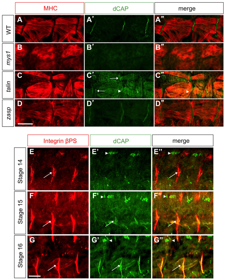
Fig. 8.

Integrin and Talin requirements for CAP localization at MASs. (A-A″) Wild-type, (B-B″) β-PS integrin, (C-C″) talin and (D-D″) Zasp mutant embryos immunostained with anti-CAP and anti-MHC. CAP localization at MASs is absent in β-PS integrin (mys1) mutants and is reduced (arrowheads) in talin mutants, which also show MAS splitting (arrow). (E-G″) Wild-type embryos of various stages stained with βPS integrin and CAP antibodies to label MASs (arrows) and scolopale cells (arrowheads). Scale bars: 10 μm.
