Abstract
Ophthalmoplegic migraine is a poorly understood neurologic syndrome characterized by recurrent bouts of head pain and ophthalmoplegia. By reviewing cases presenting to our centers in whom the phenotype has been carefully dissected, and systematically reviewing all published cases of ophthalmoplegic migraine in the magnetic resonance imaging (MRI) era, this review sets out to clearly define the syndrome and discuss possible etiologies. We found that in up to one-third of patients, the headache was not migrainous or associated with migrainous symptoms. In three-quarters of the cases involving the third nerve, there was focal nerve thickening and contrast enhancement on MRI. Observational data suggest systemic corticosteroids may be beneficial acutely. The etiology remains unclear, but may involve recurrent bouts of demyelination of the oculomotor nerve. “Ophthalmoplegic migraine” is a misnomer in that it is probably not a variant of migraine but rather a recurrent cranial neuralgia. A more appropriate name might be “ophthalmoplegic cranial neuropathy.”
Keywords: ophthalmoplegic migraine, headache, aura, neuralgia
Ophthalmoplegic migraine is a rare neurologic syndrome characterized by recurrent bouts of head pain and ophthalmoplegia. The third cranial nerve is most commonly affected, in which case mydriasis and ptosis can also be observed. Most patients recover completely within days to weeks, but a minority are left with persistent neurologic deficits.1 The disorder usually presents in children, but can persist in adulthood or persist into adulthood.2–4 Ophthalmoplegic migraine is entirely distinct from migraine with visual aura, in which patients experience transient visual phenomena before, during, or after the onset of migrainous headache.5
In the International Classification of Headache Disorders (second edition), the syndrome is reclassified under the cranial neuralgias to reflect evolution in thinking about its etiology; however, the migraine appellation was still retained.5 The International Classification of Headache Disorders (second edition) defines ophthalmoplegic migraine as at least 2 attacks characterized by a “migraine-like” headache followed within 4 days by paresis of the third, fourth, and/or sixth cranial nerves, including ophthalmoparesis, ptosis, or mydriasis.5 Structural causes of painful ophthalmoparesis, such as tumor, infection, and thrombosis, must have been excluded by appropriate imaging.
We systematically reviewed all published adult and pediatric cases of ophthalmoplegic migraine in the magnetic resonance imaging (MRI) era (since 1987) and analyzed demographic, clinical, and radiologic characteristics. We also report new cases from our centers to illustrate the salient clinical features. These data suggest that ophthalmoplegic “migraine” is a misnomer since the syndrome may not be a migraine variant but rather a recurrent cranial neuropathy. The continuing use of the appellation migraine in these patients is unhelpful as it suggests inappropriate pathophysiology and leads to unhelpful management strategies. A case is made to change the name on this basis.
Methods
PubMed was searched for all papers published in English using the search terms “ophthalmoplegic migraine” and “migraine with ophthalmoplegia” (last performed January 24, 2011). Case histories were then abstracted by one of the authors (AAG). References from these papers were also systematically reviewed to identify additional cases published in either article or abstract form.
Inclusion criteria for this systemic review were as follows: (1) The patient was reported to have experienced head pain followed by objective ophthalmoparesis, pupillary dilation, or ptosis and not merely subjective diplopia; (2) two or more episodes must have occurred; (3) 0.5-Tesla or higher MRI of the brain was performed to rule out alternative structural causes of ophthalmoparesis; cases with only CT imaging were excluded; (4) the diagnosis ascribed by the authors was ophthalmoplegic migraine and not a competing etiology, such as a tumor. There were no exclusions based on age of onset, type of head pain (did not have to be “migraine like”), or the interval from headache onset to Ophthalmoplegic onset. Headache quality was considered “migrainous” if words like “throbbing,” “pounding,” “pulsating,” or “migrainous” were used to describe the quality of pain, and considered as “not migrainous” if words such as “sharp,” “constant,” “burning,” or “dull” were reported.
The following information was abstracted for each case: (1) age at onset and age at current bout; (2) sex; (3) side of involvement; (4) cranial nerve(s) involved; (5) headache location; (6) headache quality; (7) associated symptoms: nausea, vomiting, photophobia, phonophobia; (8) duration between headache onset and ophthalmoplegia onset; (9) duration of headache; (10) duration of ophthalmoplegia; (11) MRI findings; (12) cerebrospinal fluid exam findings (if obtained); (13) complete blood count and erythrocyte sedimentation rate results (if obtained) as well as any other laboratory tests performed; (14) steroid administration and results; and (15) presence of permanent deficits.
Analysis
We tabulated the data from published cases and our own cases presented here and calculated descriptive statistics using Stata 10.1 (Statacorp, College Station, Texas).
Results
Clinical and Demographic Features
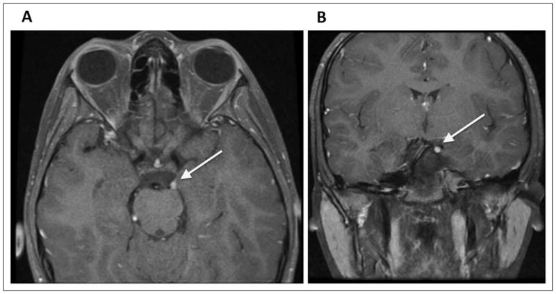
During the past 15 years (1995–2010) 4 patients with ophthalmoplegic migraine were seen at our centers and met inclusion criteria for this review. A fifth patient who was clinically diagnosed with ophthalmoplegic migraine was excluded because of the absence of an MRI scan. All 4 patients were children. Their clinical features are summarized in Table 1. Two were imaged during acute attacks and demonstrated third nerve enhancement on the affected side (see Figure 1).
Table 1.
Cases of Ophthalmoplegic Migraine From Our Centers
| Case 1 | Case 2 | Case 3 | Case 4 | |
|---|---|---|---|---|
| Age at onset (y) | 5 | 4 | 9 | 3 |
| Age at most recent episode (y) | 19 | 13 | 16 | 10 |
| Sex | M | M | F | M |
| Cranial nerve involved | 3 | 3 | 3 | 3 |
| Side | Left | Right | Right | Left |
| Headache location | Periorbital | Diffuse | Right frontal | Periorbital |
| Headache quality | Sharp, throbbing | Throbbing | Throbbing | Sharp, constant |
| Interval between headache onset and ophthalmoplegia | Several hours | 2–3 d | Hours (overnight) | 2 d |
| Headache duration | ~8 h | 2–3 d | Hours to several days | 2 d |
| Ophthalmoplegia duration | 3–7 d | – | 3–7 d | Several weeks |
| Associated symptoms | ||||
| Photophobia | Yes | Yes | No | No |
| Phonophobia | No | Yes | No | No |
| Nausea | Yes | Yes | No | Yes |
| Vomiting | – | Yes | – | Yes |
| History of other headaches | Yes | – | – | No |
| Family history of headache | Yes | Yes | Yes | No |
| Cerebrospinal fluid examination | – | – | – | Unremarkable |
| Vascular imaging | – | Normal MRA | – | Normal MRA |
| Improved with steroids | – | Yes | – | Yes |
| Persistent neurologic findings | No | No | No | Yes, partial third nerve palsy |
| MRI findings | Acute MRI showed enhancement of the cisternal portion of the third nerve Repeat MRI 1 y later was normal |
Nonacute MRI was normal | Nonacute MRI was normal (no gadolinium given) | Acute MRI showed thickening and enhancement of cisternal portion of third nerve Repeat nonacute MRI showed persistent thickening but no enhancement |
Abbreviations: F, female; M, male; MRA, magnetic resonance angiography; MRI, magnetic resonance imaging.
Figure 1.
Magnetic resonance images from Case 4: Axial (A) and coronal (B) post-gadolinium fluid-attenuated inversion recovery images showing thickening and enhancement of the cisternal portion of the left oculomotor nerve (arrows). This is the classic imaging finding of ophthalmoplegic migraine.
An additional 80 cases were identified in the published English-language literature,1,2,4,6–28 50 of which had received a 0.5 Tesla or greater brain MRI with the administration of gadolinium during an acute attack (see Table 2).
Table 2.
Compilation of Ophthalmoplegic Migraine Characteristics of Cases Published Between 1987 and 2010 and 4 Additional Cases
| Age at onset (y) (median [IQR]) (n = 63) | 8 (3, 16), range 0.58–50 |
| Age at best reported bout (y) (median [IQR]) (n = 76) | 16 (8, 32), range 1–74 |
| Sex (n = 84) | 53 (63%) female |
| Cranial nerve(s) involved (n = 84) | 65 (77%) third |
| 1 (1%) fourth | |
| 13 (15%) sixth | |
| 5 (6%) multiple | |
| Side (n = 69) | 32 (46%) left |
| 36 (52%) right | |
| 1 (1%) bilateral | |
| Headache location (n = 53) | 30 (57%) peri-/retroorbital |
| 17 (32%) ipsilateral | |
| 6 (11%) other | |
| Headache quality (n = 38) | 26 (68%) migrainous |
| 12 (32%) not migrainous | |
| Interval from headache onset to ophthalmoplegia (d) (median [IQR], range [d]) (n = 54) | 1.6 (0.5, 3), range 0–14 |
| Associated features | |
| Photophobia (n = 40) | 26 (65%) |
| Phonophobia (n = 25) | 14 (56%) |
| Nausea (n = 38) | 25 (66%) |
| Vomiting (n = 35) | 24 (69%) |
| History of headaches (n = 41) | 34 (83%) |
| Family history of headache (n = 47) | 29 (62%) |
| Cerebrospinal fluid findings (n = 32) | 30 normal (94%), 1 case had IgG index 0.87 and 1 had a single oligoclonal band |
| Vascular imaging (n = 42) | 16 (38%) conventional angiography |
| 24 (57%) MR angiography | |
| 2 (5%) CT angiography | |
| Normal in 39 (93%) | |
| Steroid response (n = 26) | 54% (14) beneficial |
| 8% (2) not beneficial | |
| 4% (1) worsened | |
| 35% (9) unclear whether beneficial | |
| Persistent findings (n = 35) | 19 (54%) |
| Subgroup analysis of those who had third nerve involvement and had an MRI with gadolinium during an acute attack (n = 52) | |
| Nerve root thickening (n = 50) | 38 (76%) |
| Nerve root enhancement (n = 52) | 39 (75%) |
Abbreviations: CT, computed tomographic; IQR, interquartile range; MR, magnetic resonance; MRI, magnetic resonance imaging.
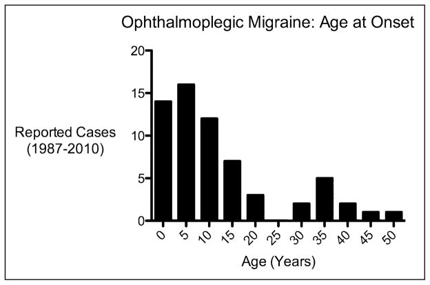
The median (interquartile range) age at the time of the first ophthalmoplegic migraine attack was 8 years (3–16 years) and ranged from 7 months to 50 years (Figure 2). Age at presentation of the most recent reported bout ranged from 1 to 74 years. Two-thirds of ophthalmoplegic migraine cases were in females and one-third in males. The site of involvement was evenly distributed between the right and left sides. The third cranial nerve was involved in the vast majority of cases (83%), 93% of which were characterized by isolated third nerve involvement. The sixth cranial nerve was involved in 20% of cases (77% of those cases having an isolated sixth). The fourth nerve was only involved in 2% of cases (with only a single case of an isolated fourth nerve deficit). Of the 5 cases (6%) with multiple cranial nerve involvement, the third nerve was involved in all, 1 patient had third and fourth nerve involvement, and 4 patients had third and sixth nerve involvement. Symptoms alternated sides on subsequent episodes in only 2 cases.14,23 There was only 1 reported case of simultaneous bilateral ophthalmoplegia.28
Figure 2.
Age of onset of ophthalmoplegic migraine.
Of the 53 cases in which headache location was reported, the majority of patients experienced peri- or retroorbital pain (57%). Less than half of the cases noted headache quality (n = 38), but among those that did, the pain was migrainous in 68% and not migrainous in 32%. In some cases involving infants or young children, the head pain component was not clearly apparent during the initial bout, and only emerged during subsequent episodes.12,16
In the 41 cases with information about the patient’s headache history, 83% had a history of headaches outside of their ophthalmoplegic migraine bouts. In the 47 cases that describe family history, 62% had a family history of headache.
Associated symptoms were reported in less than half of cases; photophobia (n = 40) was noted in 26 of 40 cases (65%), phonophobia (n = 25) in 14 of 25 (56%), nausea in 25 of 38 (66%), and vomiting in 24 of 35 (69%).
The interval between headache onset and ophthalmoparesis ranged from immediate to up to 14 days, with a median (inter-quartile range) of 1.6 days (0.5–3 days). The headache component typically lasted on the order of several days to a week, while the ophthalmoplegia tended to last longer, often on the order of 2 to 3 weeks to 2 to 3 months (data not shown).
In 35 cases, the presence or absence of persistent findings on follow-up examination was clearly noted, and in 19 of these cases (54%), persistent deficits were observed. Examples of persistent deficits included persistent mydriasis, ptosis, or tropias.
Radiologic Manifestations
Of patients who had a brain MRI with the administration of gadolinium performed during an acute attack involving the third nerve (n = 52), 75% had contrast enhancement of the third nerve and 76% had nerve thickening. Nerve enhancement of the fourth nerve was seen in one case and in the intraparenchymal portion of the sixth nerve in another case.29,30
Vascular imaging was obtained in half of the reported cases (n = 42). Abnormalities were noted in only 3 patients (7%), AND these were of unclear etiologic significance: (1) a venous angioma draining into the cavernous sinus25; (2) infundibular dilation of a perforating branch of the posterior cerebral artery just above the superior cerebellar artery that did contact the involved oculomotor nerve27; and (3) apparent contact between the affected sixth nerve and the basilar artery and ipsilateral anterior inferior cerebellar artery.31
Cerebrospinal Fluid Analysis
Cerebrospinal fluid was obtained in 38% of all reported ophthalmoplegic migraine cases (n = 32). The results were normal except in 2 patients: 1 had an elevated immunoglobulin G (IgG) index but no oligoclonal bands, and the other had a single oligoclonal band.
Laboratory Findings
Serum laboratory tests included a complete blood count in 17% of ophthalmoplegic migraine patients (n = 14) and an erythrocyte sedimentation rate in 25% (n = 21); these were normal in all such cases. Electrolytes, hemoglobin A1c, and autoimmune testing, when performed, were normal (data not shown). Testing for viral or spirochetal etiologies was never indicative of acute infection.
Treatment
Steroids were used to treat at least 1 bout in 31% (n = 26) of patients. There was a perceived benefit from corticosteroid treatment, defined as improvement within 1 to 2 days, in 14 patients (54%), with no apparent benefit or clinical worsening in 3 (12%). In 9 (35%), the effect was unclear. Treatment and evaluation was not blinded in any of these cases.
Discussion
Here we have systematically reviewed and analyzed published cases of ophthalmoplegic migraine, including both pediatric and adult cases, and added illustrative cases from our practices. The most notable findings in this review were the following: (1) in up to one-third of cases the associated head pain was not migrainous in quality nor were there associated migrainous symptoms, such as nausea or vomiting; (2) the symptoms were overwhelmingly side locked; (3) there was a marked time lag between headache onset and ophthalmoplegia extending up to 14 days; and (4) there was focal third nerve enhancement and thickening on contrast-enhanced brain MRI in more than three-quarters of cases involving the third nerve. Taken together, these data suggest that this syndrome is a not a migraine variant but rather a form of cranial neuropathy that triggers headache secondarily. Ophthalmoplegic Cranial Neuropathy may be a more suitable diagnostic term.
Lal and colleagues published a review of 62 adult patients with painful ophthalmoplegia32; the majority had only a single episode, and it is possible that some of these cases represented other etiologies. Their case mix was very different from ours, and indeed to what we have typically seen reported in the literature, with onset in adulthood and not a single case of third nerve enhancement. It seems likely the entity classically known as ophthalmoplegic migraine, and classified by the International Classification of Headache Disorders (second edition), is different from that described in that series. The difference is so striking that perhaps there is a genetically based difference seen more obviously in the Indian subcontinent.
While the pathophysiology of ophthalmoplegic migraine is unclear, these data provide some clues about what the syndrome is not. By International Classification of Headache Disorders (second edition) criteria, there can be no apparent structural lesion to account for the findings. In our analysis, only 3 cases had associated vascular findings on imaging and these were unlikely to be etiologically significant.25,27,31 If such abnormal vascular contact is functionally meaningful, it likely occurs in only a small percentage of ophthalmoplegic migraine cases. This is in contrast to trigeminal neuralgia—a prototypical cranial neuralgia characterized by much briefer and more frequent painful episodes—in which abnormal contact between vessel and nerve is a more common association.
There was no evidence to suggest a systemic inflammatory process associated with ophthalmoplegic migraine in this analysis. There was also no evidence of intrathecal inflammation in any of the cases involving the third cranial nerve. Possibly inflammatory cerebrospinal fluid abnormalities were reported in only 2 cases, which happened to be the only 2 patients with fourth nerve involvement: one with an elevated IgG index, without oligoclonal bands, and the other with a single oligoclonal band. This raises the question of whether this rarest variant of ophthalmoplegic migraine, involving the fourth nerve, may be different etiologically from typical ophthalmoplegic migraine. More data are required to clarify this issue.
Virologic studies on cerebrospinal fluid and serum were rarely reported, but there is no clear evidence to date of an association between viral infection and ophthalmoplegic migraine. A single report claiming an association between ophthalmoplegic migraine and a positive cytomegalovirus IgG, but not IgM,33 is of doubtful significance without definitive evidence of acute infection given the high prevalence of cytomegalovirus seropositivity in the general population.34 Familial cases with recurrent bouts of facial palsy and ophthalmoplegia preceded by head pain have been reported,35 and several of the ophthalmoplegic migraine cases in this analysis had personal or family histories of Bell’s palsy,1,36–38 which could suggest an abnormal response to neurotropic viral infection in these patients given the association between herpes simplex virus and varicella zoster virus with Bell’s palsy,39 but this remains to be proven. Of note, facial nerve enhancement can be seen on gadolinium-enhanced MRI in Bell’s palsy as well.40
The oculomotor nerve does not normally enhance on MRI following the administration of gadolinium, which makes the observation that up to three-quarters of all ophthalmoplegic migraine cases with third nerve involvement had nerve enhancement on MRI striking.41 The differential diagnosis for enhancement of the cisternal portion of the oculomotor nerve includes neoplastic, inflammatory, and infiltrative conditions; however, most of these would not be expected to resolve spontaneously as occurs in ophthalmoplegic migraine.20,42 One diagnostic possibility raised by the authors in several cases is an oculomotor nerve schwannoma, although there is only 1 pathology-proven case in the literature initially mistaken for ophthalmoplegic migraine.43 Two additional cases where schwannoma was implicated were not pathology proven.44,45 A schwannoma would be expected to cause a persistent or progressive nerve palsy. The observation that nerve enhancement in ophthalmoplegic migraine is often markedly diminished on follow-up imaging weeks to months later argues against schwannoma as a common etiology in ophthalmoplegic migraine.1–3,7,10,14,17,20,30,41,46
A recurrent demyelinating cranial neuropathy has gained favor in recent years as a possible explanation for ophthalmoplegic migraine, with some authors drawing the analogy to the nerve swelling seen in chronic inflammatory demyelinating polyradiculopathy.2 If demyelination is indeed on the pathophysiological pathway, however, what initiates the demyelination remains unclear. Unlike chronic inflammatory demyelinating polyradiculopathy, the cerebrospinal fluid tends to be bland in ophthalmoplegic migraine. Moreover, repeated attacks of a single unilateral cranial nerve would be highly unusual for a primary antibody-mediated attack. In several cases, the ophthalmoplegia persisted longer with subsequent bouts,1,38,47–49 which could be consistent with recurrent bouts of demyelination leading to cumulative axonal damage. Repeated episodes of demyelination with remyelination may also help explain the observation of focal enlargement of the oculomotor nerve in ophthalmoplegic migraine.41
There are no autopsy cases of ophthalmoplegic migraine from the post-MRI era. Carlow reviewed 5 historical cases that fulfill “the generally accepted clinical criteria for ophthalmoplegic migraine.”50 Excluding 1 patient who had tuberculous meningitis and granulomatous involvement of the cranial nerve, 3 of the remaining 4 cases showed thickening of the oculomotor nerve at or near the midbrain exit.
There have been no published treatment trials for ophthalmoplegic migraine, and the level of evidence for evaluating effective treatments is entirely observational. Nevertheless, oral steroids may be of possible benefit in treating acute exacerbations based on available case series. Given that ophthalmoplegic migraine is not a variant of migraine pathophysiologically, migraine-specific acute and preventive therapies probably do not have a role in treating ophthalmoplegic migraine. Injection of botulinum toxin or strabismus surgery may be considered for patients with persistent eye misalignment.51
Only cases of ophthalmoplegic migraine published in the literature could be reviewed, which is the main limitation of this type of analysis. There may be a publication bias wherein authors are more likely to submit cases with classic imaging findings, risking overestimation of characteristic MRI findings or other clinical or demographic variables.
Ophthalmoplegic migraine is currently classified as a cranial neuralgia in the International Classification of Headache Disorders (second edition).5 This analysis suggests the classification criteria, and even the name of the disorder, improved upon to allow for greater clarity and diagnostic accuracy. The findings here suggest the current classification criteria, which require the headache to be “migraine-like,” are too restrictive as the headache quality was not “migraine-like” in roughly a third of the patients, a critique also raised by other authors.26,30,33,52 In addition, while the current criteria require that ophthalmoplegia develop within 4 days of headache onset, these data indicate that ophthalmoplegia can take up to 14 days to develop following headache onset. The cases with long latencies between headache onset and cranial nerve deficits were indistinguishable from those with shorter latencies. Lastly, the term “ophthalmoplegic migraine” itself is misleading diagnostically and could encourage the inappropriate use of migraine-specific preventive therapies. A more appropriate name might be “recurrent ophthalmoplegic cranial neuropathy.” Further research is needed to understand more about the underlying biology of this unusual condition.
Acknowledgments
Cases were drawn from the Great Ormond Street center in London, as well as the Child Neurology division and Headache Center at UCSF. The analysis was performed at University of California, San Francisco (UCSF). Preliminary results were presented in poster form at the UCSF Neurology Department Fishman Conference on June 15, 2011.
Funding
The authors disclosed receipt of the following financial support for the research, authorship, and/or publication of this article: AAG receives funding from NIH/NINDS. JMG receives funding from the National Multiple Sclerosis Society. PP has, over the past 5 years, advised, lectured in meetings sponsored by, or done research in association with Bristol-Myers Squibb, Merck/MSD and National Institute for Health Research (UK). PJG has consulted for, advised, or collaborated with Advanced Bionics, Allergan, Almirall, ATI, AstraZeneca, Belgian Research Council, Boehringer-Ingelheim, BMS, Boston Scientific, Colucid, Eli-Lilly, Fidelity Foundation, GlaxoSmithKline, Johnson & Johnson, Kalypsys, Medtronic, MAP, Migraine Research Foundation, Migraine Trust, Minster, Medical Research Council-UK, MSD, NINDS, Netherlands Research Council, Neuralieve, Neuraxon, NTP, Organisation for Understanding Cluster Headache–UK and US and Pfizer.
Footnotes
Reprints and permission: sagepub.com/journalsPermissions.nav
Author Contributions
AAG was responsible for study concept and design, data collection and analysis, and wrote the first draft of the manuscript. JMG assisted with data analysis, PP submitted cases, and PJG contributed to study concept and design. All authors revised the manuscript for intellectual content.
Ethical Approval
As the literature cases are from the published literature and do not contain any protected health information, no human subjects committee review was obtained for reviewing them. Similarly for the cases from our centers, there is no protected health information included and hence human subjects review was not sought for publishing these case reports.
Declaration of Conflicting Interests
The authors declared no potential conflicts of interest with respect to the research, authorship, and/or publication of this article.
References
- 1.McMillan HJ, Keene DL, Jacob P, Humphreys P. Ophthalmoplegic migraine: inflammatory neuropathy with secondary migraine? Can J Neurol Sci. 2007;34(3):349–355. doi: 10.1017/s0317167100006818. [DOI] [PubMed] [Google Scholar]
- 2.Bharucha DX, Campbell TB, Valencia I, Hardison HH, Kothare SV. MRI findings in pediatric ophthalmoplegic migraine: a case report and literature review. Pediatr Neurol. 2007;37(1):59–63. doi: 10.1016/j.pediatrneurol.2007.03.008. [DOI] [PubMed] [Google Scholar]
- 3.Doran M, Larner AJ. MRI findings in ophthalmoplegic migraine: nosological implications. J Neurol. 2004;251(1):100–101. doi: 10.1007/s00415-004-0219-4. [DOI] [PubMed] [Google Scholar]
- 4.Shin DJ, Kim JH, Kang SS. Ophthalmoplegic migraine with reversible thalamic ischemia shown by brain SPECT. Headache. 2002;42(2):132–135. doi: 10.1046/j.1526-4610.2002.02029.x. [DOI] [PubMed] [Google Scholar]
- 5.Headache Classification Subcommittee of the International Headache Society. The International Classification of Headache Disorders. Cephalalgia. (2) 2004;24(suppl 1):9–160. doi: 10.1111/j.1468-2982.2003.00824.x. [DOI] [PubMed] [Google Scholar]
- 6.Verhagen WI, Prick MJ, van Dijk Azn R. Onset of ophthalmoplegic migraine with abducens palsy at middle age? Headache. 2003;43(7):798–800. doi: 10.1046/j.1526-4610.2003.03140.x. [DOI] [PubMed] [Google Scholar]
- 7.Carlow TJ. Oculomotor ophthalmoplegic migraine: is it really migraine? J Neuroophthalmol. 2002;22(3):215–221. doi: 10.1097/00041327-200209000-00006. [DOI] [PubMed] [Google Scholar]
- 8.Sommer M, Nomikos P, Paulus W. A case of ophthalmoplegic migraine with recurrent oculomotor nerve palsy. J Neurol. 2003;250(suppl 2):II190. (abstract P733) [Google Scholar]
- 9.Ramelli GP, Vella S, Lovblad K, Remonda L, Vassella F. Swelling of the third nerve in a child with transient oculomotor paresis: a possible cause of ophthalmoplegic migraine. Neuropediatrics. 2000;31(3):145–147. doi: 10.1055/s-2000-7532. [DOI] [PubMed] [Google Scholar]
- 10.O’Hara MA, Anderson RT, Brown D. Magnetic resonance imaging in ophthalmoplegic migraine of children. J AAPOS. 2001;5(5):307–310. doi: 10.1067/mpa.2001.118670. [DOI] [PubMed] [Google Scholar]
- 11.Mucchiut M, Valentinis L, Provenzano A, Cutuli D, Bergonzi P. Adult-onset ophthalmoplegic migraine with recurrent sixth nerve palsy: a case report. Headache. 2006;46(10):1589–1590. doi: 10.1111/j.1526-4610.2006.00616_1.x. [DOI] [PubMed] [Google Scholar]
- 12.Ostergaard JR, Moller HU, Christensen T. Recurrent ophthalmoplegia in childhood: diagnostic and etiologic considerations. Cephalalgia. 1996;16(4):276–279. doi: 10.1046/j.1468-2982.1996.1604276.x. [DOI] [PubMed] [Google Scholar]
- 13.Lane R, Davies P. Ophthalmoplegic migraine: the case for reclassification. Cephalalgia. 2010;30(6):655–661. doi: 10.1111/j.1468-2982.2009.01977.x. [DOI] [PubMed] [Google Scholar]
- 14.Lance JW, Zagami AS. Ophthalmoplegic migraine: a recurrent demyelinating neuropathy? Cephalalgia. 2001;21(2):84–89. doi: 10.1046/j.1468-2982.2001.00160.x. [DOI] [PubMed] [Google Scholar]
- 15.Hansen SL, Borelli-Moller L, Strange P, Nielsen BM, Olesen J. Ophthalmoplegic migraine: diagnostic criteria, incidence of hospitalization and possible etiology. Acta Neurol Scand. 1990;81(1):54–60. doi: 10.1111/j.1600-0404.1990.tb00931.x. [DOI] [PubMed] [Google Scholar]
- 16.Hassin H. Ophthalmoplegic migraine wrongly attributed to measles immunization. Am J Ophthalmol. 1987;104(2):192–193. doi: 10.1016/0002-9394(87)90020-1. [DOI] [PubMed] [Google Scholar]
- 17.Zafeiriou DI, Vargiami E. Childhood steroid-responsive painful opthalmoplegia: clues to opthalmoplegic migraine. J Pediatr. 2006;149(6):881. doi: 10.1016/j.jpeds.2006.08.019. [DOI] [PubMed] [Google Scholar]
- 18.Wong V, Wong WC. Enhancement of oculomotor nerve: a diagnostic criterion for ophthalmoplegic migraine? Pediatr Neurol. 1997;17(1):70–73. doi: 10.1016/s0887-8994(97)80671-6. [DOI] [PubMed] [Google Scholar]
- 19.Weiss AH, Phillips JO. Ophthalmoplegic migraine. Pediatr Neurol. 2004;30(1):64–66. doi: 10.1016/s0887-8994(03)00424-7. [DOI] [PubMed] [Google Scholar]
- 20.Mark AS, Casselman J, Brown D, et al. Ophthalmoplegic migraine: reversible enhancement and thickening of the cisternal segment of the oculomotor nerve on contrast-enhanced MR images. AJNR Am J Neuroradiol. 1998;19(10):1887–1891. [PMC free article] [PubMed] [Google Scholar]
- 21.Huang TH, Hsu WC, Yeh JH, Chiu HC, Chen WH. A case of ophthalmoplegic migraine mimicking ocular myasthenia gravis. Eur Neurol. 2005;53(4):215–217. doi: 10.1159/000086734. [DOI] [PubMed] [Google Scholar]
- 22.De Silva DA, Siow HC. A case report of ophthalmoplegic migraine: a differential diagnosis of third nerve palsy. Cephalalgia. 2005;25(10):827–830. doi: 10.1111/j.1468-2982.2005.00953.x. [DOI] [PubMed] [Google Scholar]
- 23.Choi JY, Jang SH, Park MH, Kim BJ, Lee DH. Ophthalmoplegic migraine with alternating unilateral and bilateral internal ophthalmoplegia. Headache. 2007;47(5):726–728. doi: 10.1111/j.1526-4610.2007.00795_2.x. [DOI] [PubMed] [Google Scholar]
- 24.Borade A, Prabhu AS, Kumar S, Prasad V, Rajam L. Magnetic resonance imaging findings in ophthalmoplegic migraine. J Postgrad Med. 2009;55(2):137–138. doi: 10.4103/0022-3859.52848. [DOI] [PubMed] [Google Scholar]
- 25.Berbel-Garcia A, Martinez-Salio A, Porta-Etessam J, et al. Venous angioma associated with atypical ophthalmoplegic migraine. Headache. 2004;44(5):440–442. doi: 10.1111/j.1526-4610.2004.04097.x. [DOI] [PubMed] [Google Scholar]
- 26.Crevits L, Verschelde H, Casselman J. Ophthalmoplegic migraine: an unresolved problem. Cephalalgia. 2006;26(10):1255–1259. doi: 10.1111/j.1468-2982.2006.01203.x. [DOI] [PubMed] [Google Scholar]
- 27.Vieira JP, Castro J, Gomes LB, Jacinto S, Dias A. Ophthalmoplegic migraine and infundibular dilatation of a cerebral artery. Headache. 2008;48(9):1372–1374. doi: 10.1111/j.1526-4610.2008.01179.x. [DOI] [PubMed] [Google Scholar]
- 28.Giraud P, Valade D, Lanteri-Minet M, Donnet A, Geraud G, Guégan-Massardier E Observatoire des Migraines et Céphalées of the French Headache Society. Is migraine with cranial nerve palsy an ophthalmoplegic migraine? J Headache Pain. 2007;8(2):119–122. doi: 10.1007/s10194-007-0371-1. [DOI] [PMC free article] [PubMed] [Google Scholar]
- 29.Lee TG, Choi WS, Chung KC. Ophthalmoplegic migraine with reversible enhancement of intraparenchymal abducens nerve on MRI. Headache. 2002;42(2):140–141. doi: 10.1046/j.1526-4610.2002.02031.x. [DOI] [PubMed] [Google Scholar]
- 30.van der Dussen DH, Bloem BR, Liauw L, Ferrari MD. Ophthalmoplegic migraine: migrainous or inflammatory? Cephalalgia. 2004;24(4):312–315. doi: 10.1111/j.1468-2982.2004.00669.x. [DOI] [PubMed] [Google Scholar]
- 31.Linn J, Schwarz F, Reinisch V, Straube A. Ophthalmoplegic migraine with paresis of the sixth nerve: a neurovascular compression syndrome? Cephalalgia. 2008;28(6):667–670. doi: 10.1111/j.1468-2982.2008.01563.x. [DOI] [PubMed] [Google Scholar]
- 32.Lal V, Sahota P, Singh P, Gupta A, Prabhakar S. Ophthalmoplegia with migraine in adults: is it ophthalmoplegic migraine? Headache. 2009;49(6):838–850. doi: 10.1111/j.1526-4610.2009.01405.x. [DOI] [PubMed] [Google Scholar]
- 33.Ogose T, Manabe T, Abe T, Nakaya K. Ophthalmoplegic migraine with a rise in cytomegalovirus-specific IgG antibody. Pediatr Int. 2009;51(1):143–145. doi: 10.1111/j.1442-200X.2008.02776.x. [DOI] [PubMed] [Google Scholar]
- 34.Staras SA, Dollard SC, Radford KW, Flanders WD, Pass RF, Cannon MJ. Seroprevalence of cytomegalovirus infection in the United States, 1988–1994. Clin Infect Dis. 2006;43(9):1143–1151. doi: 10.1086/508173. [DOI] [PubMed] [Google Scholar]
- 35.Lee AG, Brazis PW, Eggenberger E. Recurrent idiopathic familial facial nerve palsy and ophthalmoplegia. Strabismus. 2001;9(3):137–141. doi: 10.1076/stra.9.3.137.6758. [DOI] [PubMed] [Google Scholar]
- 36.Celebisoy N, Sirin H, Gokcay F. Ophthalmoplegic migraine: two patients, one at middle age with abducens palsy. Cephalalgia. 2005;25(2):151–153. doi: 10.1111/j.1468-2982.2004.00814.x. [DOI] [PubMed] [Google Scholar]
- 37.Prats JM, Mateos B, Garaizar C. Resolution of MRI abnormalities of the oculomotor nerve in childhood ophthalmoplegic migraine. Cephalalgia. 1999;19(7):655–659. doi: 10.1046/j.1468-2982.1999.019007655.x. [DOI] [PubMed] [Google Scholar]
- 38.Peck R. Ophthalmoplegic migraine presenting in infancy: a self-reported case. Cephalalgia. 2006;26(10):1242–1244. doi: 10.1111/j.1468-2982.2006.01181.x. [DOI] [PubMed] [Google Scholar]
- 39.Gilden DH. Bell’s palsy. Clinical Practice. N Engl J Med. 2004;351(13):1323–1331. doi: 10.1056/NEJMcp041120. [DOI] [PubMed] [Google Scholar]
- 40.Bsoul SA, Terezhalmy GT, Moore WS. Bell’s palsy. Quintessence Int. 2004;35(6):506–507. [PubMed] [Google Scholar]
- 41.Bek S, Genc G, Demirkaya S, Eroglu E, Odabasi Z. Ophthalmoplegic migraine. Neurologist. 2009;15(3):147–149. doi: 10.1097/NRL.0b013e3181870408. [DOI] [PubMed] [Google Scholar]
- 42.Mark AS, Blake P, Atlas SW, Ross M, Brown D, Kolsky M. Gd-DTPA enhancement of the cisternal portion of the oculomotor nerve on MR imaging. AJNR Am J Neuroradiol. 1992;13(5):1463–1470. [PMC free article] [PubMed] [Google Scholar]
- 43.Murakami T, Funatsuka M, Komine M, et al. Oculomotor nerve schwannoma mimicking ophthalmoplegic migraine. Neuropediatrics. 2005;36(6):395–398. doi: 10.1055/s-2005-873056. [DOI] [PubMed] [Google Scholar]
- 44.Bisdorff AR, Wildanger G. Oculomotor nerve schwannoma mimicking ophthalmoplegic migraine. Cephalalgia. 2006;26(9):1157–1159. doi: 10.1111/j.1468-2982.2006.01167.x. [DOI] [PubMed] [Google Scholar]
- 45.Kawasaki A. Oculomotor nerve schwannoma associated with ophthalmoplegic migraine. Am J Ophthalmol. 1999;128(5):658–660. doi: 10.1016/s0002-9394(99)00242-1. [DOI] [PubMed] [Google Scholar]
- 46.Romano LM, Besocke AG. Teaching video neuroimages: recurrent oculomotor neuropathy with isolated ptosis vs ophthalmoplegic migraine. Neurology. 2009;72(9):e44. doi: 10.1212/01.wnl.0000343964.79460.f3. [DOI] [PubMed] [Google Scholar]
- 47.Manzouri B, Sainani A, Plant G, Lee J, Sloper J. The aetiology and management of long-lasting sixth nerve palsy in ophthalmoplegic migraine. Cephalalgia. 2007;27(3):275–278. doi: 10.1111/j.1468-2982.2006.01252.x. [DOI] [PubMed] [Google Scholar]
- 48.Leone M, Grazzi L, Moschiano F, Bussone G. Internal ophthalmoplegia associated with migraine attacks. Cephalalgia. 1994;14(6):461–462. doi: 10.1046/j.1468-2982.1994.1406461.x. [DOI] [PubMed] [Google Scholar]
- 49.Lavin PJ, Aulino JM, Uskavitch D. “Ophthalmoplegic migraine” with reversible MRI enhancement of the cisternal sixth cranial nerve. J Neuroophthalmol. 2009;29(2):151–153. doi: 10.1097/WNO.0b013e3181a57d3a. [DOI] [PubMed] [Google Scholar]
- 50.Lee AG. Oculomotor ophthalmoplegic migraine: what really causes it? J Neuroophthalmol. 2003;23(3):240. doi: 10.1097/00041327-200309000-00010. author reply 240–241. [DOI] [PubMed] [Google Scholar]
- 51.Granado L, Guillen G. Treatment options for ophthalmoplegic migraine. J Postgrad Med. 2009;55(3):231. doi: 10.4103/0022-3859.57389. author reply 231–232. [DOI] [PubMed] [Google Scholar]
- 52.Kandt RS, Goldstein GW. Steroid-responsive ophthalmoplegia in a child. Diagnostic considerations. Arch Neurol. 1985;42(6):589–591. doi: 10.1001/archneur.1985.04060060095016. [DOI] [PubMed] [Google Scholar]


