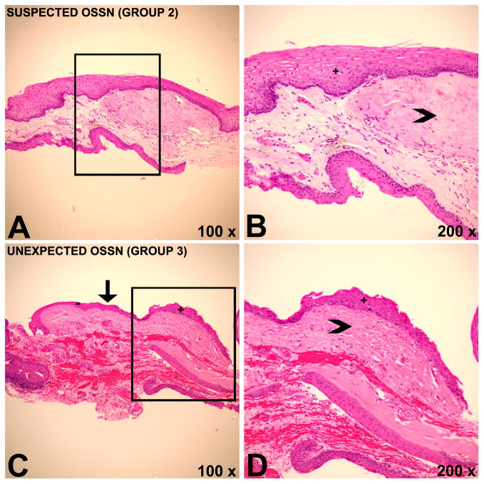
Figure 3.
Histopathology images demonstrating coexistent pterygium and ocular surface squamous neoplasia (OSSN). A,B) Histopathology of a patient belonging to group 2 with marked solar elastosis (arrowhead) and overlying epithelium with faulty maturational sequencing (+), indicating OSSN with an underlying pterygium. In this case, OSSN was anticipated by the surgeon. C, D) Histopathology of a patient belonging to group 3 demonstrating focus of OSSN (+) with a clear transition (arrow) towards unremarkable epithelium (−). Solar elastosis (arrowhead) is present, indicating a pterygium. OSSN was found incidentally in pathology and was not suspected by the surgeon. Hematoxylin & Eosin (H&E).

