Abstract
Ankyrin repeat domain-encoding genes are common in the eukaryotic and viral domains of life, but they are rare in bacteria, the exception being a few obligate or facultative intracellular Proteobacteria species. Despite having a reduced genome, the arthropod strains of the alphaproteobacterium Wolbachia contain an unusually high number of ankyrin repeat domain-encoding genes ranging from 23 in wMel to 60 in wPip strain. This group of genes has attracted considerable attention for their astonishing large number as well as for the fact that ankyrin proteins are known to participate in protein-protein interactions, suggesting that they play a critical role in the molecular mechanism that determines host-Wolbachia symbiotic interactions. We present a comparative evolutionary analysis of the wMel-related ankyrin repeat domain-encoding genes present in different Drosophila-Wolbachia associations. Our results show that the ankyrin repeat domain-encoding genes change in size by expansion and contraction mediated by short directly repeated sequences. We provide examples of intra-genic recombination events and show that these genes are likely to be horizontally transferred between strains with the aid of bacteriophages. These results confirm previous findings that the Wolbachia genomes are evolutionary mosaics and illustrate the potential that these bacteria have to generate diversity in proteins potentially involved in the symbiotic interactions.
Introduction
Wolbachia is a group of intracellular and maternally transmitted alphaproteobacteria, comprising strains with diverse symbiotic relationships with numerous arthropod as well as filarial nematode species [1], [2]. These bacteria are quite widespread in insects and crustaceans [3]. Recent screens suggest that >40% of all terrestrial arthropod species are infected, rendering Wolbachia perhaps the most abundant symbiotic microorganism of the biosphere [4].
The successful spread of Wolbachia into insect host populations has been attributed to their unique ability to act as manipulators of host reproduction in order to ensure their own transmission. Wolbachia infections have been associated with the induction of feminization, thelytokous parthenogenesis, male killing and, most commonly, cytoplasmic incompatibility (CI) [1], [2], [5]. During the last decade, several studies have shown that Wolbachia infections can affect, in addition to reproduction, several additional aspects of host biology, physiology, ecology and evolution [1], [2], [5], [6]. The molecular mechanisms that allow Wolbachia to establish symbiotic associations and to induce its extended phenotypes are yet to be unraveled.
Advances in genomics provided significant new information about Wolbachia biology. There are currently four complete genome sequences of Wolbachia, while several others are either available as permanent drafts or in progress [2], [7]–[13]. A common feature of all insect Wolbachia genomes is the high percentage of repetitive elements such as insertion sequences, group II introns, duplicated segments of prophages and multi-gene families such as the ankyrin repeat genes [14]–[16].
Ankyrin repeat domains (ANK hereafter) consist of a tandem motif of about 33 amino acids and act as scaffolds that mediate protein-protein interactions [17], [18]. Proteins with ANK domains are commonly found in eukaryotes and viruses, but they can also, although rarely, be found in the Bacteria and Archaea [19]. ANK proteins are known to be involved a multitude of functions, such as cell-cycle regulation, transcriptional regulation, cytoskeleton interactions, signal transduction, development, intracellular trafficking, sex differentiation, and they can also act as toxins [17], [18], [20]. Recent findings suggest that ANK proteins represent a new family of bacterial type IV effectors that play a major role in host-pathogen interactions and the evolution of infections [21], [22]. Indeed, it was shown that ANK proteins of certain pathogenic intracellular bacteria are secreted into the host cytoplasm and interact with host factors [22]–[25]. The intracellular pathogen Anaplasma phagocytophilum, for example, secrets AnkA through a type IV secretion system (T4SS). The protein interacts with specific regions of the host chromatin, resulting in a modulation of host gene transcription [23], [24], [26]–[28].
The potential role of ANK proteins in Wolbachia symbiosis and the manipulation of host reproduction has been investigated in Drosophila and mosquito species, but no direct correlation of the Wolbachia ankyrin gene repertoire with any bacterial phenotype has so far been established [10], [29]–[34]. It is worth noting, however, that (a) the presence of certain ANK gene variants has been associated with crossing types in Culex quinquefasciatus [32] and (b) some ANK genes are under host sex-specific regulation [31]. A recent large-scale proteomic analysis of the excretory-secretory products of the Wolbachia infected filarial parasite Brugia malayi identified the presence of two Wolbachia-encoded ankyrin proteins [35]. These are probably the first Wolbachia ANK proteins shown to be secreted. Wolbachia also carries a functional T4SS [9], [10], [13], [36], strengthening the hypothesis that the ANK genes play a role in functional and evolutionary processes of host-Wolbachia symbiosis.
Genomic analysis showed that the ANK genes account for up to 4% of the total number of genes in the insect Wolbachia strains wMel, wRi and wPip [9], [10], [13]. Additionally, comparative analyses between the ANK genes found in these genomes showed that they often evolve very rapidly, through for example gene duplication and contraction/expansion of repeated sequences within ANK domains [9], [10]. Taken together, the above led to the suggestion that the ANK proteins may play a pivotal role in the molecular interaction between host and symbiont [9], [10], [13], [30].
To learn more about the genetic diversity and the molecular mechanisms that underlie the evolution of the Wolbachia ANK gene family, we investigated the distribution of wMel ANK genes in several different Drosophila-Wolbachia associations, analyzed their genetic diversity by sequencing the identified orthologs and reconstructed their phylogenetic relationship. Our results indicate that both homologous and illegitimate recombination along with genomic flux provided by prophages and transposable elements are key factors in generating polymorphism and shaping the ANK gene pool in the Wolbachia strains studied.
Materials and Methods
Ethics Statement
N/A.
Fly Lines and Wolbachia Strains
The Drosophila lines and Wolbachia strains used in this study are listed in [37]–[45] Table 1. Flies were grown at 24°C ±1 on cornflour/sugar/yeast medium. Wolbachia strains were chosen according to their modification/rescue phenotype (mod+/resc+, mod−/resc+, mod−/resc−) and their embryonic localization pattern (posterior, anterior or uniform) as described by Veneti et al. [44]. The Wolbachia infection status of the Drosophila lines used was confirmed based on wsp and MLST typing as well as on CI properties [46]–[48].
Table 1. Drosophila lines and Wolbachia strains.
| Wolbachia supergroup | Host species | Wolbachia strain | Embryonic distribution | CI phenotype | Reference |
| A | D. melanogaster | wMel | posterior | mod+ resc+ | [38], [40], [44] |
| D. melanogaster | wMelPop | posterior | mod+ resc+ | [43], [44] | |
| D. simulans | wAu | posterior | mod− resc− | [42], [44] | |
| D. teissieri | wTei | posterior | mod− resc+ | [44], [45] | |
| D. yakuba | wYak | posterior | mod− resc+ | [44], [45] | |
| D. santomea | wSan | posterior | mod− resc+ | [44], [45] | |
| D. simulans | wRi | uniform | mod+ resc+ | [41], [44] | |
| D. simulans | wHa | uniform | mod+ resc+ | [41], Veneti pers. comm. | |
| B | D. simulans | wNo | anterior | mod+ resc+ | [41], [44] |
| D. mauritiana | wMau | anterior | mod− resc+ | [39], [44] | |
| D. simulans | wMa | anterior | mod− resc+ | [37], [44] |
Detection of ANK Genes with PCR and Southern Blot Analysis
Total genomic DNA was extracted from each Drosophila line according to the Holmes-Bonner method [49]. Specific primers for each of the 23 wMel ANK genes were designed (Table S1), based on the sequenced Wolbachia strain wMel and used to probe for the homologs of the 23 wMel ANK genes in the 11 Wolbachia strains listed in Table 1. Reaction mixtures (final volume of 20 µl) contained 1x Taq reaction buffer (750 mM Tris-Cl pH 8.8, 200 mM (NH4)2SO4, 0.1% Tween 20), 1.5 mM MgCl2, 125 µM dNTPs, 10 pmol of each primer, 1 unit Taq polymerase (Promega) and 50 ng DNA. PCRs were run on a PTC-200 thermal cycler (MJ Research Inc.) with the following cycling conditions: an initial step at 94°C for 5 min, followed by 35 cycles of 94°C for 30 sec, 30 sec at a primer dependent annealing temperature, and 72°C for 3 min, plus a final extension at 72°C for 10 min.
For Southern blot analysis, 5 µg of total genomic DNA were digested with EcoRI. The DNA fragments were separated on a 0.8% agarose gel and blotted onto Immobilon-Ny+ filters (Millipore), according to the manufacturer’s instructions and hybridized at 68°C for 18 h in Denhardt’s solution, according to standard procedures [50]. Probes for the 23 wMel ANK genes were generated by PCR in 50 µl reactions, using specific primers amplifying part of each gene (see Table S1). PCR products were purified with the QIAquick PCR purification kit (QIAGEN) and radioactively labeled with [α-32P] dATP (IZOTOP) using the Prime-a-gene labeling kit (Promega). A specific probe for the Wolbachia ftsZ cell division gene was generated (with primers ftsZ1∶5′GTATGCCGATTGCAGAGCTTG and ftsZ2∶5′GCCATGAGTATTCACTTGGCT [51]) and used as positive control.
Sequencing of wMel-like ANK Genes
The wMel-like ANK genes from the different Wolbachia strains were PCR amplified in 50 µl PCR reactions as described above, using the primers listed in Table S1. PCR products from three independent reactions were purified with the QIAquick PCR purification kit (QIAGEN), pulled together and sequenced by Macrogen (Korea). In cases of poor sequencing quality or the presence of unpredicted multiple products, PCR products were cloned into vector pGEM-T easy (Promega). Plasmid of at least three different clones was extracted from DH5α using the Qiaprep Miniprep kit (Qiagen) and directly sequenced with primers T7 and SP6. Sequence trace files from sequencing reactions were analyzed using the DNAStar 5.0 software package.
Analysis of Genetic Diversity
Multiple protein sequence alignments were performed and back-translated to nucleotide sequences with MUSCLE [52], as implemented in the Geneious package, version 4.0.3 [53]. Protein sequence alignment is usually preferable due to the larger protein alphabet and because it takes into account the redundancy of the amino acid codons resulting in more reliable nucleotide sequence alignment. Alignments were confirmed by visual inspection and edited manually. In order to avoid inflations, only unambiguously aligned sequences were used. Analysis of genetic diversity was performed with DNAsp, version 4.90.1 [54]. Substitution rates were estimated with Codeml, PAML 4.1 [55], using the codon substitution model of Goldman and Yang [56]. Ankyrin repeat domains were predicted by searching the sequences against the HMM profiles of the Pfam database (http://pfam.sanger.ac.uk/) and SMART (Simple Modular Architecture Research Tool; http://smart.embl-heidelberg.de/), using the default parameters [57], [58]. Exact direct repeats larger than 8 nt within ANK genes were identified with the program REPFIND [59], using a P-value cutoff of 0.0001.
Phylogenetic Analysis
The phylogenetic relationships of the ANK genes were estimated using the maximum-likelihood (ML) method. Datasets with strong homology (>99% at nucleotide level) between the ankyrin gene orthologs were omitted from the analysis. ANK gene sequences from the A-supergroup strain wUni and the B-supergroup strain wPip, which infect the non-Drosophila hosts Muscidifurax uniraptor (Hymenoptera) and Culex quinquefasciatus (Diptera), respectively, were also included in our datasets for comparison. Prior to ML analysis, DNA substitution model parameters were estimated using Modeltest3.7 [60] and the Akaike information criterion (AIC): K81uf (WD0292, WD0766); TIM (WD0073); HKY (WD0035, WD0291, WD0441, WD0550, WD1213); TrN+I (WD0438, WD0498, WD0596); TVM+I (WD0191); TVM+G (WD0385, WD0566, WD0633, WD0754); TrN+G (WD0636); GTR (WD0147); GTR+I (WD0637). ML heuristic searches were performed using 100 random taxon addition replicates with tree bisection and reconnection (TBR) branch swapping. Bootstrap support was inferred using 100 bootstrap replicates. Searches were performed with PAUP, version 4.0b10 [61]. The ML method was also used to study the relationships between individual ankyrin repeat domains. Finally, the congruence between tree topologies was evaluated, using the SH-test [62], as implemented in PAUP 4.0b10. The SH-test compares the likelihood of score (lnL) of a given data set across its ML tree, versus the lnL of that data set across alternative topologies, which in this case are the ML phylogenies for the other ANK gene data sets [63]. The significance in differences among the likelihood scores was evaluated with a bootstrap test, using 1,000 permutations under full optimization.
Test for Recombination
For the purpose of recombination analysis, we discarded from the ANK gene datasets all but one sequence from groups of identical sequences. Furthermore, sequences that shared less than 70% identity were also discarded in order to avoid misalignment artifacts and to minimize the probability of false positive signals. In order to increase the possibility of including potential parental or daughter sequences, ankyrin repeat gene sequences from the A-group strain wUni and the B-group strain wPip were also added to our datasets. To identify potential recombination events, we used the recombination detection program RDP, version 3b27 [64], which implements different methods for detecting recombination signals. We primarily used the MaxChi method [65], [66], but detected signals were considered significant only when they were confirmed by multiple methods, including Chimera [65], [66], Geneconv [67], RDP [68] and Bootscan [69]. The highest acceptable P value cutoff was set to 0.001, using a Bonferroni correction. Significance was evaluated with a permutation test based on 1000 permutations.
Nucleotide Sequence Accession Numbers
All ANK gene sequences generated in this study have been deposited into GenBank under accession numbers JX839306-JX839448.
Results
Distribution of wMel Like ANK Genes Using PCR and Southern Blot Analysis
The occurrence of the 23 wMel ANK genes in the eleven different Drosophila-Wolbachia associations listed in Table 1 was investigated by PCR and Southern blot analysis (Figure S1). The results of this analysis are summarized in Table S2. The data are largely in agreement with the work of Iturbe-Ormaetxe et al. [30], which examined the distribution of the 23 wMel ANK genes in nine Wolbachia strains [seven strains in common with the present study] (see Table S2). However, there are some differences, mostly between Wolbachia strains belonging to supergroup B (Table S2). Possible explanations for these variations are: (a) the different probes used in the two studies, (b) the different hybridization conditions and (c) the different primer sets used for the PCR analysis.
Only nine out of the twenty-three wMel ANK genes (WD0035, WD0191, WD0438, WD0441, WD0498, WD0636, WD0637, WD0766 and WD1213) are present in all supergroup A and B Wolbachia strains tested, based on Southern blot analysis. However, their presence was not for all of them confirmed by PCR, probably due to variability of the primer binding sites. All twenty-three wMel ANK genes were detected in the Wolbachia strains belonging to the wMel subgroup of supergroup A (wMelPop, wAu, wTei, wYak, wSan), with the exception of WD0514, which was absent from strain wAu. The distribution was different for the two more distantly related strains wRi and wHa. Both strains were tested positive for almost the same number of ANK genes (16 genes in wRi and 17 genes in wHa). Interestingly, the group of WO-A prophage-associated ANK genes (WD0285, WD0286, WD0291, WD0292, and WD0294) was found in wHa, whereas they were absent from wRi. However, the absence of these genes from wRi does not imply the absence of prophage WO-A [10], [14]. Lateral phage transfer, along with the activity of transposable elements could have resulted in the differential loss or independent acquisition of ANK genes between those strains. Although we could not detect a copy of the ANK gene WD0566, the published genome sequence of strain wRi [10], confirmed the presence of a highly diverged variant (∼52% pairwise identities at nucleotide level with wMel). Finally, except for the nine universal genes, most ANK genes could not be detected in supergroup B strains (wNo, wMau, wMa).
Sequence Analysis Revealed ANK Gene Polymorphism
Using internal and/or external primers, we obtained partial or, in some cases, full length sequence of wMel-like ANK genes from the different Wolbachia strains. The sequence analysis showed that some ANK genes display, beyond single nucleotide polymorphisms, variations in the number and organization of the ANK repeat domains, as well as structural disruptions, including ORF disruption by frame shift mutations and insertion of transposable elements (Table 2).
Table 2. Major evolutionary events shaping Wolbachia ANK genes.
| Ankyrin genesa | Variation in the ankyrin repeat architecture | Frameshift mutations | Disruption by IS | Recombination (MaxChi, P<0.001) | |
| homologs | WD0073 | Yes | Yes (wUni)b | ||
| WD0147 | Yes | ||||
| WD0294 | Yes | ||||
| WD0385 | Yes | Yes | Yes | ||
| WD0438 | Yes | Yes (wRi) | |||
| WD0514 | Yes | ||||
| WD0550 | Yes | Yes | Yes | Yes (wUni) | |
| WD0566 | Yes | ||||
| WD0633 | Yes | Yes (wAu) | |||
| WD0636 | |||||
| WD0754 | Yes | ||||
| WD0766 | Yes | Yes | Yes | Yes | |
| WD1213 | Yes | ||||
| true orthologs | WD0035 | ||||
| WD0191 | |||||
| WD0285 | |||||
| WD0286 | |||||
| WD0291 | |||||
| WD0292 | |||||
| WD0441 | |||||
| WD0498 | |||||
| WD0596 | |||||
| WD0637 |
Prophage associated ANK genes are presented in bold face.
Strains potentially involved in recombination are shown in parenthesis.
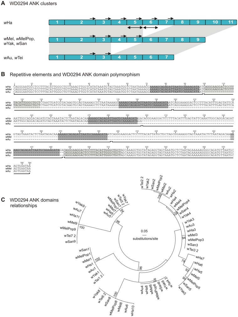
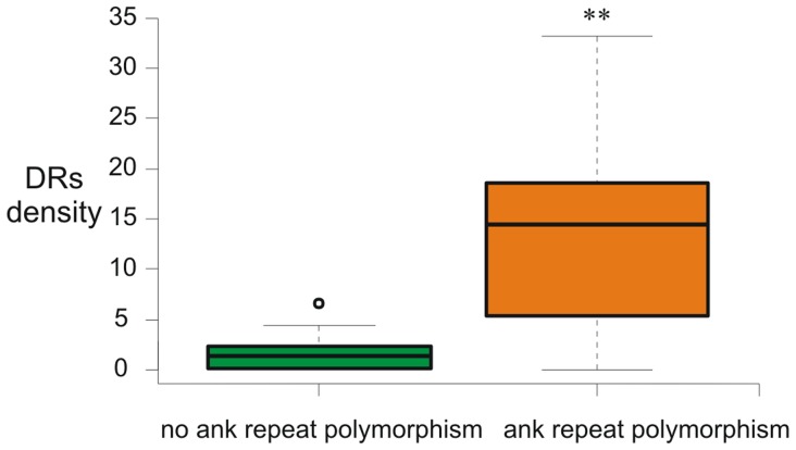
Careful visual inspection of the alignments of the wMel-related ANK genes that display domain number variations revealed that short or long identical direct repeats always flanked the duplicated and/or deleted segments (Figures 1, 2 and supporting Figure S2). For example, the identified deletions in the prophage-associated WD0294-related ANK genes are flanked by two groups of perfect repetitive elements of 26 bp (AAAAGCAGAGATTAATGCAAAAGATA) and 30 bp (CAGGGAAGGACTCCTTTACATTGGGCTGCT) long (Figure 1B). This striking observation suggests that the polymorphism of the number of ankyrin repeat domains depends on the presence of repetitive sequences scattered over the ANK clusters. In order to test the hypothesis that these short repetitive sequences are implicated in the expansion and/or contraction of the ankyrin repeat domains, the density of direct repeats (number of DRs/kb) was estimated for each ANK gene. The estimation was done on direct repeats larger than 8nt with the average length being between 10–20nt (supporting Figure S3). It was also investigated whether genes with ankyrin repeat domain number variations had greater DR density compared to genes that do not display variations. The former genes were indeed found to contain significantly more direct repeats (average of 14.3 and 1.4 repeats/kb, respectively, P<0.005, Mann-Whitney test) (Figure 3).
Figure 1. Repetitive DNA sequences and ankyrin repeat domain number polymorphism.
The example of WD0294. A) Only the ankyrin repeat domain containing regions are shown. Blue rectangles represent individual ankyrin repeat domains. The light gray shading between the ANK clusters indicates homologies between the different strains. Double arrows represent identical duplications. Small black arrows indicate direct repeats capable of engaging into illegitimate recombination. B) Detail of the nucleotide alignment around the deleted region. This region includes the ANK domains 3–8 from wHa, 3–6 from wMel and 3–4 from wAu. Gray rectangles show the position of repeated sites flanging the deletions. ANK repeats are underlined. C) ML phylogenetic relationships between individual ankyrin repeat domains.
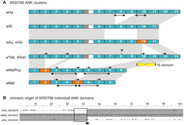
Figure 2. Repetitive DNA sequences and ankyrin repeat domain number polymorphism.
The example of WD0766. A) Only the ankyrin repeat domain containing regions are shown. Blue rectangles represent individual ankyrin repeat domains. Dark gray rectangles with dotted outline represent ankyrin repeat domain remnants. The light gray shading between the ANK clusters indicates homologies between the different strains. Orange rectangles represent putative chimeric ankyrin repeat domains, and double arrows represent identical duplications. Small black arrows indicate direct repeats capable of engaging into illegitimate recombination. The reconstructed structure of disrupted wYak and wSan ANK homologs is also presented. The asterisk and the double yellow arrows correspond to a frame shift mutation and the position of the IS5 element, respectively. B) example of chimeric origin of wMel (and wMelPop) ankyrin repeat domains 2. Identities with the parental ankyrin repeat domains 2 and 4 from wAu are shaded. Box shows the position of the repeated site between the three sequences. The vertical arrow indicates the loop between the two α-helices of the ankyrin repeat domain.
Figure 3. Density of direct repeats larger than 8nt per ANK gene.
Box plot graph showing that ANK genes display ankyrin repeat number variations have greater DR density compared with genes that do not display variations (**P<0.005, Mann-Whitney test).
Almost half of the wMel-related ANK genes studied (WD0073, WD0147, WD0294, WD0385, WD0438, WD0514, WD0550, WD0566, WD0633, WD0754 and WD0766) display variations in the architecture of the ankyrin repeat domains in at least one of the studied Wolbachia strains (Table 2). This is particularly evident in the closely wMel-related strains wMelPop, wAu, wTei, wYak and wSan, in which the wMel-related ANK genes share high sequence similarity at the nucleotide level; however display differences in the number of ankyrin repeat domains. The rest of the genes exhibit similar numbers and organization of ankyrin repeat domains in the different Wolbachia strains.
Duplications and/or deletions of ankyrin repeat domains, including shuffling between the structural elements of physically distant ankyrin repeat domains, are often the reason for copy number variations. For example, the prophage-associated WD0294-related ANK genes display high similarity at the nucleotide level in all strains which harbour it (Table S3). However, in the strains wAu and wTei, the genes code for two ankyrin repeat domains less than those present in the strains wMel, wMelPop, wYak and wSan. On the other hand, in the strain wHa the homolog of the same gene codes for two ankyrin repeat domains more (Figure 1A). Similarly, (a) the WD0514-related ANK genes present in the strains wTei, wYak and wSan code for two ankyrin repeat domains less than those found in the strains wMel and wMelPop and (b) the WD0550-related ANK genes present in the strains wAu and wMelPop code for two ankyrin repeat domains more than those of the strains wMel, wTei, wYak and wSan (data not shown).
The present study confirmed that the WD0766-related ANK genes exhibit extensive variability in both number and organization of the ankyrin repeat domains (Figure 2A) as previously reported [30], [70]. In addition, sequence analysis of the WD0766-related ANK genes in the closely related Wolbachia strains wYak and wSan showed that these genes are disrupted due to the insertion of a full-length IS5 element (a similar phenomenon was reported for the WD0385-related ANK gene in the strain wAu by Iturbe-Ormaetxe et al., 2005) [30] and the presence of a frame-shift upstream of the IS element. The reconstruction of these two ORFs showed that the WD0766-related ANK genes contain 13 complete ankyrin repeat domains, two repeats longer than the WD0766-related ANK gene of the closely related strain wTei and that of the Wolbachia strain wAu. It is worth noting that the 5′ and the 3′ regions of the WD0766-related ANK gene, including the first and the last ankyrin repeat domains, share high conservation between all Wolbachia strains studied. However, extensive duplications and deletions of the internal ankyrin repeat domains interrupt this homology.
Inspection of the multiple alignment of WD0766-related ANK gene sequences reveals that deletion events within the ANK cluster may have resulted in the shuffling of physically distant ankyrin repeat domains. One such putative case is the formation of the second ankyrin repeat domain of the wMel and wMelPop genes. According to the alignment, this ankyrin repeat domain is chimeric. The first 42 bp, which encode the first α-helix of the ankyrin repeat domain (amino acid residues 1–14), are very similar to the corresponding region of the second ankyrin repeat domain of the WD0766-related ANK genes of strains wAu, wTei, wRi and wHa, while the last 57 bp, which encode for the second α-helix (amino acid residues 15–33), are very similar to the fourth ankyrin repeat domain of the same strains (Figure 2B). Similarly, the 10th ankyrin repeat domain of the wAu and wTei WD0766-related ANK genes is probably the result of shuffling between the 10th and the 12th ankyrin repeat domains of the corresponding wYak and wSan genes, while the 5th ankyrin repeat domain of the wMel gene (WD0766) seems to be the result of a shuffling event between the 5th and the 6th ankyrin repeat of the wMelPop WD0766-related ankyrin gene (Figure 2A).
The WD0147-, WD0438-, WD0566- and WD0754-related ANK genes present in the Wolbachia wMel subgroup display no ankyrin-repeat polymorphism. It is also worth noting that the partial sequences of the WD0385-related ANK genes of the wMel subgroup strains, which correspond to the last two ankyrin repeat domains, have identical sequences. However, the corresponding five genes of the wRi strain display extensive variations in number and/or organization of the ankyrin repeat domains, suggesting that they have undergone extensive duplications and rearrangements [10].
The prophage-associated WD0636-related ANK genes present in Wolbachia strains wAu and wTei contain a premature stop codon, due to the insertion of G between position 283 and 284 of the wMel sequence, eliminating the last ankyrin repeat domain of the gene together with the 3′-end of the gene (data not shown). The WD1213-related ANK genes present in the strains wNo, wMau and wMa (all B supergroup strains) are identical to each other; however, they carry eleven indels, three of which cause frame shifts, compared to the WD1213 ANK gene in wMel (data not shown).
Based on the above, the studied wMel-related ANK genes were classified into two major groups: (a) the true orthologs, which are of the same length and have the same number of ankyrin repeat domains and (b) the homologs which differ in the length, copy number and the organization of the ankyrin repeat domains and may also carry structural disruptions, such as frame shifts, deletions and insertions.
Genetic Diversity of ANK Genes
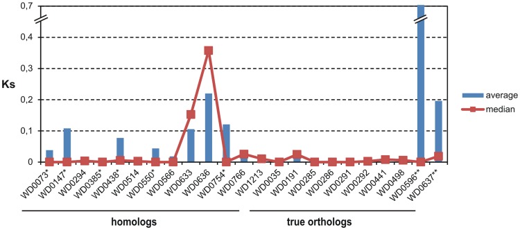
Table S3 summarizes the features of the ANK genes detected in the studied Wolbachia strains, the great majority of which belong to supergroup A. The G+C content ranged from 30.8% (chromosomal ANK gene WD0438) to 42% (phage ANK gene WD0636), with an average of 35%. Overall, the genetic diversity analysis revealed high conservation at the nucleotide level across the ANK genes, as indicated by the synonymous substitutions (Ks) (Figure 4 and Table S3). In general, the majority of the ANK genes of wMel-subgroup strains (wMel, wMelPop, wAu, wTei, wYak and wSan) are highly conserved (almost 100% identity at the nucleotide level) compared to the corresponding genes of the more distantly related strains wRi and/or wHa (distantly related based on MLST analysis [46], [47]), which display the highest degree of genetic variability and in some cases act as outliers.
Figure 4. Comparative analysis of genetic diversity.
The graph represents the patterns of synonymous substitutions within A-supergroup strains. (*: wRi is an outlier, **: wHa is an outlier).
It is worth noting that although the WD0294, WD0514, WD0550 and WD0766 homologs differ in ankyrin repeat domain architecture, they show limited sequence polymorphism (SNPs). However, the WO-B prophage-associated homologs WD0633 and WD0636 exhibit significant levels of genetic diversity (Ks>0.05) (Figure 4 and Table S3), including the two copies of the WD0636 gene detected in the Wolbachia strain wSan (95% identity at nucleotide level - Ks = 0.082).
The sequences of only five (WD0441, WD0498, WD0636, WD0637 and WD01213) of the nine universally occurring ANK genes were retrieved from the three B-supergroup Wolbachia strains wNo, wMau and wMa (Table S2) and showed almost 100% identity. The prophage-associated ANK gene WD0636 was again the exception, exhibiting a high level of genetic diversity (Ksavg = 0.07535) (Table S3). Interestingly, two copies of the WD0636 gene were detected in the strain wNo, which were 89% identical at nucleotide level (Ks = 0.15). It should also be noted that we sequenced a partial fragment (356 bp) of a highly diverged WD0191 ortholog from strain wMau, which exhibited 67.2% identity to A-supergroup orthologs. Overall, there is a greater degree of genetic divergence between supergroups A and B, as indicated by the patterns of synonymous substitutions (Ks) ranging from Ksavg = 0.238 (WD0636) to Ksavg = 0.495 (WD0498; Table S3).
Phylogenetic Analysis
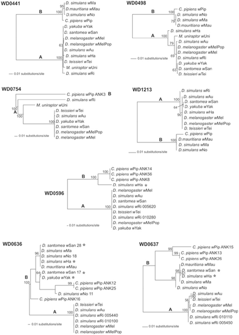
The relationships of the ANK genes were also studied with ML phylogenetic analysis. ANK orthologs with almost 100% identity at nucleotide level (like the WO-A prophage ankyrin genes WD0285 and WD0286) were omitted from the analysis. Most trees clearly reflect the evolutionary divergence of the two major Wolbachia supergroups A and B; however, branch lengths separating the two supergroups are often in disagreement between the different ANK gene-based trees (Figure 5 and supporting Figure S4). The discordance suggests that the rate of evolution varies for different genes, and this is supported by the different levels of genetic diversity observed between these genes. Within supergroup A, the topologies were not significantly different between the datasets, placing all strains in a single clade. However, in some cases the more distantly related strains wRi and wHa branched at different positions, being separated by long genetic distances (WD0498 and WD0754 gene-based trees in Figure 5). This is supported by the highly heterogeneous pattern of genetic diversity observed in different ANK genes in different Wolbachia strains and could reflect different evolutionary rates or even different times of gene acquisition.
Figure 5. Phylogeny of four chromosomal- (WD0441, WD0498, WD0754 and WD1213) and three prophage-associated (WD0596, WD0636, WD0637) ANK genes.
The trees are midpoint-rooted and inferred using maximum likelihood. ML bootstrap support values inferred from 100 replicates are also presented. Bootstrap values lower than 50 are omitted. The discordant positions of strains wYak, wSan and wHa between the chromosomal- and prophage-associated ANK gene phylogenies are highlighted with asterisks. Evolutionary model parameters were estimated with Modeltest under the Akaike Information Criterion: HKY (WD0441,WD1213); TrN+I (WD0498, WD0596); TVM+G (WD0754); TrN+G (WD0636); GTR+I (WD0637).
Perhaps the most interesting observation was that the two WO-B prophage-associated ANK genes WD0636 and WD0637 showed significant topological conflicts with the chromosomal ANK genes and also with other WO-B prophage-associated ankyrin genes (Figure 5). The WD0636 and WD0637 orthologs-based phylogenetic trees strongly support grouping of the two A-group strains wYak and wSan with wHa and the B-supergroup strains wNo, wMau and wMa. To statistically evaluate the topological incongruence, the ML phylogenies of the two prophage-associated ANK genes were compared with the ML phylogenies of the chromosomal ANK genes (WD0441, WD0498 and WD1213), using the SH test. The analysis was restricted to eight Wolbachia strains (wMel, wAu, wTei, wSan, wRi, wHa, wNo and wMau). The likelihood-based SH test for significance of topological differences supports the discordances among topologies of the WD0636 and WD0637 gene-based phylogenies with the topologies of chromosomal ANK genes (Table 3). Interestingly, the phylogenies of WD0636 and WD0637 also showed topological incongruence (SH test P<0.05, data not shown) with other WO-B prophage-associated ANK genes like WD0596 and WD0633. That could be explained either by the presence of multiple prophage elements in the genomes of wYak and wSan, harbouring a different ANK gene repertoire, or due to recombination events between different prophages.
Table 3. Shimodaira-Hasegawa test for the statistical significance of the topological incongruence between alternative chromosomal- and prophage-associated ankyrin genes.
| Data Set | |||||
| Topology | WD0441 | WD0498 | WD0636 | WD0637 | WD1213 |
| WD0441 | 1228.38 | 1054.82 | 497.64*** | 1599.62*** | 1196.26 |
| WD0498 | 1228.56 | 1046.17 | 454.18*** | 1365.17*** | 1200.87 |
| WD0636 | 1624.91*** | 1141.92*** | 399.14 | 1118.92 | 1665.61*** |
| WD0637 | 1624.91*** | 1145.01*** | 409.40 | 1091.34 | 1653.28*** |
| WD1213 | 1228.38 | 1054.82 | 497.64*** | 1565.64*** | 1188.06 |
Values are the likelihood score (-ln L) of a given data set across its own ML tree (boldface), as well as across the alternative tree topologies. Significance levels are based on full optimization.
P<0.0001.
Recombination within ANK Genes
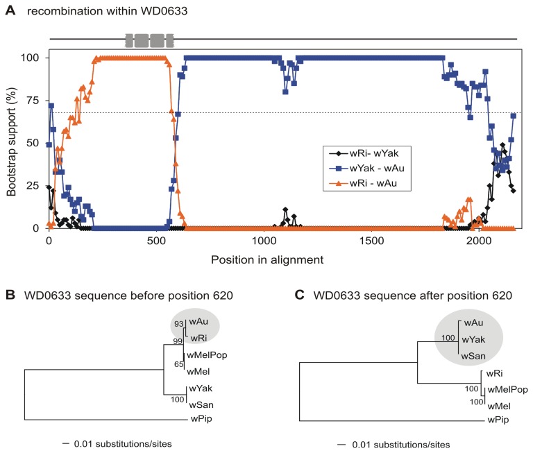
The role of recombination in the evolution of Wolbachia ANK genes was investigated with the program MaxChi [65], [66]. Statistically significant recombination signals were also confirmed by the programs Chimera [65], [66], Geneconv [67], RDP [68] and Bootscan [69]. As summarized in Table 2, five ANK genes, the prophage-associated ANK gene WD0633 and the chromosomal ANK genes WD0073, WD0438, WD0550 and WD0766, exhibited significant evidence of intragenic recombination (MaxChi, P<0.0001 based on 1000 permutations). We present in detail only a single example, that of the prophage-associated ANK gene WD0633. A significant recombination event was detected by MaxChi, as well as by Chimera, Geneconv, RDP, and Bootscan (P<<0.001 based on 1000 permutations), at position 620 of the nucleotide sequence alignment. The breakpoint detected by MaxChi divides the alignment into two parts, the first of which encodes the 3rd and the 4th ankyrin repeat domains, demonstrating exchange of the entire ANK clusters between Wolbachia strains wRi and wAu (Figure 6A). The sequence of the wAu homolog 5′ region with respect to the predicted breakpoint is more similar to the wRi (as well as to the wMel and wMelPop) homologs, while the sequence of the 3′ region of the breakpoint is more similar to the wYak and wSan homologs. Phylogenetic trees reconstructed separately for the two regions (Figure 6B & C) clearly show the shifted position of strain wAu.
Figure 6. Recombination within the prophage-associated ANK gene WD0633.
A) The relative bootstrap support values (1000 bootstrap resampling) are shown, calculated for a moving 200 bp window with a 10bp step size across the alignment of the WD0633 homologs. For each alignment-window, nucleotide distances and phylogenetic trees were produced using the neighbor joining method. The dotted line indicates 70% cutoff. Gray rectangles represent the positions of the ankyrin repeats in the alignment. B, C) ML phylogeny of Wolbachia strains reconstructed separately for the 5′ and 3′ regions of WD0633 supports the group shift of the putative recombinant strain (wAu).
Our results indicate that Wolbachia ANK genes undergo recombination rather frequently, as it was possible to detect significant recombination signals in our small dataset. The possibility that more ANK genes have been involved in recombination events could not be excluded, as the detection of recombination signals might have been masked by extensive sequence variability and the sampling bias. This is supported by the fact that some ANK genes displayed local variations in nucleotide divergences between different strains. For example, the chromosomal ANK gene WD0441 displayed in a comparison between A- and B-supergroup strains, local conservation in the 3′ region of the gene (including the two ankyrin repeat domains) with ∼90% identity, while the 5′ region of the gene displayed extensive variability with less than 50% identity (Figure S5).
Discussion
ANK proteins are involved in numerous and diverse processes and have been suggested to play an important role in host-symbiont interactions [17], [70]–[73]. This study investigated the presence, diversity and evolution of ANK genes in different Drosophila-Wolbachia associations. Our data show that the Wolbachia ANK genes form a rapidly evolving gene family and the plausible mechanisms of their evolution are classical homologous recombination, illegitimate recombination and genomic flux mediated by prophages. These data further confirm that recombination represents a powerful mechanism that accelerates and shapes genomic evolution [74]–[79].
Sequence and phylogenetic analysis of the Wolbachia ANK genes revealed that one of the major causes for the observed sequence polymorphism is recombination, both homologous recombination and illegitimate recombination. Intragenic recombination was observed in both chromosomal and prophage-associated ANK genes. In several cases, intragenic recombination events resulted in the exchange of entire ANK clusters.
An important finding of the present study was the positive correlation of the presence of short DRs scattered over the ankyrin repeat domains with ankyrin repeat domain number polymorphism providing an example of the mechanisms affecting ANK gene evolution. Such short repetitive sequence elements are known to play a major role in DNA deletion and duplication events in both prokaryotes and eukaryotes. Illegitimate recombination events are independent of RecA and are thought to occur by at least two different mechanisms: replication slippage and single-strand annealing [80], [81]. The presence of DRs in the same location with respect to ankyrin repeat domains is important in order to maintain reading frame, structure and hence the function of the ankyrin repeats. Thus, the direct repeats involved in illegitimate recombination, duplication, deletion as well as recombination between distant ankyrin domains, likely play an important role in evolution of Wolbachia ANK genes. Recombination events mediated by short repeated sequences scattered over genes may have resulted in gene deterioration and the production of species-specific orphans, including a putative ANK gene, in the closely related Rickettsia species, R. conori, R. rickettsii and R. montana [82].
It is also important to note that stress-response genes in prokaryotes are known to have a higher than average number of short or large repeats capable of engaging in recombination, probably as a strategy to cope with unstable environmental conditions [83]. One could speculate that the ANK genes may have a similar role in Wolbachia or they may determine the host range and/or tissue tropism, like the ANK proteins of eukaryotic viruses [84]. In addition, it has been suggested that the evolutionary success of the eukaryotic ANK protein family is in part due to their ability to bind to multiple targets by adapting their binding sites through duplication, deletion and shuffling, as a result of alternative exon splicing [71], [72], [85], [86]. It is also believed that modular proteins, like the ANK proteins, evolve faster than non-modular proteins through recombination [72], [86]. Illegitimate recombination, as well as homologous recombination, can act within one genome (including the ANK genes) providing a source of genetic variability needed for Wolbachia strains to rapidly adapt to new environmental conditions.
Bacteriophage Flux and Evolution of Wolbachia ANK Genes
Bacteriophages are major determinants in bacterial genome evolution [87]–[89]. Wolbachia prophage elements are abundant and widespread and can laterally transfer between different Wolbachia strains co-inhabiting the same arthropod host [15], [63], [90]–[93]. A recent study reported that the prophage-associated ANK genes form one of the most divergent groups of Wolbachia genes [14]. Our analysis supports this finding and indicates that the ANK gene “cargo” of a given prophage may differ between strains, as is the case for the five WO-A associated ANK genes that seem to be an independent acquisition in wHa and the wMel-like strains. Furthermore, there is an absence of congruence between the phylogenies of prophage WO-B associated ANK genes not only with the chromosomal but also with other WO-B associated ANK genes. This incongruence is indicative of an active phage, which is able to move horizontally between different Wolbachia strains sharing a common host. This mechanism of genetic exchange was previously suggested for Wolbachia strains including wHa and is known as the “intracellular arena” hypothesis [63], [91]. Lateral phage transfer coupled to intra- and intergenic recombination events could account for this rapid exchange and spreading of ANK genes.
Multiple Phages vs Multiple Infections
Our analysis of the 23 wMel-like ANK genes in different Drosophila-Wolbachia associations revealed striking differences between the strains, as well as between the genes studied. This was particularly evident for the prophage-associated ANK genes, suggesting that the two ANK groups (chromosomal and phage) have different evolutionary histories. However, conclusions about the evolutionary history of some genes may not be easily drawn. For example, the Wolbachia strains wSan (supergroup A) and wNo (supergroup B) contain two copies of the prophage-associated ANK gene WD0636. It is not clear if the two copies are the products of a gene duplication event or belong to different copies of the WO-B prophage. The existence of multiple WO-B prophages has already been described in strain wRi, which harbors two identical copies of a WO-B-like prophage [10], as well as in strain wPip, which harbors five WO-B-like prophage regions [9].
It was recently shown that another prophage-associated gene, the DNA adenine methylatrasnferase gene met2, is present in two copies in the symbiotic association between D. teissieri and wTei; wTei being a strain closely related to wSan. Molecular analysis indicated that the two met2 gene copies are present in two different Wolbachia strains which co-exist in the host D. teissieri [94]. Coinfection may thus explain multiple copies of prophage-associated ANK gene WD0636 in the hosts D. santomea and in D. simulans Noumea. Although the presence of a double infection is rather a speculation for D. santomea (but see also below), it may indeed be the case for D. simulans Noumea. This host was originally doubly infected with two Wolbachia strains, wNo and wHa, before it was established as a wNo mono-infected line through selection [95]. However, recent sequencing of the Wolbachia strain(s) infecting D. simulans Noumea also suggested the presence of wHa in low quantities (supergroup A) [Ellegaard, Klasson, Näslund, Bourtzis and Andersson, unpublished data]. A PCR analysis may thus report genes present in wHa but not in wNo as present in D. simulans Noumea because of the slight contamination with wHa. Indeed, the identification of WD0285 in wNo (Table S2) is an artifact of the presence of this gene in wHa, which is co-infecting at a low concentration.
Riegler et al. [70] recently proposed that polymorphic variable number tandem repeats and ANK genes can be used as a new diagnostic tool for genotyping Wolbachia strains. They also suggested two variable ANK genes (WD0766 and WD0550) for fingerprinting and discrimination between closely related Wolbachia strains belonging to supergroup A, including strains analyzed in the present study. Although the results of the two studies are largely in agreement, two differences deserve clarification. According to Riegler and colleagues, the WD0766-like ANK gene in the wTei strain is disrupted by an IS5 element, as is also the case for strains wYak and wSan. This observation is based on PCR amplicon size similarities between the three stains. Only the wSan gene copy was sequenced. Our sequence analysis clearly demonstrates that the WD0766-like ANK gene of wTei is identical to that of wAu. Furthermore, the number of ankyrin repeat domains of the WD0550-like ANK gene of wSan differs between the two studies (eight ankyrin repeats in Riegler et al.vs six in the present study). While the first difference in WD0766 between the two studies was confirmed, it was raised that for WD0550 wSan was erroneously listed with 8 ankyrin repeat domains in Riegler et al. [70] when it only has 6 (personal communication M Riegler, I Iturbe-Ormaetxe, WJ Miller). There are two possible explanations for the discrepancy regarding WD0776. First, the presence of hidden multiple infections with different infection levels in the Drosophila stocks could account for these differences. As discussed above, there is evidence supporting the presence of more than one Wolbachia strain in D. santomea, as recently documented for the closely related species D. teissieri [94]. An alternative explanation could be different evolutionary events in the two stocks, which could account for the observed differences. Also, Wolbachia IS elements are quite active, and frequent IS5 polymorphisms have been documented for wMel strains [96], wPip strains [97] and across a range of different A-group Wolbachia strains [30], which could also play a role [30], [96]–[98]. These hypotheses can be tested by further analyzing the WD0766-like ankyrin gene in wTei and related strains used in both studies.
Wolbachia Ankyrin Genes: Unknown Origin and Function
Earlier studies also highlighted the genetic diversity between Wolbachia ANK genes, but restricted themselves, however, to an attempt to correlate the observed genetic variability with different CI patterns [30], [32]. According to our results, a correlation between Wolbachia phenotypes and distribution or genetic polymorphism of the different ANK genes is not obvious. Papafotiou et al. [31] showed that two ANK genes, WD0438 and WD1213, show higher expression levels in testes than in ovaries; however, the authors did not detect any evidence that this sex-specific expression is related to CI.
The origin of the Wolbachia ANK genes remains unclear. It has been suggested that prokaryotic ANK genes were acquired from a eukaryotic host rather than evolved independently [99]. However, the discovery of ANK genes in archaea and free-living bacterial species suggests a more ancient origin [19]. The largest numbers of ANK genes in prokaryotes were found in the genomes of Coxiella burnetii, Legionella pneumophila, Rickettsia bellii, Rickettsia felis, Orientia tsutsugamushi and sponge symbiotic bacteria residing within eukaryotic host cells [100]–[106].The presence of a large number of ANK proteins within the genomes of these bacteria may be related to their unique lifestyle. This may also be the case for Wolbachia. Despite the fact that several studies investigated the direct or indirect association of the ANK genes with Wolbachia-induced reproductive phenotypes, mainly with cytoplasmic incompatibility, a functional correlation remains elusive [29]–[32], [70]. The present study does not shed more light on this either. However, our results strongly indicate that phage transfer, homologous recombination and illegitimate recombination have provided Wolbachia with a unique repertoire of ANK genes. Their role for the lifestyle of Wolbachia remains to be established.
Supporting Information
Example of Southern blot analysis. Each membrane was hybridized simultaneously with two probes: a probe specific for the ANK gene under study and a probe specific for the ftsZ gene, which was used as a positive control. A) WD0441 and B) WD0294. 1: wMel, 2: wMelPop, 3: wAu, 4: wTei, 5: wYak, 6: wSan, 7: wRi, 8: wHa, 9: wNo, 10: wMa, 11: wMau.
(TIFF)
Repetitive DNA sequences. Alignments of partial fragments of ANK genes present ankyrin repeat domain polymorphism (A) WD0385, (B) WD0514 and (C) WD0550. Gray rectangles show the position of repeated sites flanging the deletions. ANK repeats are underlined.
(TIF)
Abundance of DRs within ANK genes.
(TIF)
Phylogeny of the ANK genes. The trees are midpoint rooted and inferred using maximum likelihood. ML bootstrap support values inferred from 100 replicates are also presented. Bootstrap values lower than 50 are omitted. Evolutionary model parameters were estimated with Modeltest under the Akaike Information Criterion: K81uf (WD0292, WD0766); TIM (WD0073); HKY (WD0035, WD0291, WD0550); TrN+I (WD0438); TVM+I (WD0191); TVM+G (WD0385, WD0566, WD0633); GTR (WD0147).
(TIF)
Local variation in nucleotide divergences within WD0441-like ANK gene sequences. A sliding window analysis of genetic distance between A- and B- supergroup strains indicates that the two supergroups share more similarities in the 3′ end, which includes the ankyrin repeat domains.
(TIFF)
Primers used in this study.
(DOCX)
Distribution of w Mel-like ANK genes in different Drosophila-Wolbachia associations.
(DOCX)
Comparison of Wolbachia ankyrin repeat genes.
(XLSX)
Acknowledgments
The authors thank Dr. Stefan Oehler for his critical comments on an earlier version of the manuscript. The authors also thank two anonymous reviewers for valuable comments and suggestions which greatly improved the manuscript.
Funding Statement
This research was supported in part by the European Community’s Seventh Framework Programme CSA-SA_REGPROT-2007-1 (under grant agreement no 203590 (KB). LK, SA, HRB and KB benefitted by travel grants in the frame of the EU COST Action FA0701 “Arthropod Symbiosis: from fundamental studies to pest and disease management”. The funders had no role in study design, data collection and analysis, decision to publish, or preparation of the manuscript.
References
- 1. Saridaki A, Bourtzis K (2010) Wolbachia: more than just a bug in insects genitals. Curr Opin Microbiol 13: 67–72. [DOI] [PubMed] [Google Scholar]
- 2. Werren JH, Baldo L, Clark ME (2008) Wolbachia: master manipulators of invertebrate biology. Nat Rev Microbiol 6: 741–751. [DOI] [PubMed] [Google Scholar]
- 3. Cordaux R, Pichon S, Ben Afia Hatira H, Doublet V, Grève P, et al. (2012) Widespread Wolbachia infection in terrestrial isopods and other crustaceans. Zookeys 176: 123–131. [DOI] [PMC free article] [PubMed] [Google Scholar]
- 4. Zug R, Hammerstein P (2012) Still a host of hosts for Wolbachia: Analysis of recent data suggests that 40% of terrestrial arthropod species are infected. PLoS One 7: e38544. [DOI] [PMC free article] [PubMed] [Google Scholar]
- 5. Serbus LR, Casper-Lindley C, Landmann F, Sullivan W (2008) The Genetics and cell biology of Wolbachia-host interactions. Annu Rev Genet 42: 683–707. [DOI] [PubMed] [Google Scholar]
- 6. Cordaux R, Bouchon D, Grève P (2011) The impact of endosymbionts on the evolution of host sex-determination mechanisms. Trends Genet 27: 332–341. [DOI] [PubMed] [Google Scholar]
- 7.Doudoumis V, Alam U, Aksoy E, Abd-Alla AMM, Tsiamis G, et al. (2012) Tsetse-Wolbachia symbiosis: Comes of age and has great potential for pest and disease control. J Invertebr Pathol. [DOI] [PMC free article] [PubMed]
- 8. Foster J, Ganatra M, Kamal I, Ware J, Makarova K, et al. (2005) The Wolbachia genome of Brugia malayi: Endosymbiont evolution within a human pathogenic nematode. PLoS Biol 3: 599–614. [DOI] [PMC free article] [PubMed] [Google Scholar]
- 9. Klasson L, Walker T, Sebaihia M, Sanders MJ, Quail MA, et al. (2008) Genome evolution of Wolbachia strain wPip from the Culex pipiens group. Mol Biol Evol 25: 1877–1887. [DOI] [PMC free article] [PubMed] [Google Scholar]
- 10. Klasson L, Westberg J, Sapountzis P, Näslund K, Lutnaes Y, et al. (2009) The mosaic genome structure of the Wolbachia wRi strain infecting Drosophila simulans . Proc Natl Acad Sci U S A 106: 5. [DOI] [PMC free article] [PubMed] [Google Scholar]
- 11.Salzberg SL, Hotopp JCD, Delcher AL, Pop M, Smith DR, et al. (2005) Serendipitous discovery of Wolbachia genomes in multiple Drosophila species. Genome Biol 6. [DOI] [PMC free article] [PubMed]
- 12. Salzberg SL, Puiu D, Sommer DD, Nene V, Lee NH (2009) Genome sequence of the Wolbachia endosymbiont of Culex quinquefasciatus JHB. J Bacteriol 191: 1725–1725. [DOI] [PMC free article] [PubMed] [Google Scholar]
- 13. Wu M, Sun LV, Vamathevan J, Riegler M, Deboy R, et al. (2004) Phylogenomics of the reproductive parasite Wolbachia pipientis wMel: A streamlined genome overrun by mobile genetic elements. PLoS Biol 2: 327–341. [DOI] [PMC free article] [PubMed] [Google Scholar]
- 14. Ishmael N, Dunning Hotopp JC, Ioannidis P, Biber S, Sakamoto J, et al. (2009) Extensive genomic diversity of closely related Wolbachia strains. Microbiology 106: 5725–5730. [DOI] [PMC free article] [PubMed] [Google Scholar]
- 15. Kent BN, Bordenstein SR (2010) Phage WO of Wolbachia: lambda of the endosymbiont world. Trends Microbiol 18: 173–181. [DOI] [PMC free article] [PubMed] [Google Scholar]
- 16. Leclercq S, Giraud I, Cordaux R (2011) Remarkable abundance and evolution of mobile group II Introns in Wolbachia bacterial endosymbionts. Mol Biol Evol 28: 685–697. [DOI] [PubMed] [Google Scholar]
- 17. Mosavi LK, Cammett TJ, Desrosiers DC, Peng ZY (2004) The ankyrin repeat as molecular architecture for protein recognition. Protein Science 13: 1435–1448. [DOI] [PMC free article] [PubMed] [Google Scholar]
- 18. Sedgwick SG, Smerdon SJ (1999) The ankyrin repeat: a diversity of interactions on a common structural framework. Trends Biochem Sci 24: 311–316. [DOI] [PubMed] [Google Scholar]
- 19. Al-Khodor S, Price CT, Kalia A, Abu Kwaik Y (2010) Functional diversity of ankyrin repeats in microbial proteins. Trends Microbiol 18: 132–139. [DOI] [PMC free article] [PubMed] [Google Scholar]
- 20. Bork P (1993) Hundreds of ankyrin-like repeats in functionally diverse proteins - mobile modules that cross phyla horizontally. Proteins 17: 363–374. [DOI] [PubMed] [Google Scholar]
- 21. Habyarimana F, Al-Khodor S, Kalia A, Graham JE, Price CT, et al. (2008) Role for the Ankyrin eukaryotic-like genes of Legionella pneumophila in parasitism of protozoan hosts and human macrophages. Environ Microbiol 10: 1460–1474. [DOI] [PubMed] [Google Scholar]
- 22. Pan X, Luhrmann A, Satoh A, Laskowski-Arce MA, Roy CR (2008) Ankyrin repeat proteins comprise a diverse family of bacterial Type IV effectors. Science 320: 1651–1654. [DOI] [PMC free article] [PubMed] [Google Scholar]
- 23. Lin MQ, den Dulk-Ras A, Hooykaas PJJ, Rikihisa Y (2007) Anaplasma phagocytophilum AnkA secreted by type IV secretion system is tyrosine phosphorylated by Abl-1 to facilitate infection. Cell Microbiol 9: 2644–2657. [DOI] [PubMed] [Google Scholar]
- 24. Park J, Kim KJ, Choi K, Grab DJ, Dumler JS (2004) Anaplasma phagocytophilum AnkA binds to granulocyte DNA and nuclear proteins. Cell Microbiol 6: 743–751. [DOI] [PubMed] [Google Scholar]
- 25. Rikihisa Y, Lin M (2010) Anaplasma phagocytophilum and Ehrlichia chaffeensis type IV secretion and Ank proteins. Curr Opin Microbiol 13: 59–66. [DOI] [PMC free article] [PubMed] [Google Scholar]
- 26. Caturegli P, Asanovich KM, Walls JJ, Bakken JS, Madigan JE, et al. (2000) ankA: an Ehrlichia phagocytophila group gene encoding a cytoplasmic protein antigen with ankyrin repeats. Infect Immun 68: 5277–5283. [DOI] [PMC free article] [PubMed] [Google Scholar]
- 27. Garcia-Garcia JC, Rennoll-Bankert KE, Pelly S, Milstone AM, Dumler JS (2009) Silencing of host cell CYBB gene expression by the nuclear effector AnkA of the intracellular pathogen Anaplasma phagocytophilum . Infect Immun 77: 2385–2391. [DOI] [PMC free article] [PubMed] [Google Scholar]
- 28. Ijdo JW, Carlson AC, Kennedy EL (2007) Anaplasma phagocytophilum AnkA is tyrosine-phosphorylated at EPIYA motifs and recruits SHP-1 during early infection. Cell Microbiol 9: 1284–1296. [DOI] [PubMed] [Google Scholar]
- 29. Duron O, Boureux A, Echaubard P, Berthomieu A, Berticat C, et al. (2007) Variability and expression of ankyrin domain genes in Wolbachia variants infecting the mosquito Culex pipiens . J Bacteriol 189: 4442–4448. [DOI] [PMC free article] [PubMed] [Google Scholar]
- 30. Iturbe-Ormaetxe I, Burke GR, Riegler M, O’Neill SL (2005) Distribution, expression, and motif variability of ankyrin domain genes in Wolbachia pipientis . J Bacteriol 187: 5136–5145. [DOI] [PMC free article] [PubMed] [Google Scholar]
- 31. Papafotiou G, Oehler S, Savakis C, Bourtzis K (2011) Regulation of Wolbachia ankyrin domain encoding genes in Drosophila gonads. Res Microbiol 162: 764–772. [DOI] [PubMed] [Google Scholar]
- 32. Sinkins SP, Walker T, Lynd AR, Steven AR, Makepeace BL, et al. (2005) Wolbachia variability and host effects on crossing type in Culex mosquitoes. Nature 436: 257–260. [DOI] [PubMed] [Google Scholar]
- 33.Walker T, Klasson L, Sebaihia M, Sanders MJ, Thomson NR, et al. (2007) Ankyrin repeat domain-encoding genes in the wPip strain of Wolbachia from the Culex pipiens group. BMC Biol 5. [DOI] [PMC free article] [PubMed]
- 34. Yamada R, Iturbe-Ormaetxe I, Brownlie JC, O’Neill SL (2010) Functional test of the influence of Wolbachia genes on cytoplasmic incompatibility expression in Drosophila melanogaster . Insect Mol Biol 20: 75–85. [DOI] [PubMed] [Google Scholar]
- 35. Bennuru S, Semnani R, Meng Z, Ribeiro JMC, Veenstra TD, et al. (2009) Brugia malayi excreted/secreted proteins at the host/parasite interface: stage- and gender-specific proteomic profiling. PLoS Negl Trop Dis 3: e410. [DOI] [PMC free article] [PubMed] [Google Scholar]
- 36. Félix C, Pichon S, Braquart-Varnier C, Braig H, Chen L, et al. (2008) Characterization and transcriptional analysis of two gene clusters for type IV secretion machinery in Wolbachia of Armadillidium vulgare . Res Microbiol 159: 481–485. [DOI] [PubMed] [Google Scholar]
- 37. Bourtzis K, Dobson SL, Braig HR, O’Neill SL (1998) Rescuing Wolbachia have been overlooked. Nature 391: 852–853. [DOI] [PubMed] [Google Scholar]
- 38. Bourtzis K, Nirgianaki A, Onyango P, Savakis C (1994) A prokaryotic dnaA sequence in Drosophila melanogaster: Wolbachia infection and cytoplasmic incompatibility among laboratory strains. Insect Mol Biol 3: 131–142. [DOI] [PubMed] [Google Scholar]
- 39. Giordano R, O’Neill SL, Robertson HM (1995) Wolbachia infections and the expression of cytoplasmic incompatibility in Drosophila sechellia and D. Mauritiana . Genetics 140: 1307–1317. [DOI] [PMC free article] [PubMed] [Google Scholar]
- 40. Hadfield SJ, Axton JM (1999) Germ cells colonized by endosymbiotic bacteria. Nature 402: 482. [DOI] [PubMed] [Google Scholar]
- 41. Hoffmann AA, Turelli M, Simmons GM (1986) Unidirectional incompatibility between populations of Drosophila simulans . Evolution 40: 692–701. [DOI] [PubMed] [Google Scholar]
- 42. Hoffmann AA, Clancy D, Duncan J (1996) Naturally-occurring Wolbachia infection in Drosophila simulans that does not cause cytoplasmic incompatibility. Heredity (Edinb) 76: 1–8. [DOI] [PubMed] [Google Scholar]
- 43. McGraw EA, Merritt DJ, Droller JN, O’Neill SL (2001) Wolbachia-mediated sperm modification is dependent on the host genotype in Drosophila . Proc R Soc Lond B Biol Sci 268: 2565–2570. [DOI] [PMC free article] [PubMed] [Google Scholar]
- 44. Veneti Z, Clark ME, Karr TL, Savakis C, Bourtzis K (2004) Heads or tails: Host-parasite Interactions in the Drosophila-Wolbachia system. Appl Environ Microbiol 70: 5366–5372. [DOI] [PMC free article] [PubMed] [Google Scholar]
- 45. Zabalou S, Charlat S, Nirgianaki A, Lachaise D, Mercot H, et al. (2004) Natural Wolbachia infections in the Drosophila yakuba species complex do not induce cytoplasmic incompatibility but fully rescue the wRi modification. Genetics 167: 827–834. [DOI] [PMC free article] [PubMed] [Google Scholar]
- 46. Baldo L, Hotopp JCD, Jolley KA, Bordenstein SR, Biber SA, et al. (2006) Multilocus sequence typing system for the endosymbiont Wolbachia pipientis . Appl Environ Microbiol 72: 7098–7110. [DOI] [PMC free article] [PubMed] [Google Scholar]
- 47. Paraskevopoulos C, Bordenstein SR, Wernegreen JJ, Werren JH, Bourtzis K (2006) Toward a Wolbachia multilocus sequence typing system: Discrimination of Wolbachia strains present in Drosophila species. Curr Microbiol 53: 388–395. [DOI] [PubMed] [Google Scholar]
- 48. Zabalou S, Apostolaki A, Pattas S, Veneti Z, Paraskevopoulos C, et al. (2008) Multiple rescue factors within a Wolbachia strain. Genetics 178: 2145–2160. [DOI] [PMC free article] [PubMed] [Google Scholar]
- 49. Holmes DS, Bonner J (1973) Preparation, molecular-weight, base composition, and secondary structure of giant nuclear ribonucleic-acid. Biochemistry 12: 2330–2338. [DOI] [PubMed] [Google Scholar]
- 50.Sambrook SL FE, Maniatis T (1989) Molecular Cloning - A Laboratory Manual (2nd. Ed.): Cold Spring Harbor Laboratory Press, Cold Spring Harbor, NY.
- 51. Holden PR, Brookfield JFY, Jones P (1993) Cloning and characterization of an ftsZ homolog from a bacterial symbiont of Drosophila melanogaster . Mol Gen Genet 240: 213–220. [DOI] [PubMed] [Google Scholar]
- 52. Edgar RC (2004) MUSCLE: multiple sequence alignment with high accuracy and high throughput. Nucleic Acids Res 32: 1792–1797. [DOI] [PMC free article] [PubMed] [Google Scholar]
- 53.Drummond AJ, Cheung M, Heled J, Kearse M, Moir R, Stones-Havas S, Thierer T, Wilson A (2008) Geneious v4.0. Available from: http://www.geneious.com.
- 54. Rozas J, Sanchez-DelBarrio JC, Messeguer X, Rozas R (2003) DnaSP, DNA polymorphism analyses by the coalescent and other methods. Bioinformatics 19: 2496–2497. [DOI] [PubMed] [Google Scholar]
- 55. Yang Z (2007) PAML 4: Phylogenetic Analysis by Maximum Likelihood. Mol Biol Evol 24: 1586–1591. [DOI] [PubMed] [Google Scholar]
- 56. Goldman N, Yang ZH (1994) Codon-based model of nucleotide substitution for protein-coding DNA-sequences. Mol Biol Evol 11: 725–736. [DOI] [PubMed] [Google Scholar]
- 57. Finn RD, Tate J, Mistry J, Coggill PC, Sammut SJ, et al. (2008) The Pfam protein families database. Nucleic Acids Res 36: D281–D288. [DOI] [PMC free article] [PubMed] [Google Scholar]
- 58. Schultz J, Milpetz F, Bork P, Ponting CP (1998) SMART, a simple modular architecture research tool: Identification of signaling domains. Proc Natl Acad Sci U S A 95: 5857–5864. [DOI] [PMC free article] [PubMed] [Google Scholar]
- 59. Betley JN, Frith MC, Graber JH, Choo S, Deshler JO (2002) A ubiquitous and conserved signal for RNA localization in chordates. Curr Biol 12: 1756–1761. [DOI] [PubMed] [Google Scholar]
- 60. Posada D, Crandall KA (1998) MODELTEST: testing the model of DNA substitution. Bioinformatics 14: 817–818. [DOI] [PubMed] [Google Scholar]
- 61.Swofford DL (2002) PAUP*. Phylogenetic Analysis Using Parsimony (*and Other Methods). Version 4.: Sinauer Associates, Sunderland, Massachusetts.
- 62. Shimodaira H, Hasegawa M (1999) Multiple comparisons of log-likelihoods with applications to phylogenetic inference. Mol Biol Evol 16: 1114–1116. [Google Scholar]
- 63. Bordenstein SR, Wernegreen JJ (2004) Bacteriophage flux in endosymbionts (Wolbachia): Infection frequency, lateral transfer, and recombination rates. Mol Biol Evol 21: 1981–1991. [DOI] [PubMed] [Google Scholar]
- 64. Martin DP, Williamson C, Posada D (2005) RDP2: recombination detection and analysis from sequence alignments. Bioinformatics 21: 260–262. [DOI] [PubMed] [Google Scholar]
- 65. Posada D, Crandall KA (2001) Evaluation of methods for detecting recombination from DNA sequences: Computer simulations. Proc Natl Acad Sci U S A 98: 13757–13762. [DOI] [PMC free article] [PubMed] [Google Scholar]
- 66. Smith JM (1992) Analyzing the mosaic structure of genes. J Mol Evol 34: 126–129. [DOI] [PubMed] [Google Scholar]
- 67. Padidam M, Sawyer S, Fauquet CM (1999) Possible emergence of new geminiviruses by frequent recombination. Virology 265: 218–225. [DOI] [PubMed] [Google Scholar]
- 68. Martin D, Rybicki E (2000) RDP: detection of recombination amongst aligned sequences. Bioinformatics 16: 562–563. [DOI] [PubMed] [Google Scholar]
- 69. Salminen MO, Carr JK, Burke DS, McCutchan FE (1995) Identification of breakpoints in intergenotypic recombinants of HIV type 1 by bootscanning. AIDS Res Hum Retroviruses 11: 1423–1425. [DOI] [PubMed] [Google Scholar]
- 70. Riegler M, Iturbe-Ormaetxe I, Woolfit M, Miller W, O’Neill S (2012) Tandem repeat markers as novel diagnostic tools for high resolution fingerprinting of Wolbachia . BMC Microbiol 12: S12. [DOI] [PMC free article] [PubMed] [Google Scholar]
- 71. Kobe B, Kajava AV (2000) When protein folding is simplified to protein coiling: the continuum of solenoid protein structures. Trends Biochem Sci 25: 509–515. [DOI] [PubMed] [Google Scholar]
- 72. Marcotte EM, Pellegrini M, Yeates TO, Eisenberg D (1998) A census of protein repeats. J Mol Biol 293: 151–160. [DOI] [PubMed] [Google Scholar]
- 73. McGraw EA, O’Neill SL (2004) Wolbachia pipientis: intracellular infection and pathogenesis in Drosophila . Curr Opin Microbiol 7: 67–70. [DOI] [PubMed] [Google Scholar]
- 74. Baldo L, Bordenstein S, Wernegreen JJ, Werren JH (2006) Widespread recombination throughout Wolbachia genomes. Mol Biol Evol 23: 437–449. [DOI] [PubMed] [Google Scholar]
- 75. Baldo L, Lo N, Werren JH (2005) Mosaic nature of the Wolbachia surface protein. J Bacteriol 187: 5406–5418. [DOI] [PMC free article] [PubMed] [Google Scholar]
- 76.Ioannidis P, Hotopp JCD, Sapountzis P, Siozios S, Tsiamis G, et al. (2007) New criteria for selecting the origin of DNA replication in Wolbachia and closely related bacteria. BMC Genomics 8. [DOI] [PMC free article] [PubMed]
- 77. Jiggins FM (2002) The rate of recombination in Wolbachia bacteria. Mol Biol Evol 19: 1640–1643. [DOI] [PubMed] [Google Scholar]
- 78. Posada D, Crandall KA, Holmes EC (2002) Recombination in evolutionary genomics. Annu Rev Genet 36: 75–97. [DOI] [PubMed] [Google Scholar]
- 79. Werren JH, Bartos JD (2001) Recombination in Wolbachia . Curr Biol 11: 431–435. [DOI] [PubMed] [Google Scholar]
- 80. Bzymek M, Lovett ST (2001) Instability of repetitive DNA sequences: The role of replication in multiple mechanisms. Proc Natl Acad Sci U S A 98: 8319–8325. [DOI] [PMC free article] [PubMed] [Google Scholar]
- 81. Rocha EPC (2003) An appraisal of the potential for illegitimate recombination in bacterial genomes and its consequences: From duplications to genome reduction. Genome Res 13: 1123–1132. [DOI] [PMC free article] [PubMed] [Google Scholar]
- 82. Amiri H, Davids W, Andersson SGE (2003) Birth and death of orphan genes in Rickettsia . Mol Biol Evol 20: 1575–1587. [DOI] [PubMed] [Google Scholar]
- 83. Rocha EPC, Matic I, Taddei F (2002) Over-representation of repeats in stress response genes: a strategy to increase versatility under stressful conditions? Nucleic Acids Res 30: 1886–1894. [DOI] [PMC free article] [PubMed] [Google Scholar]
- 84. Werden SJ, McFadden G (2008) The role of cell signaling in poxvirus tropism: The case of the M-T5 host range protein of myxoma virus. Biochim Biophys Acta 1784: 228–237. [DOI] [PubMed] [Google Scholar]
- 85.Cai X, Zhang Y (2006) Molecular evolution of the ankyrin gene family. Mol Biol Evol. [DOI] [PubMed]
- 86. Tripp KW, Barrick D (2004) The tolerance of a modular protein to duplication and deletion of internal repeats. J Mol Biol 344: 169–178. [DOI] [PubMed] [Google Scholar]
- 87. Casjens S (2003) Prophages and bacterial genomics: what have we learned so far? Mol Microbiol 49: 277–300. [DOI] [PubMed] [Google Scholar]
- 88. Moran NA, Degnan PH, Santos SR, Dunbar HE, Ochman H (2005) The players in a mutualistic symbiosis: Insects, bacteria, viruses, and virulence genes. Proc Natl Acad Sci U S A 102: 16919–16926. [DOI] [PMC free article] [PubMed] [Google Scholar]
- 89. Wagner PL, Waldor MK (2002) Bacteriophage control of bacterial virulence. Infect Immun 70: 3985–3993. [DOI] [PMC free article] [PubMed] [Google Scholar]
- 90. Chafee ME, Funk DJ, Harrison RG, Bordenstein SR (2010) Lateral phage transfer in obligate intracellular bacteria (Wolbachia): verification from natural populations. Mol Biol Evol 27: 501–505. [DOI] [PMC free article] [PubMed] [Google Scholar]
- 91. Gavotte L, Henri H, Stouthamer R, Charif D, Charlat S, et al. (2006) A survey of the bacteriophage WO in the endosymbiotic bacteria Wolbachia . Mol Biol Evol 24: 427–435. [DOI] [PubMed] [Google Scholar]
- 92. Masui S, Kamoda S, Sasaki T, Ishikawa H (2000) Distribution and evolution of bacteriophage WO in Wolbachia, the endosymbiont causing sexual alterations in arthropods. J Mol Evol 51: 491–497. [DOI] [PubMed] [Google Scholar]
- 93. Metcalf JA, Bordenstein SR (2012) The complexity of virus systems: the case of endosymbionts. Curr Opin Microbiol 15: 546–552. [DOI] [PMC free article] [PubMed] [Google Scholar]
- 94. Saridaki A, Sapountzis P, Harris HL, Batista PD, Biliske JA, et al. (2011) Wolbachia prophage DNA adenine methyltransferase genes in different Drosophila-Wolbachia associations. PLoS One 6: e19708. [DOI] [PMC free article] [PubMed] [Google Scholar]
- 95. Merçot H, Llorente B, Jacques M, Atlan A, Montchamp-Moreau C (1995) Variability within the Seychelles cytoplasmic incompatibility system in Drosophila simulans . Genetics 141: 1015–1023. [DOI] [PMC free article] [PubMed] [Google Scholar]
- 96. Riegler M, Sidhu M, Miller WJ, O Neill SL (2005) Evidence for a global Wolbachia replacement in Drosophila melanogaster . Curr Biol : CB 15: 1428–1433. [DOI] [PubMed] [Google Scholar]
- 97. Duron O, Lagnel J, Raymond M, Bourtzis K, Fort P, et al. (2005) Transposable element polymorphism of Wolbachia in the mosquito Culex pipiens: evidence of genetic diversity, superinfection and recombination. Mol Ecol 14: 1561–1573. [DOI] [PubMed] [Google Scholar]
- 98. Cordaux R, Pichon S, Ling A, Pérez P, Delaunay C, et al. (2008) Intense transpositional activity of insertion sequences in an ancient obligate endosymbiont. Mol Biol Evol 25: 1889–1896. [DOI] [PMC free article] [PubMed] [Google Scholar]
- 99. Ponting CP, Aravind L, Schultz J, Bork P, Koonin EV (1999) Eukaryotic signalling domain homologues in archaea and bacteria. Ancient ancestry and horizontal gene transfer. J Mol Biol 289: 729–745. [DOI] [PubMed] [Google Scholar]
- 100. Cazalet C, Rusniok C, Bruggemann H, Zidane N, Magnier A, et al. (2004) Evidence in the Legionella pneumophila genome for exploitation of host cell functions and high genome plasticity. Nat Genet 36: 1165–1173. [DOI] [PubMed] [Google Scholar]
- 101. Chien MC, Morozova I, Shi SD, Sheng HT, Chen J, et al. (2004) The genomic sequence of the accidental pathogen Legionella pneumophila . Science 305: 1966–1968. [DOI] [PubMed] [Google Scholar]
- 102. Nakayama K, Yamashita A, Kurokawa K, Morimoto T, Ogawa M, et al. (2008) The whole-genome sequencing of the obligate intracellular bacterium Orientia tsutsugamushi revealed massive gene amplification during reductive genome evolution. DNA Res 15: 185–199. [DOI] [PMC free article] [PubMed] [Google Scholar]
- 103. Ogata H, La Scola B, Audic S, Renesto P, Blanc G, et al. (2006) Genome sequence of Rickettsia bellii illuminates the role of amoebae in gene exchanges between intracellular pathogens. PLoS Genet 2: 733–744. [DOI] [PMC free article] [PubMed] [Google Scholar]
- 104. Ogata H, Renesto P, Audic S, Robert C, Blanc G, et al. (2005) The genome sequence of Rickettsia felis identifies the first putative conjugative plasmid in an obligate intracellular parasite. PLoS Biol3: 1391–1402. [DOI] [PMC free article] [PubMed] [Google Scholar]
- 105. Seshadri R, Paulsen IT, Eisen JA, Read TD, Nelson KE, et al. (2003) Complete genome sequence of the Q-fever pathogen Coxiella burnetii . Proc Natl Acad Sci U S A 100: 5455–5460. [DOI] [PMC free article] [PubMed] [Google Scholar]
- 106. Thomas T, Rusch D, DeMaere MZ, Yung PY, Lewis M, et al. (2010) Functional genomic signatures of sponge bacteria reveal unique and shared features of symbiosis. ISME J 4: 1557–1567. [DOI] [PubMed] [Google Scholar]
Associated Data
This section collects any data citations, data availability statements, or supplementary materials included in this article.
Supplementary Materials
Example of Southern blot analysis. Each membrane was hybridized simultaneously with two probes: a probe specific for the ANK gene under study and a probe specific for the ftsZ gene, which was used as a positive control. A) WD0441 and B) WD0294. 1: wMel, 2: wMelPop, 3: wAu, 4: wTei, 5: wYak, 6: wSan, 7: wRi, 8: wHa, 9: wNo, 10: wMa, 11: wMau.
(TIFF)
Repetitive DNA sequences. Alignments of partial fragments of ANK genes present ankyrin repeat domain polymorphism (A) WD0385, (B) WD0514 and (C) WD0550. Gray rectangles show the position of repeated sites flanging the deletions. ANK repeats are underlined.
(TIF)
Abundance of DRs within ANK genes.
(TIF)
Phylogeny of the ANK genes. The trees are midpoint rooted and inferred using maximum likelihood. ML bootstrap support values inferred from 100 replicates are also presented. Bootstrap values lower than 50 are omitted. Evolutionary model parameters were estimated with Modeltest under the Akaike Information Criterion: K81uf (WD0292, WD0766); TIM (WD0073); HKY (WD0035, WD0291, WD0550); TrN+I (WD0438); TVM+I (WD0191); TVM+G (WD0385, WD0566, WD0633); GTR (WD0147).
(TIF)
Local variation in nucleotide divergences within WD0441-like ANK gene sequences. A sliding window analysis of genetic distance between A- and B- supergroup strains indicates that the two supergroups share more similarities in the 3′ end, which includes the ankyrin repeat domains.
(TIFF)
Primers used in this study.
(DOCX)
Distribution of w Mel-like ANK genes in different Drosophila-Wolbachia associations.
(DOCX)
Comparison of Wolbachia ankyrin repeat genes.
(XLSX)


