

Abstract
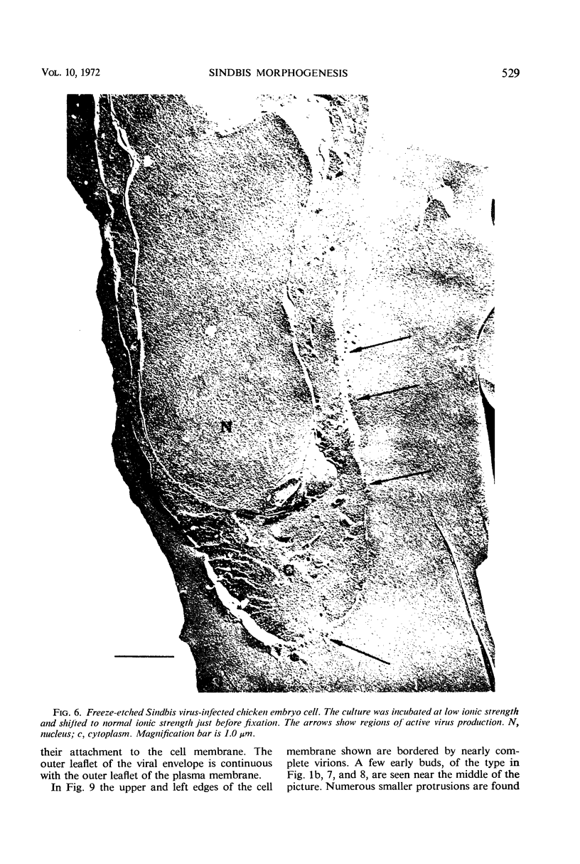
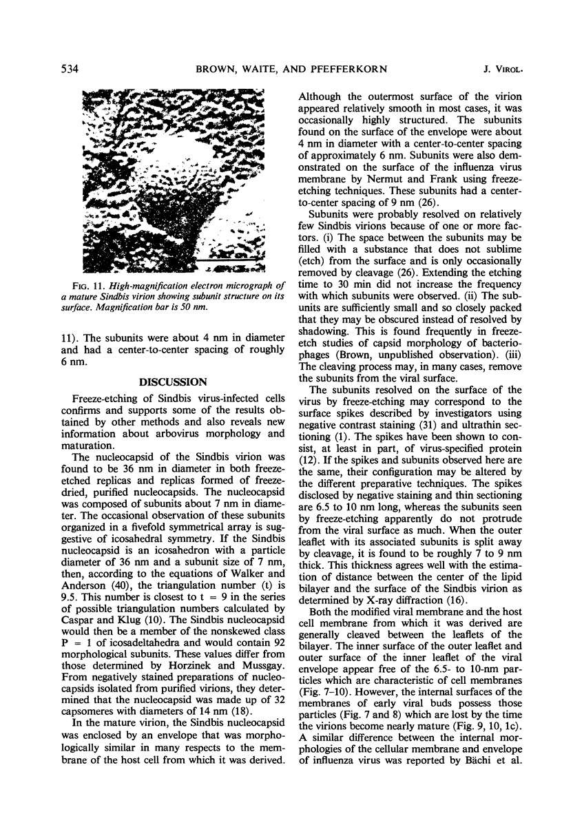
Freeze-etch electron microscope studies of the morphogenesis and morphology of Sindbis virus confirmed results obtained by other workers employing thin-sectioning techniques. The 68-nm virion was found to have a nucleocapsid 36 nm in diameter surrounded by a double-layered, unit membrane. The membranous envelope is acquired as the capsid buds through the plasma membrane of the infected cell. The freeze-etch technique also provided the following new information. (i) At any one time, budding occurs in patches rather than evenly over the cell surface. (ii) The nucleocapsid is composed of capsomers 7 nm in diameter. (iii) The capsid interacts strongly with the membrane, both prior to budding and after maturation. (iv) The 7- to 10-nm particles characteristic of the internal faces of plasma membranes, which presumably represent host membrane proteins, are present in early stages of budding but disappear as morphogenesis progresses. (v) Fusion of the cell membrane at the base of the budding virion is a two-step process; the inner leaflet fuses into a sphere before the outer one. (vi) The outer surface of the viral envelope is covered with 4-nm subunits with a center-to-center spacing of 6 nm.
Full text
PDF


Images in this article


Selected References
These references are in PubMed. This may not be the complete list of references from this article.
- Acheson N. H., Tamm I. Replication of Semliki Forest virus: an electron microscopic study. Virology. 1967 May;32(1):128–143. doi: 10.1016/0042-6822(67)90261-9. [DOI] [PubMed] [Google Scholar]
- Bayer M. E., Remsen C. C. Bacteriophage T2 as seen with the freeze-etching technique. Virology. 1970 Mar;40(3):703–718. doi: 10.1016/0042-6822(70)90215-1. [DOI] [PubMed] [Google Scholar]
- Brown D. T., Brown N. C., Burlingham B. T. Morphology and physical properties of Staphylococcus bacteriophage P11-M15. J Virol. 1972 Apr;9(4):664–671. doi: 10.1128/jvi.9.4.664-671.1972. [DOI] [PMC free article] [PubMed] [Google Scholar]
- Burge B. W., Strauss J. H., Jr Glycopeptides of the membrane glycoprotein of Sindbis virus. J Mol Biol. 1970 Feb 14;47(3):449–466. doi: 10.1016/0022-2836(70)90314-1. [DOI] [PubMed] [Google Scholar]
- Buschmann R. J., Taylor A. B. Extraction of absorbed lipid (linoleic acid-1-14C) from rat intestinal epithelium during processing for electron microscopy. J Cell Biol. 1968 Jul;38(1):252–255. doi: 10.1083/jcb.38.1.252. [DOI] [PMC free article] [PubMed] [Google Scholar]
- Bächi T., Gerhard W., Lindenmann J., Mühlethaler K. Morphogenesis of influenza A virus in Ehrlich ascites tumor cells as revealed by thin-sectioning and freeze-etching. J Virol. 1969 Nov;4(5):769–776. doi: 10.1128/jvi.4.5.769-776.1969. [DOI] [PMC free article] [PubMed] [Google Scholar]
- CASPAR D. L., KLUG A. Physical principles in the construction of regular viruses. Cold Spring Harb Symp Quant Biol. 1962;27:1–24. doi: 10.1101/sqb.1962.027.001.005. [DOI] [PubMed] [Google Scholar]
- Clark A. W., Branton D. Fracture faces in frozen outer segments from the guinea pig retina. Z Zellforsch Mikrosk Anat. 1968;91(4):586–603. doi: 10.1007/BF00455276. [DOI] [PubMed] [Google Scholar]
- Compans R. W. Location of the glycoprotein in the membrane of Sindbis virus. Nat New Biol. 1971 Jan 27;229(4):114–116. doi: 10.1038/newbio229114a0. [DOI] [PubMed] [Google Scholar]
- Dobos P., Faulkner P. Molecular weight of Sindbis virus ribonucleic acid as measured by polyacrylamide gel electrophoresis. J Virol. 1970 Jul;6(1):145–147. doi: 10.1128/jvi.6.1.145-147.1970. [DOI] [PMC free article] [PubMed] [Google Scholar]
- Grimley P. M., Berezesky I. K., Friedman R. M. Cytoplasmic structures associated with an arbovirus infection: loci of viral ribonucleic acid synthesis. J Virol. 1968 Nov;2(11):1326–1338. doi: 10.1128/jvi.2.11.1326-1338.1968. [DOI] [PMC free article] [PubMed] [Google Scholar]
- Holland J. J., Kiehn E. D. Influenza virus effects on cell membrane proteins. Science. 1970 Jan 9;167(3915):202–205. doi: 10.1126/science.167.3915.202. [DOI] [PubMed] [Google Scholar]
- Horzinek M., Mussgay M. Studies on the nucleocapsid structure of a group A arbovirus. J Virol. 1969 Oct;4(4):514–520. doi: 10.1128/jvi.4.4.514-520.1969. [DOI] [PMC free article] [PubMed] [Google Scholar]
- LUFT J. H. Improvements in epoxy resin embedding methods. J Biophys Biochem Cytol. 1961 Feb;9:409–414. doi: 10.1083/jcb.9.2.409. [DOI] [PMC free article] [PubMed] [Google Scholar]
- Laver W. G., Webster R. G. The structure of influenza viruses. IV. Chemical studies of the host antigen. Virology. 1966 Sep;30(1):104–115. doi: 10.1016/s0042-6822(66)81014-0. [DOI] [PubMed] [Google Scholar]
- Moody M. F. Structure of the sheath of bacteriophage T4. I. Structure of the contracted sheath and polysheath. J Mol Biol. 1967 Apr 28;25(2):167–200. doi: 10.1016/0022-2836(67)90136-2. [DOI] [PubMed] [Google Scholar]
- Nermut M. V., Frank H. Fine structure of influenza A2 (Singapore) as revealed by negative staining, freeze-drying and freeze-etching. J Gen Virol. 1971 Jan;10(1):37–51. doi: 10.1099/0022-1317-10-1-37. [DOI] [PubMed] [Google Scholar]
- PFEFFERKORN E. R., CLIFFORD R. L. THE ORIGIN OF THE PROTEIN OF SINDBIS VIRUS. Virology. 1964 Jun;23:217–223. doi: 10.1016/0042-6822(64)90285-5. [DOI] [PubMed] [Google Scholar]
- STEERE R. L. Electron microscopy of structural detail in frozen biological specimens. J Biophys Biochem Cytol. 1957 Jan 25;3(1):45–60. doi: 10.1083/jcb.3.1.45. [DOI] [PMC free article] [PubMed] [Google Scholar]
- Scheele C. M., Pfefferkorn E. R. Virus-specific proteins synthesized in cells infected with RNA+ temperature-sensitive mutants of Sindbis virus. J Virol. 1970 Mar;5(3):329–337. doi: 10.1128/jvi.5.3.329-337.1970. [DOI] [PMC free article] [PubMed] [Google Scholar]
- Simpson R. W., Hauser R. E. Basic structure of group A arbovirus strains Middelburg, Sindbis, and Semliki Forest examined by negative staining. Virology. 1968 Feb;34(2):358–361. doi: 10.1016/0042-6822(68)90248-1. [DOI] [PubMed] [Google Scholar]
- Strauss J. H., Jr, Burge B. W., Darnell J. E. Carbohydrate content of the membrane protein of Sindbis virus. J Mol Biol. 1970 Feb 14;47(3):437–448. doi: 10.1016/0022-2836(70)90313-x. [DOI] [PubMed] [Google Scholar]
- Strauss J. H., Jr, Burge B. W., Pfefferkorn E. R., Darnell J. E., Jr Identification of the membrane protein and "core" protein of Sindbis virus. Proc Natl Acad Sci U S A. 1968 Feb;59(2):533–537. doi: 10.1073/pnas.59.2.533. [DOI] [PMC free article] [PubMed] [Google Scholar]
- Tan K. B. Electron microscopy of cells infected with Semliki forest virus temperature-sensitive mutants: correlation of ultrastructural and physiological observations. J Virol. 1970 May;5(5):632–638. doi: 10.1128/jvi.5.5.632-638.1970. [DOI] [PMC free article] [PubMed] [Google Scholar]
- Waite M. R., Brown D. T., Pfefferkorn E. R. Inhibition of Sindbis virus release by media of low ionic strength: an electron microscope study. J Virol. 1972 Sep;10(3):537–544. doi: 10.1128/jvi.10.3.537-544.1972. [DOI] [PMC free article] [PubMed] [Google Scholar]
- Waite M. R., Pfefferkorn E. R. Effect of altered osmotic pressure on the growth of Sindbis virus. J Virol. 1968 Jul;2(7):759–760. doi: 10.1128/jvi.2.7.759-760.1968. [DOI] [PMC free article] [PubMed] [Google Scholar]
- Waite M. R., Pfefferkorn E. R. Inhibition of Sindbis virus production by media of low ionic strength: intracellular events and requirements for reversal. J Virol. 1970 Jan;5(1):60–71. doi: 10.1128/jvi.5.1.60-71.1970. [DOI] [PMC free article] [PubMed] [Google Scholar]
- Walker D. H., Jr, Anderson T. F. Morphological variants of coliphage P1. J Virol. 1970 Jun;5(6):765–782. doi: 10.1128/jvi.5.6.765-782.1970. [DOI] [PMC free article] [PubMed] [Google Scholar]