
Figure 2.
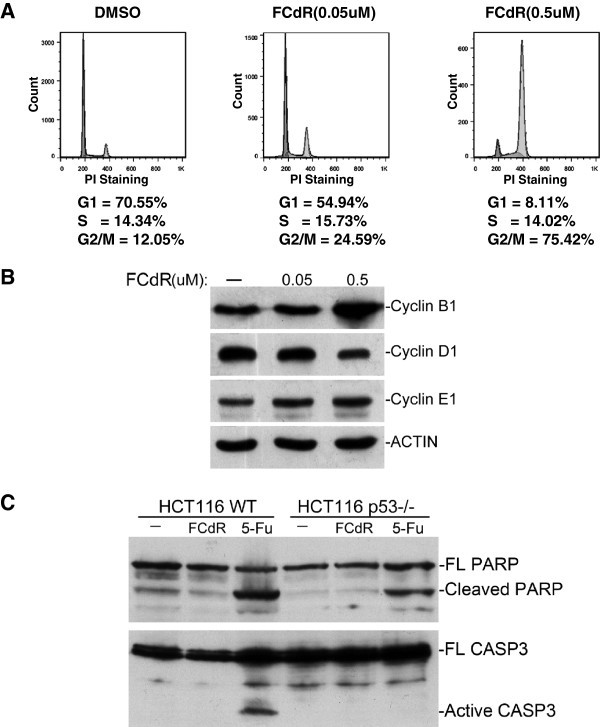
FCdR arrested HCT116 cells at G2/M phase.A. HCT116 cells were treated with 0.05 μM or 0.5 μM FCdR for 48 h and cell cycle was analyzed by flow cytometry. Compared with control, percentage of cells at G2/M phase increased significantly after FCdR treatment. B. HCT116 cells were treated with indicated amount of FCdR for 48 h and the cyclins checked by western blot. CyclinB1, which accumulates at G2/M phase, significantly increased after FCdR treatment. C. HCT116 p53+/+ and p53−/− cells were treated with 0.5 μM FCdR or 375 μM 5-Fu for 48 h and apoptosis assayed by PARP and CASP3 western blot. 5-Fu treatment caused cleavage of PARP and CASP3, but not FCdR.