Abstract
Squamous cell carcinoma of the gallbladder is rare and constitutes only 0.5-3% of all malignancies of this organ. Most of the reported cases have had a component of adenocarcinoma. We report a 70-year-old man who presented with acute onset right upper quadrant pain. He operated on based on a presumptive diagnosis of acute cholecystitis according to clinical and ultrasonographic findings. Histopathological examination of the infiltrating mass of the gallbladder revealed well differentiated keratinized squamous cell carcinoma invading full wall thickness. Thorough evaluations revealed no other primary site for the tumor. Pure primary squamous cell carcinoma of the gallbladder is rarely reported. Clinicians and pathologists must be aware of its vague clinical presentations.
Key Words: Gallbladder, Squamous cell carcinoma, Acute cholecystitis
Introduction
Carcinoma of the gallbladder is more common in women and usually seen in patients older than 50 years of age. It is more common in the white population than the black and in western countries than the Mediterranean.1 The relation between gallstone and gallbladder carcinoma remain controversial. Adenocarcinoma is the most common malignant neoplasm of the gallbladder.1 Although areas of squamous differentiation is seen in adenocarcinoma, pure primary squamous cell carcinoma is rarely reported and accounts for less than 1% of all gallbladder malignancies.1
The histogenesis of squamous cell carcinoma of the gallbladder has not been well understood. Some researchers have stated that squamous cell carcinoma originates from pre-existing squamous metaplasia of the gallbladder epithelium, while others concluded that it originates from squamous differentiation of neoplastic cells of adenocarcinoma.1,2
We present a pure case of squamous cell carcinoma with some areas of squamous metaplasia in the vicinity of the invasive tumor. Therefore, the former theory is more compatible with this case. Another purpose of this case presentation was to emphasize on the vague clinical presentation of gallbladder carcinoma.
Case Description
A 70-year-old man presented with acute onset right upper quadrant abdominal pain and fever since two days prior to admission. The patient was relatively well except for a controlled essential hypertension. On physical examination he was acutely ill and mildly icteric without respiratory distress. He was also febrile with an orally obtained temperature of 38.5°C. His pulse rate and blood pressure were 100 /min and 150/90 mmHg, respectively. The abdomen was tender but there was no physical sign of peritonitis. Examination of heart and lungs were unremarkable. Laboratory data showed leukocytosis and neutrophilia, with a shift to the left. The erythrocyte sedimentation rate was 55 mm/hr. Liver function tests showed total protein: 7.2 g/dL, Alb: 4.1 g/dL, ALT:40 IU, AST: 38 IU, Alkaline phosphatase: 150 IU, total bilirubin: 2.3 mg/dL, and direct bilirubin: 1.8 mg/dL. Other serum chemistry profiles were unremarkable.
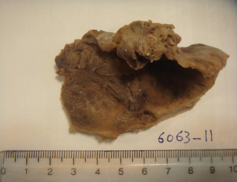
Abdominal ultrasonography showed thickened gallbladder wall without gallstone in favor of acute acalculus cholecystitis. With the presumptive diagnosis of acute cholecystitis, the patient received supportive care and antibiotics. However, he finally underwent cholecystectomy. The patient’s condition was well three days after operation. Gross examination of the gallbladder revealed an ill-defined infiltrating creamy white mass in the body of the gallbladder measuring 3×2×2 cm with focal exophytic configurations (figure 1).
Figure 1.
Gross appearance of the squamous cell carcinoma shows the infiltrative tumor and a focal fungating configuration.
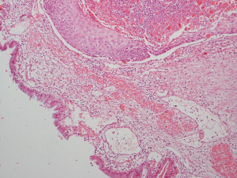
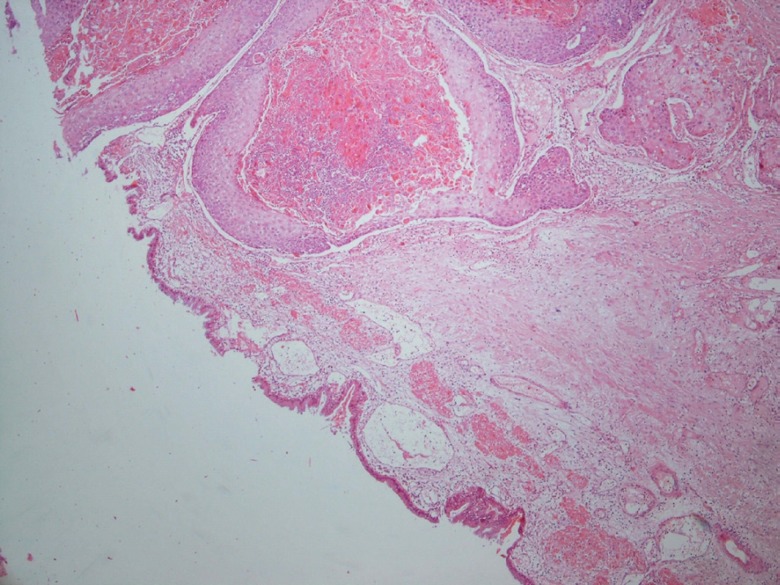
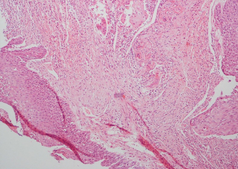
There was no hemorrhage or necrosis. The cystic duct was partially oblitrated by the tumor. Microscopic examination of the mass showed well differentiated keratinized squamous cell carcinoma invading full wall thickness to the serosal surface (figures 2 and 3). The keratinization was extensive with numerous keratohyalin pearls and dyskeratotic cells. No lymph node or liver tissue was submitted for pathological examination. The mucosa showed mature squamous metaplasia in the vicinity of the tumor (figure 4). The surgical resected margin of the cystic duct was involved by the tumor. The tumor lacked any glandular differentiation. In the follow-up visits all examinations were negative for the primary origin of the squamous cell carcinoma and the patient was well in a follow-up period of 6 months.
Figure 2.
This figure shows well differentiated keratinized squamous cell carcinoma is invading through the wall of the gallbladder (H&E×100).
Figure 3.
This figure shows areas of extensive keratinization is shown in invasive squamous cell carcinoma (H&E×400).
Figure 4.
This figure shows mature squamous metaplasia of the gallbladder mucosa is shown in the vicinity of the tumor (H&E×400).
Discussion
Adenocarcinoma is the most common histological subtype of gallbladder cancer constituting about 90-95% of the cases. Although areas of squamous differentiation are seen in some reported cases, pure squamous cell carcinoma of the gallbladder is very rare.1 Traditionally it has been said that adenosquamous and squamous cell carcinomas have very poor prognosis. However, currently it is important to differentiate adenocarcinoma with squamous differentiation from pure squamous cell carcinoma because of the better prognosis of the latter.
Pure squamous cell carcinoma of the gallbladder grows slowly, is usually localized and rarely metastasized. On the other hand adenosquamous carcinoma is aggressive and metastasizes widely.1,2 Roa and colleagues studied 606 carcinomas of the gallbladder. 34 cases showed squamous differentiation and only 1% had pure squamous cell carcinoma. The female/male ratio was 3.8%, similar to adenocarcinoma. The mean age of the patients with squamous cell carcinoma was 65 years and only 13% of patients were suspected for carcinoma preoperatively.1
The etiology and pathogenesis of squamous cell carcinoma is not well understood; however, two important presumptive causative possibilities are gallstones and parasitic infestation.2,3 Another important pathogenetic clue for squamous cell carcinoma is the metaplasia-dysplasia-carcinoma sequence. Most of the cases with squamous cell carcinoma show some degrees of atypical epithelial change adjacent to the invasive tumor.3,4 Gupta and co-workers reported a case of primary squamous cell carcinoma presenting as acute cholecystitis. The patient was operated on after 12 hours and cholecystectomy showed wall thickening with multiple gallstones.5 Other researchers reported a case of primary squamous cell carcinoma of the gallbladder in an elderly lady with infiltration to the adjacent hepatic parenchyma.6 Rai and colleagues concluded that pure squamous cell carcinoma of the gallbladder was less aggressive than adenocarcinoma. This type of carcinoma should be suspected when the lesion reaches a large size without metastasis.7
We presented a 70-year-old man diagnosed as having acute cholecystitis based on clinical examinations and ultrasonographic findings. The patient lacked general signs of malignancy such as weight loss or apparent jaundice. He was completely asymptomatic except for the presentation of acute cholecystitis.
In our case the subtle clinical presentation could be in favor of the less aggressive behavior of pure squamous cell carcinoma of the gallbladder in comparison with adenocarcinoma or adenosquamous variants, which was reported in some previous studies.
Conclusion
Pure primary squamous cell carcinoma of the gallbladder is rarely reported. Clinicians and pathologists must be aware of its vague clinical presentations.
Conflict of Interest: None declared
References
- 1.Roa JC, Tapia O, Cakir A, Basturk O, Dursun N, Akdemir D, et al. Squamous cell and adenosquamous carcinomas of the gallbladder: clinicopathological analysis of 34 cases identified in 606 carcinomas. Mod Pathol. 2011;24:1069–78. doi: 10.1038/modpathol.2011.68. doi: 10.1038/modpathol.2011.68. PubMed PMID: 21532545. [DOI] [PubMed] [Google Scholar]
- 2.Roppongi T, Takeyoshi I, Ohwada S, Sato Y, Fujii T, Honma M, et al. Minute squamous cell carcinoma of the gallbladder: a case report. Jpn J Clin Oncol. 2000;30:43–5. doi: 10.1093/jjco/hyd010. doi: 10.1093/jjco/hyd010. PubMed PMID: 10770570. [DOI] [PubMed] [Google Scholar]
- 3.Karasawa T, Itoh K, Komukai M, Ozawa U, Sakurai I, Shikata T. Squamous cell carcinoma of gallbladder--report of two cases and review of literature. Acta Pathol Jpn. 1981;31:299–308. PubMed PMID: 7257770. [PubMed] [Google Scholar]
- 4.Mingoli A, Brachini G, Petroni R, Antoniozzi A, Cavaliere F, Simonelli L, et al. Squamous and adenosquamous cell carcinomas of the gallbladder. J Exp Clin Cancer Res. 2005;24:143–50. PubMed PMID: 15943044. [PubMed] [Google Scholar]
- 5.Gupta S, Gupta SK, Aryya NC. Primary squamous cell carcinoma of gallbladder presenting as acute cholecystitis. Indian J Pathol Microbiol. 2004;47:231–3. PubMed PMID: 16295480. [PubMed] [Google Scholar]
- 6.Rekik W, Ben FadhelC, Boufaroua AL, Mestiri H, Khalfallah MT, Bouraoui S, et al. Case report: Primary pure squamous cell carcinoma of the gallbladder. J Visc Surg. 2011;148:e149–51. doi: 10.1016/j.jviscsurg.2011.03.004. doi: 10.1016/j.jviscsurg.2011.03.004. PubMed PMID: 21497148. [DOI] [PubMed] [Google Scholar]
- 7.Rai A, Ramakant , Kumar S, Pahwa HS, Kumar S. Squamous cell carcinoma of the gallbladder: an unusual presentation. The Internet Journal of Surgery. 2009:22. [Google Scholar]


