Abstract
The purposes of this study were to compare lower-extremity kinematics during a 45° and 90° cutting maneuver and to examine the relationships between lower-extremity rotations during these maneuvers. The hypotheses tested were that greater internal hip and knee rotation angles would be observed during the cutting maneuver at a 90° angle (90° cut) compared with the maneuver performed at a 45° angle (45° cut) and that the increased internal hip and knee rotation would be related to increased knee abduction measures. Nineteen athletes from women’s soccer teams (17.6 ± 2.1 yr, 165.6 ± 8.2 cm, 60.2 ± 5.6 kg) were instructed to jump across a line and cut at the appropriate angle (either 45° or 90° side-step cut) and in the appropriate direction. Lower-extremity kinematic measures were taken at peak force during the stance phase. Hip internal rotation and knee internal rotation (p = 0.008) were increased during the 90° cut compared with the 45° cut. Mean hip flexion (p < 0.001) was also greater in the 90° cut. The only significant predictor of knee abduction during both tasks was hip adduction (R = 0.49). The findings indicate that the mechanisms underlying increased knee abduction measures in athletic women during cutting tasks were primarily coronal plane motions at the hip. Trunk and hip focused strength neuromuscular training may improve the ability of athletic women to increase control of lower-extremity alignment. Therefore, these women may decrease dangerous knee loads that result from increased hip adduction during dynamic tasks, thus decreasing anterior cruciate ligament injury risk.
Keywords: female sports, knee injury prevention, high risk biomechanics, reaction cutting, agility
Introduction
Anterior cruciate ligament (ACL) injuries have significant traumatic short-term (i.e., 1–5 yr) effects on academic achievement, sports performance, and psychosocial well being (12), as well as long-term (i.e., 5–50 yr) consequences to the health of the knee joint (21,33). Long-term knee problems are detrimental to overall health and activity of the individual, specifically with regard to the development of chronic osteoarthritis (33). Athletic women demonstrate a 3- to 3.6-fold higher rate of sustaining an ACL injury relative to athletic men competing in the same sports (1). With this increased risk of injury for women athletes as well as the ever increasing amount of women of all ages competing in sports, the current literature has been focused to investigate the multiple etiologies of the ACL injury. One potential underlying mechanism of the ACL rupture is associated with lower-extremity motions in the coronal, sagittal, and transverse planes (4,35). However, it appears that motions and torques in the coronal plane may be more sensitive and specific to predict increased risk of noncontact ACL injury risk factors and the inciting mechanism as compared with other planes (15,18,24,35).
Two common maneuvers, landing from a jump and pivoting with a rapid change in direction while running (i.e., cutting), may be related in part to high-risk coronal plane mechanics and resultant ACL injury (4,8,11,35). Hewett et al. (15) demonstrated that increased measures of lower-extremity valgus, specifically knee abduction, during landing were related to increased risk of future ACL injury. Similar predictive models during cutting maneuvers have not yet been identified. When performing cutting maneuvers, young women use increased knee valgus kinematics when compared with young men (25). These data lead to the possibility that prospective testing of cutting techniques, combined with landing evaluations, may provide a more comprehensive screen for athletic young women at high risk for ACL injury (11,15,25). A combination of testing measures that includes landing and cutting may increase the sensitivity and specificity of predictive models for ACL injury and increase their general use to a wider range of athletes (15).
Rotations at the hip may also contribute to dangerous coronal plane kinematics that lead to knee ligament injuries. Decreased hip abduction strength may be related to injury status in athletes (20). Athletic men demonstrate greater hip abduction angles than athletic women during sport maneuvers that challenge core stability (20). Compared with running tasks, cutting maneuvers increase lower-extremity coronal and transverse plane loads that can place the knee ligaments at increased risk of injury in young men (3). Athletic women, however, demonstrate greater maximum hip adduction angles, torques, and excursions during agility and landing maneuvers compared with young men (10,13) The increased external hip adduction moment may indicate that young women have difficulty controlling coronal plane motion of the hip, especially resisting adduction, during dynamic sports movements. Thus, considering the dynamic relationship between segments of the kinetic chain, particularly the hip and knee, asymmetry of abductor/adductor muscle activation at the hip may lead to the dynamic valgus position at the knee exhibited in landing patterns of young women (3,7,8,11,16,17,25).
Although the evaluation of coronal plane motion is important for the determination of relative ACL injury risk in athletes, it also appears important for the determination of the related motions in the sagittal and transverse planes. For example, Olsen and colleagues (35) reported that a non-contact cutting maneuver with a forceful valgus motion, coupled with lower-extremity rotation, was the most common mechanism of ACL injury in their study cohort of athletic women. Markolf et al. (3,15,35) demonstrated that valgus moments combined with anterior tibial force in the flexed knee increased ACL loading and potential injury risk. In addition, coupled lower-extremity rotations, especially those that increase during cutting in the coronal and transverse plane, may underlie resultant motions and loads associated with ACL injury risk during dynamic tasks. An enhanced understanding of the associated coupled lower-extremity joint motions in all planes of movement may help to determine distinct movement strategies that can predict increased ACL injury risk in athletic women. The determination of those athletic women at high risk for ACL injury may allow them to be directed to interventions that can reduce the potential for risk of future injury.
The current literature indicates that unanticipated maneuvers, in comparison with the anticipated tasks, may increase the risk of injury by inducing greater varus/valgus and internal/external rotation moments applied to the knee (2). However, it is unknown whether the cutting angle of unanticipated maneuver induces different hip or knee mechanics. Pollard et al. (37) reported that men and women collegiate soccer players showed no difference in hip and knee mechanics except that young women had less peak hip abduction during an unanticipated 45° cutting task. It is not clear whether this sex difference is greater at higher cutting angles. Thus, the purposes of this study were to compare lower-extremity kinematics during a 45° and 90° cut and to examine the relationship between increased cut angles on resultant lower-extremity kinematics. The first hypothesis tested was that greater internal hip and internal knee rotation angles would be observed during the performed cutting maneuver at a 90° angle (90° cut) compared with the maneuver at a 45° angle (45° cut). The second hypothesis tested in this study was that the increased internal hip and knee rotations induced by the 90° cut would be related to increased knee abduction measures.
Methods
Experimental Approach to the Problem
The only prospective study (15) to demonstrate the predictive measures for ACL injury using whole-body biomechanical analysis techniques used outcome test measures obtained during a box drop vertical jump. Prior investigations indicate that the 2 main mechanisms of ACL injury in young women during sport occur when landing from a jump and when performing a deceleration and cut maneuver (4,35). Because it is not clear whether cutting angle is related to increased “high-risk” biomechanics (dynamic valgus) (15), we chose to test young women athletes performing cutting angles similar to those achieved during sports competition. On the basis of experimental data of valgus-angle differences between cutting maneuvers (25) and internal rotation differences between cutting maneuvers (19), 2 different power analyses were performed to verify appropriate sample size for the current project. These power analyses indicated that a minimum of 13 (25) to 16 (19) participants were required to achieve 90% statistical power in the current study, with an exploratory alpha level of 0.05.
Subjects
Nineteen local area high school and college female soccer players were recruited for three-dimensional biomechanical testing. All athletes were competing at the varsity level, either high school or college, with varying degrees of strength training experience. They were tested during off-season training. Subjects with a prior history of knee injury or surgery were excluded from the study, and all participants indicated no lower-extremity injury at the time of data collection. After signing a consent approved by the institutional review board, each participant’s age, height, and weight were assessed (17.6 ± 2.1 yr, 165.6 ± 8.2 cm, 60.2 ± 5.6 kg).
Procedures
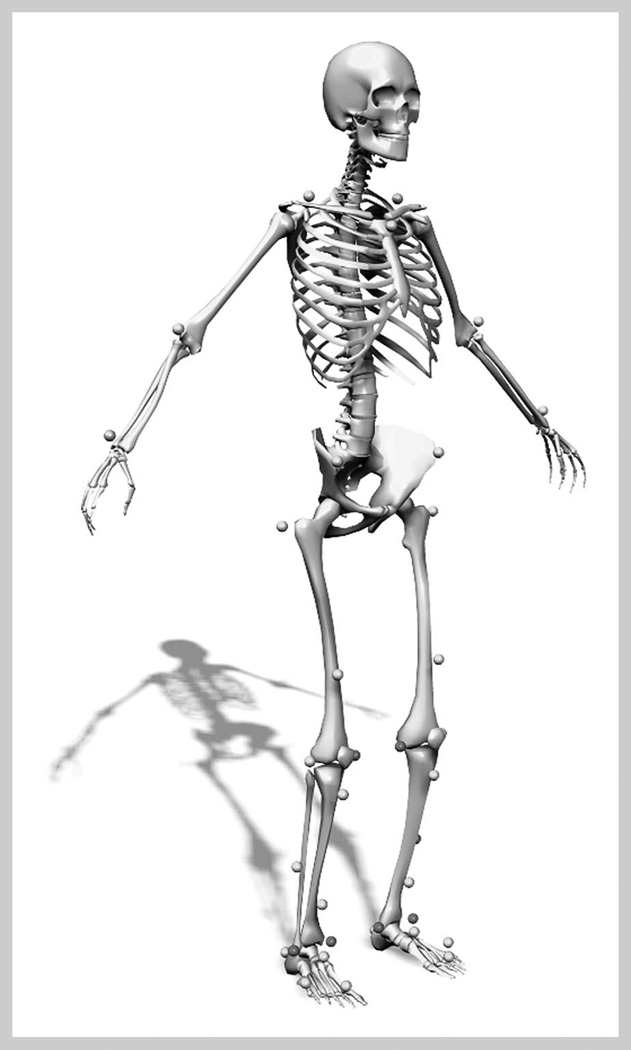
The same investigator placed the 37 retroreflective markers on each athlete on the sacrum, left posterior superior iliac spine (PSIS), and sternum and then bilaterally on the shoulder, elbow, wrist, anterior superior iliac spine (ASIS), greater trochanter, mid thigh, medial and lateral knee, tibial tubercle, mid shank, distal shank, medial and lateral ankle, heel, dorsal surface of the midfoot, lateral foot (5th metatarsal), and toe (between 2nd and 3rd metatarsals) (Figure 1). A static trial was first collected as the participant was instructed to stand still and was aligned as closely with the laboratory coordinate system as possible. This postition was designated as the participant’s neutral (zero) alignment, and subsequent kinematic measures were related back to this position.
Figure 1.
Static trial conducted to align participant with laboratory coordinate system. This alignment was designated as participant’s neutral position, and subsequent kinematic measures were related back to this position.
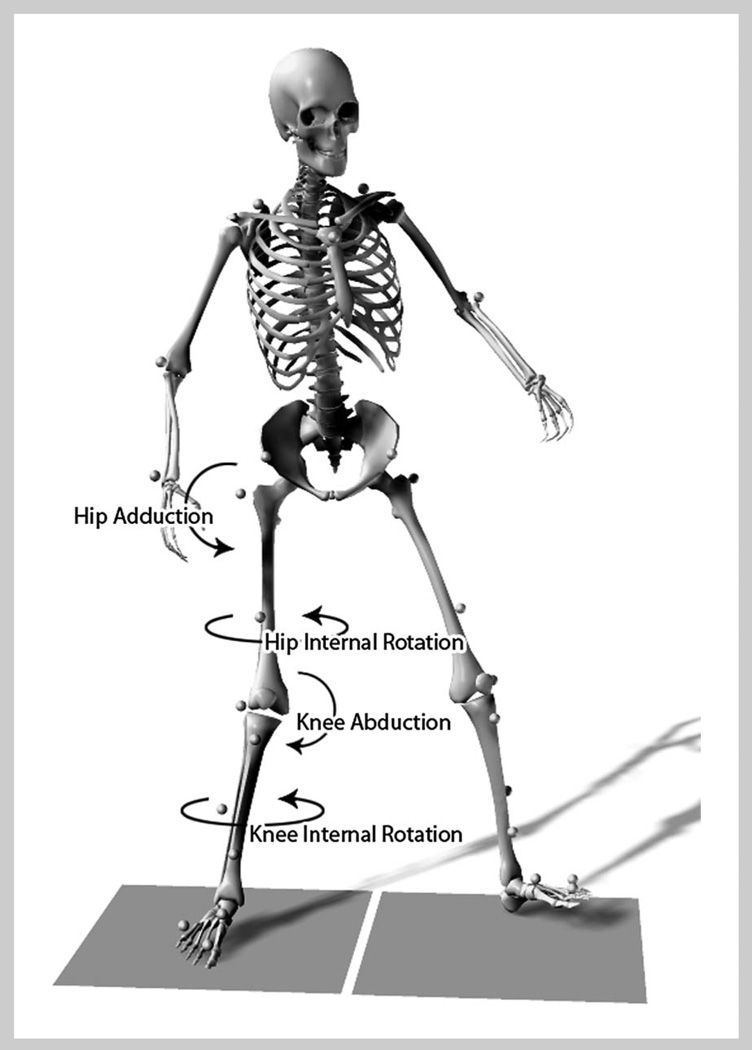
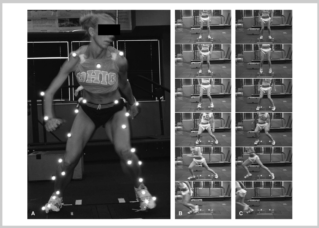
Each participant was then shown the 2 maneuvers (the 45° cut and the 90° cut) and allowed to practice each movement. Figure 2 presents the joint rotation profile during the cutting maneuvers. The athlete was instructed to line up behind a black line with feet 36 cm apart in an athletic position. At this time, they were informed as to which randomly selected cut to make, the 45° or the 90° cut. A custom computer program (KRF) simulated a stoplight on a monitor (red, yellow, green) and was used to cue the participant when to initiate the forward jump (0.4 m) (Figure 3, A and B). The same computer monitor displayed a randomized unanticipated arrow, shown 0.3 seconds after initiation of jump, directing the participant to cut in the appropriate direction. The athlete jumped across the designated line when signaled by the stoplight and then completed the appropriate side-step cut in the direction according to the arrow. The 2 force plates were situated beyond the black line and 8 cm apart so that each foot would contact a different platform during the tasks.
Figure 2.
Joint rotation profile during cutting maneuvers.
Figure 3.
Unanticipated reaction to directional cueing device.
After performing each maneuver, the participant was to run as quickly as possible at the predetermined angle through a 61 cm gate 2.5 m away. The course was chosen to control the cutting technique for all subjects and was restricted by the motion area capture volume. The athlete was instructed to complete these tasks as quickly as possible to achieve the fastest potential time through the ending gate as to simulate game situations. The cutting direction and the angle of the maneuvers were randomized over 12 trials (3 in each direction and cut). Trials were excluded if the participant performed a crossover cut, if the entire foot did not land on the force plate, or if the athlete cut in the wrong direction. Prior reliability analyses indicate that similar biomechanical testing techniques provide reliable data (9).
Data Analysis
An 8-camera, high-speed motion analysis system (Eagle, Motion Analysis Corp., Santa Rosa, CA, USA) with a force platform (AMTI) was used for data collection. Video and force data were time synchronized and collected at 240 Hz and 1,200 Hz, respectively. The kinematic data were low-pass filtered with a cubic smoothing spline at a 15 Hz cut-off frequency (39). A kinematic model was defined from a standing static trial using Mocap Solver (Motion Analysis Corp.) (26). The model consisted of 9 skeletal segments including the pelvis and bilateral foot, talus, shank, and thigh segments. The kinematic analysis used in this study incorporated a global least-squares optimization approach and had been previously detailed elsewhere (22). Sagittal, coronal, and transverse plane joint rotations at the hip and knee were calculated and expressed relative to a neutral position where all segment axes were aligned.
The vertical ground reaction force (VGRF) data were used to calculate initial contact (IC) with the ground immediately after the participant landed from the jump across the line. The IC was defined when VGRF first exceeded 10 N. Toe off (TO) was subsequently calculated after IC when the VGRF fell below 10 N. Kinematic data were normalized to 100% of stance phase (between IC and TO). The following discrete dependent variables were calculated at peak VGRF during landing phase for each trial: knee flexion-extension, knee abduction-adduction, knee internal-external rotation, hip flexion-extension, hip abduction-adduction, and hip internal-external rotation. The described kinematic variables demonstrate reliable measures when quantified during similar dynamic task (9).
Statistical Analyses
A repeated measures multivariate analysis of variance (MANOVA) was used to determine the effect of cutting angles on the dependent variables (p < 0.05). A post hoc univariate analysis was then performed to determine which dependent variables were significantly different between cuts. Stepwise multiple linear regression was used to determine the biomechanical variables knee flexion-extension, knee internal-external rotation, hip flexion-extension, hip abduction-adduction, and hip internal-external rotation and if they significantly predicted knee abduction during both the 90° and 45° cut (criteria F < 0.100). Statistical analyses were conducted in SPSS (SPSS, Version 12.0, Chicago, IL, USA).
Results
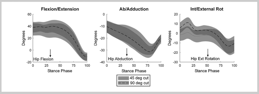
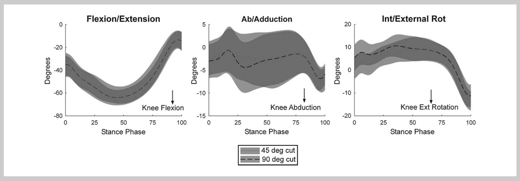
A significant difference was measured between the 45° cut and the 90° when all the dependent variables within the repeated measure MANOVA were examined (F(6,32) = 21.7, p < 0.001). The kinematic motions during stance phase for all 3 axes of rotation at the knee and hip for both cutting maneuvers are shown in Figures 4 and 5. Both hip and knee internal rotation angles at peak VGRF were significantly higher at the 90° cut as compared with the 45° cut (F(1,37) = 87.0, p < 0.001 and F(1,37) = 8.0, p = 0.008, respectively) (Figures 4 and 5). Mean hip flexion angle during the 90° cut was significantly greater than the 45° cut at peak force (F(1,37) = 52.34, p < 0.001) (Figure 5).
Figure 4.
Kinematic stance curves for all 3 axes of motion at hip during both cutting maneuvers.
Figure 5.
Kinematic stance curves for all 3 axes of motion at knee for both cutting maneuvers.
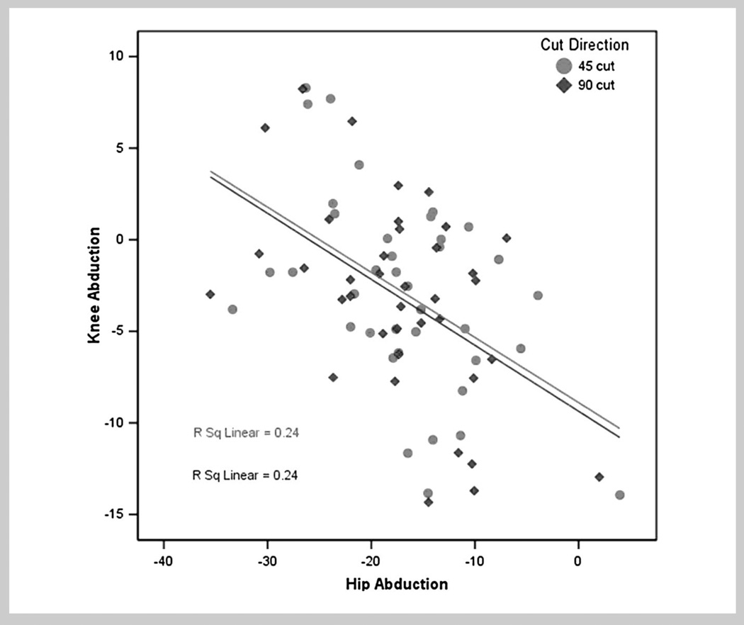
A stepwise multiple linear regression model was used to examine the potential biomechanical predictors of knee abduction (Figure 6). The sole significant predictor of knee abduction was hip adduction at peak force for both cutting maneuvers. Figure 6 shows the scatter plot of the relationship between knee and hip abduction for both cutting maneuvers. Hip adduction alone predicted 25% of knee abduction angles in the 45° cutting maneuver (R2 = 0.240, SEE = 4.732) as well as 25% in the 90° cutting maneuver (R2 = 0.242, SEE = 4.718). The increased internal hip rotation was not associated with increased abduction at the knee. Internal and external rotation of the hip was not a significant predictor of abduction angle at the knee, and knee abduction/adduction did not differ between the 45° and 90° cutting maneuvers. There were no significant differences observed in the other kinematic measures.
Figure 6.
Linear regression between knee abduction and hip adduction during each cut. Hip adduction alone predicted approximately 25% of variance in knee abduction angles in 45° cut (dotted line) as well as 25% in 90° cut (solid line).
Discussion
The purposes of this study were to determine whether there are differences in lower-extremity kinematics during a 45° and 90° cutting maneuver and to examine the relationship between lower-extremity motions during these maneuvers. The study findings supported the hypothesis that greater internal hip and internal knee rotation angles would be observed during the cutting maneuver at a 90° compared with the 45° angle. Previous authors have hypothesized that increased internal rotation may be related to increased knee abduction, resultant increased force on the ACL, and increased risk of injury (15,18,23,35). The increased transverse plane rotation at the hip and knee may initiate high-load knee positions when athletes perform cutting movements at sharper angles. These increased rotations and resultant knee loads may increase the risk for injury. However, knee abduction did not increase with the 90° cut. Thus, these findings did not support our second hypothesis that the increased transverse plane rotation induced by the 90° cut would relate to increased knee abduction. Knee abduction did not correlate with hip internal rotation; however, hip adduction was significantly correlated to knee abduction positions during both the 90° and 45° cutting tasks.
The correlation of hip adduction to knee abduction is an important finding given that knee abduction is a common mechanism of ACL injury and is related to increased risk of ACL injury in athletic women (15,18,35). As supported by the current study findings, when the hip adducted, knee abduction increased during both 45° and 90° unanticipated cuts. The sole significant predictor of knee abduction motion during both of these cuts was coronal plane hip adduction. Therefore, hip adduction increases a potential inciting mechanism (knee abduction) for increased knee load and noncontact ACL injury during side-step cutting maneuvers. Previous studies have also shown that knee abduction is an important measurement for predicting injury risk during landing (15,18,35). For example, Chaudhari and Andriacchi (6) suggested that a non-neutral alignment places the participant at increased risk of knee injury. In addition, Leetun et al. (20) reported that athletes who demonstrated significantly greater hip abduction strength were less likely to sustain a lower-extremity injury.
Recent investigations that have indicated a potential association of hip abduction strength to dynamic valgus alignments that increase ACL injury risk underline the importance of hip abduction strength (15,34,36,38). Brent and colleagues (5) recently demonstrated that hip abduction strength relative to body mass in boys is increased as they age during adolescence. In contrast, hip abduction strength showed no similar change in young women across the same age groups (5). The absence of increased hip abduction strength in adolescent girls as they mature may be related to their emergent increased risk of ACL during adolescence (14,15). These findings may indicate that strength and conditioning protocols that include hip abduction strengthening should be implemented for young girls beginning at age 12 to 13, when the divergence between girls and boys begin to occur. Hip abduction strengthening can be achieved through a variety of methods, including dynamic balance exercises, as well as resistance training exercises specifically aimed at strengthening the muscles primarily responsible for hip abduction (27). Future research is needed to determine the direct relationship between hip abduction strength measures and ACL injury incidence and to isolate the best training methods for increasing hip abduction strength in athletic women.
The current findings indicate that lower-extremity kinematics in the transverse plane are not associated with knee abduction during cutting activities. Although the transverse plane rotations increase as the cut angle increases, there were no associated increases in knee abduction angles. Because increased hip rotation may not correlate with increased knee abduction, transverse plane kinematics at the hip may not play a central role in predictive models of noncontact ACL injury risk.
A potential limitation to this study is that only women soccer players were included in the participant population. Since there is a sex difference in the relative rate of ACL rupture, inclusion of young men into our study design may have delineated more generalized sex differences in hip abduction measures related to cutting angle. In addition, including athletes from different sports in the current study may have helped to show sports-specific associations with increased risk of ACL tears. These data may have provided more insight into the high-risk cutting techniques demonstrated by athletic women. Another potential limitation to this study is the lack of joint moment (torque) and electromyographic data to examine the relationship between relative torque and muscle recruitment of the prime movers at each joint. These studies are ongoing in our laboratory.
Future study directions should include examination of movements that increase hip adduction, using combined measures of hip abduction and external rotation strength, along with electromyography measurements to examine muscular recruitment patterns. Longitudinal studies could determine how growth and development affect the association between proximal and distal kinematics and kinetics. Future investigations should also aim to use the cutting maneuvers outlined in the current study to identify potential “high-risk” athletic women from their knee abduction and hip adduction measures during dynamic tasks (11,16). When identified, the goal should be to target the “high-risk” young women with the most appropriate neuromuscular training for her specific deficits (27,30). Pilot work indicates that athletic women categorized as “high risk” on the basis of previous coupled biomechanical and epidemiologic studies (15) are more responsive to neuromuscular training (29). Thus, if future research could identify the high-risk women athletes, then coaches and practitioners could use neuromuscular training that incorporates plyometric and dynamic stabilization training components to target both hip adduction and knee abduction to reduce their potential risk of noncontact ACL injury (16,30).
Practical Applications
The results of this study indicate that hip and knee internal rotations were significantly greater during a 90° cut compared with a 45° cut. The findings also indicate that the mechanisms underlying knee abduction angles demonstrated by athletic women during cutting tasks are not primarily associated with lower-extremity transverse plane rotations. On the contrary, coronal plane motions at the hip (adduction) were the sole significant predictors of knee abduction. Strategies to prevent knee abduction in athletic women should focus in part on the control of hip adduction with training protocols that include plyometrics, dynamic stabilization, and trunk focused neuromuscular training (27,28,31,32).
Acknowledgments
The authors would like to acknowledge funding support from National Institutes of Health/NIAMS GrantsR01=AR049735, R01=AR05563, and R01=AR056259.
References
- 1.Agel J, Arendt EA, Bershadsky B. Anterior cruciate ligament injury in national collegiate athletic association basketball and soccer: a 13-year review. Am J Sports Med. 2005;33:524–530. doi: 10.1177/0363546504269937. [DOI] [PubMed] [Google Scholar]
- 2.Besier TF, Lloyd DG, Ackland TR, Cochrane JL. Anticipatory effects on knee joint loading during running and cutting maneuvers. Med Sci Sports Exerc. 2001;33:1176–1181. doi: 10.1097/00005768-200107000-00015. [DOI] [PubMed] [Google Scholar]
- 3.Besier TF, Lloyd DG, Cochrane JL, Ackland TR. External loading of the knee joint during running and cutting maneuvers. Med Sci Sports Exerc. 2001;33:1168–1175. doi: 10.1097/00005768-200107000-00014. [DOI] [PubMed] [Google Scholar]
- 4.Boden BP, Dean GS, Feagin JA, Garrett WE. Mechanisms of anterior cruciate ligament injury. Orthopedics. 2000;23:573–578. doi: 10.3928/0147-7447-20000601-15. [DOI] [PubMed] [Google Scholar]
- 5.Brent JL, Myer GD, Ford KR, Hewett TE. The effect of gender and age on isokinetic hip abduction. Med Sci Sports Exerc. 2006;38:255. [Google Scholar]
- 6.Chaudhari AM, Andriacchi TP. The mechanical consequences of dynamic frontal limb alignment for non-contact ACL injury. J Biomech. 2006;39:330–338. doi: 10.1016/j.jbiomech.2004.11.013. [DOI] [PubMed] [Google Scholar]
- 7.Chimera NJ, Swanik KA, Swanik CB, Straub SJ. Effects of plyometric training on muscle-activation strategies and performance in female athletes. J Athl Train. 2004;39:24–31. [PMC free article] [PubMed] [Google Scholar]
- 8.Ford KR, Myer GD, Hewett TE. Valgus knee motion during landing in high school female and male basketball players. Med Sci Sports Exerc. 2003;35:1745–1750. doi: 10.1249/01.MSS.0000089346.85744.D9. [DOI] [PubMed] [Google Scholar]
- 9.Ford KR, Myer GD, Hewett TE. Reliability of landing using 3D motion analysis techniques: implications for longitudinal analyses. Med Sci Sports Exerc. 2007;39:2021–2028. doi: 10.1249/mss.0b013e318149332d. [DOI] [PubMed] [Google Scholar]
- 10.Ford KR, Myer GD, Smith RL, Vianello RM, Seiwert SL, Hewett TE. A comparison of dynamic coronal plane excursion between matched male and female athletes when performing single leg landings. Clin Biomech. 2006;21:33–40. doi: 10.1016/j.clinbiomech.2005.08.010. [DOI] [PubMed] [Google Scholar]
- 11.Ford KR, Myer GD, Toms HE, Hewett TE. Gender differences in the kinematics of unanticipated cutting in young athletes. Med Sci Sports. 2005;37:124–129. [PubMed] [Google Scholar]
- 12.Freedman KB, Glasgow MT, Glasgow SG, Bernstein J. Anterior cruciate ligament injury and reconstruction among university students. Clin Orthop Related Res. 1998;356:208–212. doi: 10.1097/00003086-199811000-00028. [DOI] [PubMed] [Google Scholar]
- 13.Hewett TE, Ford KR, Myer GD, Wanstrath K, Scheper M. Gender differences in hip adduction motion and torque during a single leg agility maneuver. J Orthop Res. 2006;24:416–421. doi: 10.1002/jor.20056. [DOI] [PubMed] [Google Scholar]
- 14.Hewett TE, Myer GD, Ford KR. Decrease in neuromuscular control about the knee with maturation in female athletes. J Bone Joint Surg Am. 2004;86-A:1601–1608. doi: 10.2106/00004623-200408000-00001. [DOI] [PubMed] [Google Scholar]
- 15.Hewett TE, Myer GD, Ford KR, Heidt RS, Jr, Colosimo AJ, McLean SG, van den Bogert AJ, Paterno MV, Succop P. Biomechanical measures of neuromuscular control and valgus loading of the knee predict anterior cruciate ligament injury risk in female athletes: a prospective study. Am J Sports Med. 2005;33:492–501. doi: 10.1177/0363546504269591. [DOI] [PubMed] [Google Scholar]
- 16.Hewett TE, Stroupe AL, Nance TA, Noyes FR. Plyometric training in female athletes. Decreased impact forces and increased hamstring torques. Am J Sports Med. 1996;24:765–773. doi: 10.1177/036354659602400611. [DOI] [PubMed] [Google Scholar]
- 17.Kernozek TW, Torry MR, Cowley H, Tanner S. Gender differences in frontal and sagittal plane biomechanics during drop landings. Med Sci Sports Exerc. 2005;37:1003–1013. [PubMed] [Google Scholar]
- 18.Krosshaug T, Nakamae A, Boden BP, Engebretsen L, Smith G, Slauterbeck JR, Hewett TE, Bahr R. Mechanisms of anterior cruciate ligament injury in basketball: video analysis of 39 cases. Am J Sports Med. 2006;21:41–42. doi: 10.1177/0363546506293899. [DOI] [PubMed] [Google Scholar]
- 19.Landry SC, McKean KA, Hubley-Kozey CL, Stanish WD, Deluzio KJ. Neuromuscular and lower limb biomechanical differences exist between male and female elite adolescent soccer players during an unanticipated side-cut maneuver. Am J Sports Med. 2007;35:1888–1900. doi: 10.1177/0363546507300823. [DOI] [PubMed] [Google Scholar]
- 20.Leetun DT, Ireland ML, Willson JD, Ballantyne BT, Davis IM. Core stability measures as risk factors for lower extremity injury in athletes. Med Sci Sports Exerc. 2004;36:926–934. doi: 10.1249/01.mss.0000128145.75199.c3. [DOI] [PubMed] [Google Scholar]
- 21.Lohmander LS, Englund PM, Dahl LL, Roos EM. The long-term consequence of anterior cruciate ligament and meniscus injuries: osteoarthritis. Am J Sports Med. 2007;35:1756–1769. doi: 10.1177/0363546507307396. [DOI] [PubMed] [Google Scholar]
- 22.Lu TW, O’Connor JJ. Bone position estimation from skin marker co-ordinates using global optimisation with joint constraints. J Biomech. 1999;32:129–134. doi: 10.1016/s0021-9290(98)00158-4. [DOI] [PubMed] [Google Scholar]
- 23.Markolf KL, Burchfield DM, Shapiro MM, Shepard MF, Finerman GA, Slauterbeck JL. Combined knee loading states that generate high anterior cruciate ligament forces. J Orthop Res. 1995;13:930–935. doi: 10.1002/jor.1100130618. [DOI] [PubMed] [Google Scholar]
- 24.McLean SG, Huang X, Su A, van den Bogert AJ. Sagittal plane biomechanics cannot injure the ACL during sidestep cutting. Clin Biomech. 2004;19:828–838. doi: 10.1016/j.clinbiomech.2004.06.006. [DOI] [PubMed] [Google Scholar]
- 25.McLean SG, Lipfert SW, van den Bogert AJ. Effect of gender and defensive opponent on the biomechanics of sidestep cutting. Med Sci Sports Exerc. 2004;36:1008–1016. doi: 10.1249/01.mss.0000128180.51443.83. [DOI] [PubMed] [Google Scholar]
- 26.McLean SG, Su A, van den Bogert AJ. Development and validation of a 3-D model to predict knee joint loading during dynamic movement. J Biol Chem. 2003;125:864–874. doi: 10.1115/1.1634282. [DOI] [PubMed] [Google Scholar]
- 27.Myer GD, Brent JL, Ford KR, Hewett TE. A pilot study to determine the effect of trunk and hip focused neuromuscular training on hip and knee isokinetic strength. Br J Sports Med. 2008;42:614–619. doi: 10.1136/bjsm.2007.046086. [DOI] [PMC free article] [PubMed] [Google Scholar]
- 28.Myer GD, Chu DA, Brent JL, Hewett TE. Trunk and hip control neuromuscular training for the prevention of knee joint injury. Clin Sports Med. 2008;27:425–448. doi: 10.1016/j.csm.2008.02.006. [DOI] [PMC free article] [PubMed] [Google Scholar]
- 29.Myer GD, Ford KR, Brent JL, Hewett TE. Differential neuromuscular training effects on ACL injury risk factors in “high-risk” versus “low-risk” athletes. BMC Musculoskelet Disord. 2007;8:1–7. doi: 10.1186/1471-2474-8-39. [DOI] [PMC free article] [PubMed] [Google Scholar]
- 30.Myer GD, Ford KR, McLean SG, Hewett TE. The effects of plyometric versus dynamic stabilization and balance training on lower extremity biomechanics. Am J Sports Med. 2006;34:490–498. doi: 10.1177/0363546505281241. [DOI] [PubMed] [Google Scholar]
- 31.Myer GD, Ford KR, Brent JL, Hewett TE. The effects of plyometric versus dynamic balance training on power, balance, and landing force in female athletes. J Strength Cond Res. 2006;20:345–353. doi: 10.1519/R-17955.1. [DOI] [PubMed] [Google Scholar]
- 32.Myer GD, Ford KR, Palumbo JP, Hewett TE. Neuromuscular training improves performance and lower-extremity biomechanics in female athletes. J Strength Cond Res. 2005;19:51–60. doi: 10.1519/13643.1. [DOI] [PubMed] [Google Scholar]
- 33.Myklebust G, Bahr R. Return to play guidelines after anterior cruciate ligament surgery. Br J Sports Med. 2005;39:127–131. doi: 10.1136/bjsm.2004.010900. [DOI] [PMC free article] [PubMed] [Google Scholar]
- 34.Nyland J, Kuzemchek S, Parks M, Caborn DN. Femoral anteversion influences vastus medialis and gluteus medius EMG amplitude: composite hip abductor EMG amplitude ratios during isometric combined hip abduction-external rotation. J Electromyogr Kinesiol. 2004;14:255–261. doi: 10.1016/S1050-6411(03)00078-6. [DOI] [PubMed] [Google Scholar]
- 35.Olsen OE, Myklebust G, Engebretsen L, Bahr R. Injury mechanisms for anterior cruciate ligament injuries in team handball: a systematic video analysis. Am J Sports Med. 2004;32:1002–1012. doi: 10.1177/0363546503261724. [DOI] [PubMed] [Google Scholar]
- 36.Padua DA, Marshall SW, Beutler AI, DeMaio M, Boden BP, Yu B, Garrett WE. Predictors of knee valgus angle during a jump-landing task. Med Sci Sports Exerc. 2005;37:S398. [Google Scholar]
- 37.Pollard CD, Davis IM, Hamill J. Influence of gender on hip and knee mechanics during a randomly cued cutting maneuver. Clin Biomech. 2004;19:1022–1031. doi: 10.1016/j.clinbiomech.2004.07.007. [DOI] [PubMed] [Google Scholar]
- 38.Powers CM. The influence of altered lower-extremity kinematics on patellofemoral joint dysfunction: a theoretical perspective. J Orthop Sports Phys Ther. 2003;33:639–646. doi: 10.2519/jospt.2003.33.11.639. [DOI] [PubMed] [Google Scholar]
- 39.Woltring HJ, Huiskes R, de Lange A, Veldpaus FE. Finite centroid and helical axis estimation from noisy landmark measurements in the study of human joint kinematics. J Biomech. 1985;18:379–389. doi: 10.1016/0021-9290(85)90293-3. [DOI] [PubMed] [Google Scholar]


