Abstract
Bacillus cereus is a Gram-positive spore-forming bacterium causing food poisoning and serious opportunistic infections. These infections are characterized by bacterial accumulation despite the recruitment of phagocytic cells. We have previously shown that B. cereus Haemolysin II (HlyII) induces macrophage cell death by apoptosis. In this work, we investigated the regulation of the hlyII gene. We show that HlyIIR, the negative regulator of hlyII expression in B. cereus, is especially active during the early bacterial growth phase. We demonstrate that glucose 6P directly binds to HlyIIR and enhances its activity at a post-transcriptional level. Glucose 6P activates HlyIIR, increasing its capacity to bind to its DNA-box located upstream of the hlyII gene, inhibiting its expression. Thus, hlyII expression is modulated by the availability of glucose. As HlyII induces haemocyte and macrophage death, two cell types that play a role in the sequestration of nutrients upon infection, HlyII may induce host cell death to allow the bacteria to gain access to carbon sources that are essential components for bacterial growth.
Introduction
The Bacillus cereus group is composed of highly related pathogenic species, including B. thuringiensis, an insect pathogen, B. anthracis, the etiological agent of anthrax and B. cereus. B. cereus is an emerging human food-borne pathogen classified as the 3rd most important cause of collective food-borne infections in Europe, after Salmonella and Staphylococcus [1]. B. cereus infection generally causes mild disease characterised by gastroenteritis, but bloody diarrhea and emetic poisoning leading to some fatal cases have been reported [2]. B. cereus is also associated with severe local and systemic human infections, posing a public health problem [3]. Infections such as endophthalmitis, pneumonia and meningitis, particularly in neonates in which they may cause death of the infant within days, have been attributed to B. cereus [4], [5], [6], [7].
In the early stationary phase, B. cereus produces several extracellular compounds (degradative enzymes, cytotoxic factors and cell surface proteins) that might contribute to virulence [8], [9], [10], [11], [12]. However, the mechanisms leading to the various pathologies of B. cereus are not completely elucidated. The non-gastrointestinal infections are characterized by bacteremia despite the accumulation of inflammatory cells at the site of infection [13]. This implies that the bacteria have developed means to resist the activity of inflammatory cells and thus the host immune system. Our previous work has consistently shown that B. cereus is able to circumvent the host immune response. B. cereus spores survive, germinate and multiply in contact with macrophages [14], eventually leading to the production of toxins [15]. Among these toxins, the haemolysin HlyII has been shown to be responsible for macrophage death [15], [16]. HlyII is a member of the oligomeric ß-barrel pore-forming toxins, which include the α-toxin of Staphylococcus aureus, the ß-toxin of Clostridium perfringens and the B. cereus Cytotoxin K (CytK) [17], [18], [19]. HlyII is a secreted protein that induces pore formation in the membrane of various eukaryotic cells [20], [21], [22]. HlyII has haemolytic properties [20], [23] and induces apoptosis of host monocytes and macrophages in vivo [15]. When expressed in B. subtilis, HlyII induces haemolysis and virulence in a crustacean infection model [24]. We have also demonstrated the importance of HlyII for B. cereus virulence in insects and mice [15]. The importance of HlyII has been strengthened by the fact that the hlyII gene is present in several clinical isolates of B. cereus [25].
A precise knowledge of the conditions governing the expression of a gene improves our understanding of its role in the functioning of the bacterial cell, but also during pathogenesis. The regulation of hlyII expression has so far been exclusively studied in the heterologous systems E. coli and B. subtilis. In those systems, it has been shown that hlyII expression is negatively regulated by the transcriptional regulator HlyIIR [26]. The hlyII and hlyIIR genes are situated in the same chromosomal locus (Bc 3523 and Bc 3522 in the ATCC 14579 strain, respectively) but are not organized in an operon. In vitro analyses have shown that recombinant HlyIIR regulates hlyII expression by specific binding, as two dimers, to a 44 perfect inverted DNA repeat (22 bp x 2) centred 48 bp upstream of the hlyII transcription initiation point [27].
In this study, we investigated the regulatory process of hlyII expression and of its repressor hlyIIR. We highlight the major role of glucose 6P as a post-transcriptional activator of HlyIIR, and show that glucose 6P binds to and activates HlyIIR to inhibit hlyII expression.
Materials and Methods
Bacterial Strains
The B. thuringiensis 407 Cry_ strain was used as a model for B. cereus. This sequenced strain was originally described as a B. thuringiensis strain, but cured of its plasmid it is acrystalliferous and shows high phylogenic similarity with the B. cereus reference strain ATCC 14579 [28]. The Bt407 strain was used as the wild type background and transformed as previously described [29].
Plasmid and Mutant Strain Constructions
The hlyIIR gene was disrupted as follows. HindIII-XbaI (1024 bp) and SphI-BamHI (964 bp) DNA fragments corresponding to upstream and downstream regions of the hlyIIR gene were generated from the Bt407 chromosome by PCR using the primer pairs:
hlyIIR-1 (5′-CCCAAGCTTGTACCAGCTTTAACAGGCTATGG -3′),
hlyIIR-2 (5′- GCTCTAGACGTCTGCTCTCGAGATTTCCCC-3′), and
hlyIIR-3 (5′-ACATGCATGCGGATGCCAGAAGACTTAGTGG -3′),
hlyIIR-4 (5′-CGGGATCCCAGGCTCTAAGTTGGATAAGGGG -3′).
A TetR cassette carrying a tet gene was purified from pHTS1 [30] as a 1.6 Kb (XbaI and SphI) fragment. The amplified DNA fragments were digested with the appropriate enzymes and inserted between the HindIII and BamHI sites of pRN5101, with the selection marker cassette cloned in between the up- and downstream chromosomal fragments of the hlyIIR gene. The resulting plasmid was introduced into Bt407 by electroporation [31] and the hlyIIR gene was deleted by a double crossover event as previously described [32]. Chromosomal allele exchange was confirmed by PCR with oligonucleotide primers located upstream from hlyIIR-1: hlyIIR-5 (5′-GGGCCAAATGCCGATGCAAGTATCACAGGTAGC-3′) and downstream from hlyIIR-4: hlyIIR-6 (5′-CCAAGCGTTCAGCAAGTTGGTCTCATGATGTGG-3′), and in the TetR cassette (5′-CGGGTCGGTAATTGGGTTTG-3′), (5′-GCAGCTGCACCAGCCCCTTG-3′). The mutant strain was designated Bt407ΔhlyIIR.
Transcriptional hlyII-lacZ and hlyIIR-lacZ fusions were constructed using DNA fragments corresponding to the promoter regions of hlyII and hlyIIR, generated by PCR using the primer pair PhlyII-1 (5′- AAAACTGCAGCCAGCTGTGTTGACAGAACTGGC-3′) and PhlyII-2 (5′-GCTCTAGACGGACGCTACCGCAACGCATTTAGC -3′), and hlyIIR-1 and hlyIIR-2, respectively. The PCR fragments were digested with the appropriate enzymes and inserted between the XbaI and PstI sites of pHT304-18Z [33]. The recombinant plasmids, designated pHT-PhlyII’Z and pHT-PhlyIIR’Z were introduced into Bt407 and into the mutant strain Bt407ΔhlyIIR by electroporation. Transformants were named Bt407 [pHT-PhlyII’Z], Bt407 [pHT-PhlyIIR’Z] and Bt407ΔhlyIIR [pHT-PhlyII’Z].
Purification of the HlyIIR Protein
The plasmid pGEX6P1-GST-HlyIIR was constructed as follows. The hlyIIR gene was amplified from the Bt407 chromosome by PCR using the primer pairs hlyIIR-GST-1 (5′-CGGGATCCATGGGGAAATCTCCAGAGCAGACGA-3′) and hlyIIR-GST-2 (5′-TCCCCCGGGCCCGTATGCAAATCGAAGAGCTTAT-3′). The corresponding DNA fragment was inserted between the BamHI and SmaI sites of plasmid pGEX6P1 (GE Healthcare), and the resulting plasmid was introduced into E. coli M15hellopREP4] (Qiagen). E. coli M15hellopREP4] strain harboring the plasmid pGEX6P1-GST-HlyIIR was grown in minimal M9 medium [34] with 3% arabinose as unique carbon source, at 37°C until OD600 0.8 was reached and protein expression was induced by addition of 1 mM IPTG. Growth was continued for 4 h after IPTG induction. Bacteria were then collected by centrifugation at 7700 g for 10 min. Pellets were lyzed using 1% triton X-100 and sonication in PBS. After centrifugation, the tagged HlyIIR contained in the supernatant was added on a Bulk GST Purification module (GE Healthcare). The HlyIIR protein was purified by cleavage of the GST tag using the Precission protease according to the manufacturer’s instructions. Protein concentration was calculated by Bradford staining and purity was assessed on SDS-Page gel.
Relative Quantification of hlyII Gene Expression by RT-qPCR
Preparation of the RNA samples
Bt407 and Bt407ΔhlyIIR were cultivated aerobically in LB at 37°C with agitation.
1 ml samples were taken every hour between t0−2 h (t−2) and t0+4 h (t4), t0 h (t0) corresponding to the transition state between the exponential and the stationary phases of the bacterial culture. The bacterial cells were collected by a short centrifugation and immediately placed at −80°C. Total RNA of these cell pellets was extracted as described in [25]. Briefly, cells were thawed and lysed in Tri-reagent (Ambion), RNA was purified using the RNeasy kit (Qiagen) and finally treated with DNase (TURBO DNA-free kit; Ambion). Analyses of RNA solutions prepared according to this protocol have shown that they are of a high purity grade (OD260 nm/OD280 nm >1,95 and RIN >9) and free of DNA traces (data not shown).
RT-qPCR experiments
Target and reference gene mRNA abundance was measured by one-step RT-qPCR with the QuantiFast SYBR green RT-PCR kit, following the manufacturer’s instructions (Qiagen). We used the ssu gene, encoding 16S RNA, as endogenous reference, as its expression level in Bt407 was stable between t-2 and t4 (data not shown). RT-qPCR samples contained 1 ng of RNA and 500 nM or 1 µM of each primer in a final volume of 25 µL and were run in a LightCycler 480 (Roche). The following primer pairs were used: F-hlyII-q (5′-CTGGAAAAACCATCAAGTTACTC-3′); R-hlyII-dq (5′-TCACCATTTACAAAGATACC-3′) [25] and LC-16S-F (5′-GGTAGTCCACGCCGTAAACG-3′); LC-16S-R (5′-GACAACCATGCACCACCTG-3′) [35]. The efficiency of both primer pairs in the experiment conditions was above 95% (data not shown). Expression levels of the target gene relative to the endogenous standard were calculated using the basic relative quantification method (Roche) and the LightCycler 480 software. The specificity of each amplification reaction was verified with the melting curve profile: a single peak was obtained, attesting to the amplification of a unique product.
ß-galactosidase Assays
Strains harboring plasmid transcriptional lacZ fusions were grown in LB medium at 37°C under shaking. Samples were taken every hour from t−2 to t4. Determination of ß-galactosidase activity was achieved as previously described [36], [37]. When indicated, 0.3% glucose or arabinose was added to the medium at t−1.
Molecular Docking
The crystal structure of B. cereus HlyIIR was retrieved from the Protein Data Bank (PDB entry 2fx0) and hydrogen atoms added with the Biopolymer module of the SYBYL-X 1.3 package (Tripos Assoc. Inc, St.Louis, USA). The starting three-dimensional structure of glucose-6-Phosphate (glucose 6P) was built from the SYBYL fragment library. Docking of the ligand to the HlyIIR cavity was realized using two independent docking tools (PLANTS, GOLD).
PLANTS [38] was used in its 1.2 version with default settings. The geometric center of the HlyIIR cavity was used to define the binding site including any protein atom within 15 Å of these coordinates. Poses were scored with the ChemPLP function and up to 10 solutions were saved for the ligand.
GOLD v5.1 [39] was used to confirm previous PLANTS results, using the same binding site definition as for PLANTS. Default docking settings were applied to dock the ligand and 10 poses were saved.
Microcalorimetry
Isothermal titration calorimetry (ITC) measures heat generated or absorbed upon the binding of two molecules [40], and yields in a single experiment the thermodynamic parameters allowing the calculation of a dissociation constant (Kd). Isothermal titration calorimetry experiments were performed at 20°C with an ITC200 isothermal titration calorimeter (Microcal®). The interaction between HlyIIR and glucose 6P was tested. The protein concentration in the microcalorimeter cell was set at 28 µM. glucose 6P was resuspended at 0,9 mM in the protein storage buffer. A total of 20 injections of 2 µl of glucose 6P were made at intervals of 180 s while stirring at 800 rpm. Arabinose at 0.9 mM was used as control. We then tested the interaction between DNA and HlyIIR in the absence and presence of glucose 6P (or arabinose). The DNA probe (220 bp) was generated from the Bt407 chromosome by PCR using the following primer pair: 5′-GGAAATAATGTATCTGAATATAGGCTG-3′ and 5′-CACATGTTTCACTAATCTTCCTCTCAC-3′, and containing the HlyIIR binding region upstream of the hlyII gene [27]. The HlyIIR concentration in the microcalorimeter cell was set at 28 µM without or with glucose 6P at 1.7%. A total of 20 additions of 2 µl of DNA at 1,25 µM were made by injection at intervals of 180 s while stirring at 800 rpm.
The data were integrated to generate curves in which the areas under the injection peaks were plotted against the ratio of injected sample to cell content. Analysis of the data was performed using MicroCal Origin provided by the manufacturer according to the one-binding-site model. Changes in the free energy and entropy upon binding were calculated from determined equilibrium parameters using the equation:
where R is the universal gas constant (1.9872 cal mol-1K-1), T is the temperature in Kelvin degrees, Ka is the association constant, ΔG is the change in Gibbs free energy, ΔH is the change in enthalpy and ΔS is the change in entropy. The binding constant of each interaction is expressed as 1/Ka = Kd (in mol l−1).
Results
HlyIIR Negatively Regulates hlyII Expression in B. cereus
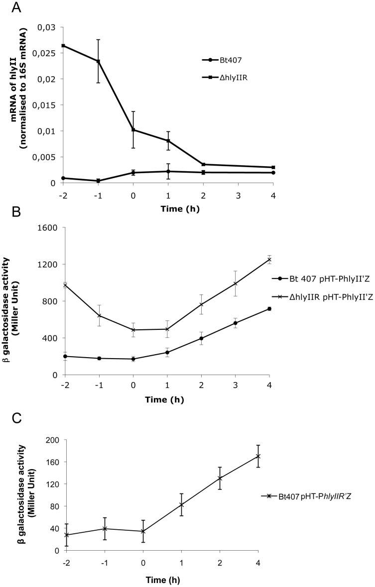
The transcription of hlyII is independent of the pleiotropic regulator PlcR [41]. Several studies using heterologous hosts have shown that HlyIIR is a negative regulator of hlyII gene transcription. However, no study is so far describing hlyII expression regulation by HlyIIR from the B. cereus group. We therefore investigated the expression of hlyII in a Bt 407 hlyIIR deficient mutant. Quantitative RT-PCR assays showed that hlyII expression was significantly higher in the Bt407ΔhlyIIR mutant than in the wild type (wt) strain (Figure 1A). This difference in expression was most pronounced during exponential phase. The ratio of hlyII expression in the ΔhlyIIR mutant compared to the wt was 30 fold at t−2, dropped to 5 fold at t0 and stayed around 2 fold after t2. Thus, HlyIIR is a negative regulator of hlyII expression in B. cereus and is especially active during the early phase of bacterial growth.
Figure 1. HlyIIR negatively regulates hlyII expression in B. cereus.
(A) Expression levels of the target hlyII gene relative to the endogenous standard 16S RNA were measured by RT-qPCR throughout bacterial growth in the wild type Bt407 (circle) and the Bt407ΔhlyIIR (square) strains. Data are expressed as the ratio of hlyII mRNA normalized to 16S RNA. Values are means of two independent experiments. (B) The specific β-galactosidase activity (Miller unit) of strains Bt407 and Bt407 ΔhlyIIR harboring the transcriptional pHT-PhlyII’Z fusion were measured from bacteria grown in LB medium at 37°C from 2 h before the culture entry into stationary phase (t−2) to 4 h after (t4). Results represent mean values of at least three independent experiments. (C) The specific β-galactosidase activity (Miller unit) of strain Bt407 harboring the pHT-PhlyIIR’Z fusion was measured from bacteria grown in LB medium at 37°C from 2 h before the culture entered into stationary phase (t−2) to 4 h after (t4). Results represent mean values of at least three independent experiments.
The results obtained by qRT PCR were confirmed by ß–galactosidase assays (Figure 1B). The tendency observed was the same, with an increased effect of HlyIIR at the early growth phase (t−2 and t−1), although the ratio measured by ß–galactosidase assay was lower (5 fold) than the one observed by qRT-PCR. However, the ß–galactosidase technique is less precise than qRT-PCR to measure gene expression in the early growth phase due to a small amount of bacteria. After t0, hlyII expression was 2 fold higher in the ΔhlyIIR mutant than in the wild type strain.
To determine whether the strong effect of HlyIIR on hlyII expression during exponential growth phase was due to an increased expression of the regulator itself during this period, we assessed hlyIIR expression using a β–galactosidase assay (Figure 1C). hlyIIR was expressed during vegetative growth and its expression increased at the onset of stationary phase. These data suggest that HlyIIR-dependent repression of hlyII expression during exponential growth phase does not depend on hlyIIR gene expression, but likely relies on the activation of the repressor at the protein level.
Glucose Activates HlyIIR at the Post-transcriptional Level to Inhibit hlyII Expression
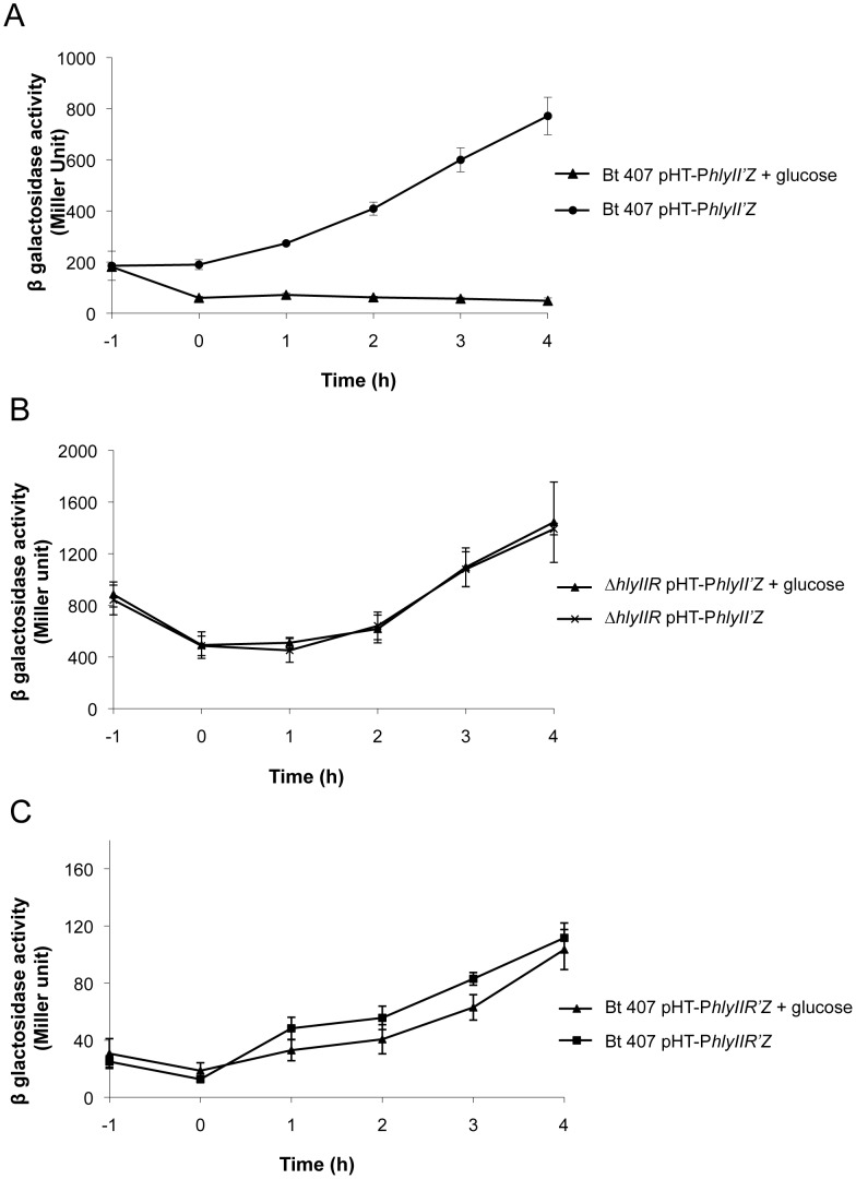
HlyIIR negatively regulates hlyII expression, especially during early steps of bacterial growth. We thus investigated whether a co-factor, present during exponential phase and decreasing throughout bacterial growth, could activate HlyIIR. Glucose is a carbon source that is commonly used and consumed by bacteria during bacterial growth. To assess the potential role of glucose on the activation of HlyIIR, we first tested whether glucose had an effect on hlyII expression by β–galactosidase assay (Figure 2A). Expression of hlyII was completely abolished when glucose was added to the culture, showing that glucose inhibited hlyII expression. Not all sugars displayed this activity, as addition of arabinose had no effect on hlyII expression (data not shown).
Figure 2. Glucose inhibits hlyII expression through activation of the HlyIIR repressor.
The bacterial strains Bt407 (A, C) and Bt407ΔhlyIIR (B) harboring the transcriptional pHT-PhlyII’Z (A, B) or pHT-PhlyIIR’Z (C) fusions were grown at 37°C in either LB medium or LB medium supplemented at t−1 with 0.3% glucose. Specific β-galactosidase activity (Miller units) was measured at the indicated time points. Values are expressed as the mean of three independent experiments.
To determine whether glucose inhibitory effect on hlyII expression acted through the repressor HlyIIR, expression of hlyII was assessed in the presence or absence of glucose in the ΔhlyIIR mutant (Figure 2B). The expression of hlyII in the ΔhlyIIR mutant was similar in the presence and absence of glucose. Thus, in the absence of HlyIIR, glucose had no effect on hlyII expression, showing that glucose acted on hlyII expression through its repressor HlyIIR.
To assess whether glucose controls hlyIIR transcription, hlyIIR expression was measured in the presence and absence of glucose (Figure 2C). Addition of glucose to the culture did not significantly alter hlyIIR expression. These data show that glucose is not involved in the control of hlyIIR expression and strongly suggest that glucose affects hlyII expression by activating HlyIIR at a post-transcriptional or a post-translational level.
Virtual Docking of Glucose 6P in the HlyIIR Ligand Pocket
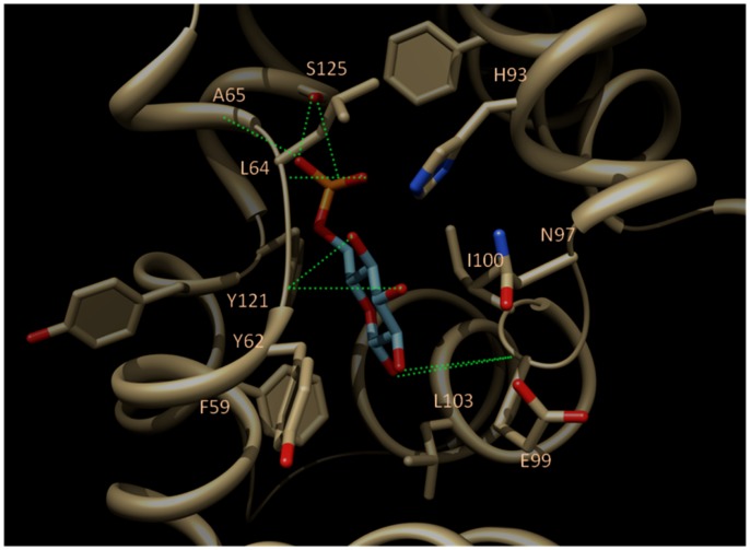
The crystal structure of HlyIIR reveals a large internal hydrophobic cavity that could accommodate a ligand with a molecular mass of up to 500 Da, suggesting a co-factor-dependent activation for the inhibition of hlyII expression [42]. When glucose is added to the bacterial culture, it is transported inside the bacteria through the complex sugar transporting phosphotransferase system (PTS) [43]. Glucose is then rapidly transformed into glucose 6P, which is thus likely to be the actual effector. To check whether glucose 6P may bind to the above-defined ligand cavity, the compound was docked using two independent state-of-the-art docking algorithms [38], [39]. Interestingly, both programs agreed to unambiguously dock glucose 6P in a single binding mode (Figure 3): an extensive network of 8 hydrogen-bonds to the receptor is found between hydroxyl groups of glucose 6P and main chain carbonyl atoms of HlyIIR (Y62, E99), as well as between the terminal phosphate moiety and the receptor residues (L64, A65, S125). Apolar contacts between the cyclic ring of the ligand and several apolar receptor side chains (F59, Y62, I100, L103) ensure the tight fit of glucose 6P within the binding pocket.
Figure 3. Predicted binding mode common to PLANTS and GOLD docking of glucose-6P (cyan sticks) to the ligand-binding cavity of HlyIIR (yellow ribbons, yellow sticks).
Main receptor-interacting residues are labelled at their C-αatoms. Intermolecular hydrogen bonds are displayed as green broken lines. Nitrogen and oxygen atoms are coloured in blue and red, respectively.
Direct Binding of Glucose 6P to HlyIIR
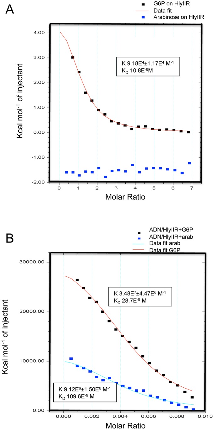
We then used isothermal titration calorimetry (ITC) to confirm and quantitatively characterize the interaction of glucose 6P with HlyIIR (Figure 4A). The HlyIIR protein was produced and purified from a medium lacking glucose or one of its derivatives, and the interaction with glucose 6P was assessed. The analysis of the integrated titration curve showed that the glucose 6P-HlyIIR interaction was characterized by a Kd of about 10.8 µM with an apparent stoichiometry of 1∶1 (N = 1.09±0.00844). These data demonstrate that glucose 6P directly binds to HlyIIR and that one molecule of glucose 6P binds to one monomer of HlyIIR. In sharp contrast, arabinose that we had previously demonstrated having no activation effect on HlyIIR (β-galactosidase assay, not shown), did not either bind HlyIIR in our affinity-binding assay (Figure 4A). This result strongly fosters our hypothesis of glucose specificity within the sugar family of molecules for HlyIIR activation.
Figure 4. Glucose 6P binds to HlyIIR to enhance its DNA binding capacity.
(A) Isothermal titration calorimetry (ITC) assays were performed to measure heat variations generated upon binding between glucose 6P (G6P) and HlyIIR (red line) or arabinose and HlyIIR (blue squares). (B) ITC assays were performed to measure binding between a DNA fragment containing the HlyIIR binding site and HlyIIR supplemented either with glucose 6P (red line) or with arabinose (blue line).
Binding of Glucose 6P to HlyIIR Increases its Affinity to its Target DNA in the hlyII Promoter Region
The N-terminal part of HlyIIR presents sequence similarity to regulators of the TetR repressor family which share common features such as a highly conserved helix-turn-helix motif implicated in DNA binding, dependence of cofactors for activity regulation, involvement in the adaptation to a changing environment and acting as homodimers. In view of the 3-D structure of HlyIIR and analogy with its structural homologues TetR, it has been proposed that binding of the appropriate ligand to HlyIIR could change the orientation of the DNA-binding domains leading to a different affinity of the ligand-bound form towards the specific DNA site [42].
We used ITC experiments to characterize whether the DNA binding affinity of HlyIIR is influenced by the presence or absence of glucose 6P binding. Calorimetric titrations were performed and revealed that the HlyIIR- glucose 6P complex binds to the specific DNA target with a Kd of about 28.7 nM (Figure 4B). In the absence of glucose 6P (not shown) or in the presence of arabinose, HlyIIR binds to its target DNA with a Kd of about 110 nM. Thus, in the absence of glucose 6P, HlyIIR was still able to bind to its target DNA but its affinity was increased around 4 times in the presence of glucose 6P.
So far, the HlyIIR-binding ligand was unknown. Thus, to our knowledge, we present the first evidence that HlyIIR trigger its repression activity on hlyII expression through its activation by binding glucose 6P.
Discussion
In this study, we show that Glucose 6P binds and activates HlyIIR, the negative transcriptional regulator of hlyII expression in B. cereus. Glucose has been shown to be one of the preferred carbon sources during the initial growth phase of B. subtilis [44]. At this stage, glucose derivative, Glucose 6P, will activate HlyIIR probably by binding to its ligand cavity. Glucose 6P has been described as an important phosphate donor, leading to activation of various regulators mainly implicated in glycolysis and/or virulence [45], [46]. However, the ability of glucose 6P to bind as a whole into a specific ligand pocket or a regulator implicated in virulence has, to the best of our knowledge, never been described. Thus, we highlight a new mechanism of activation of a transcriptional regulator involved in the regulation of a virulence gene. Other examples have been described of sugars binding to and modulating the activity of repressors implicated in the regulation of genes involved in carbon metabolism, stress response and pathogenesis [47], [48]. For example, in the presence of arabinose, AraC activates the transcription from the promoters of the catabolic operons of various genes, linking the mechanism of glycolysis with virulence [49]. For B. anthracis, glucose is a signalling molecule linking environmental sensing to virulence factor production by the way of the important transcription regulator of Gram-positive bacteria, CcpA [50]. CcpA-mediated glucose sensing is also involved in the expression of the nhe and hbl operons, two major enterotoxins of B. cereus, but not in hlyII expression [51]. The discovery and characterization of transcription factors mediating the glucose response demonstrate that glucose, like fatty acids and other key nutrients, can directly control gene expression in response to glycaemic variation [52].
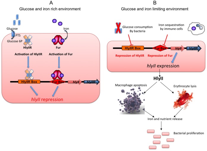
Taken together, we have shown that HlyIIR regulates hlyII gene expression through its post-transcriptional activation by glucose 6P. It has also been shown that hlyII expression depends on iron via activation of the global regulator Fur [53]. Indeed, a Fur box was found in the promoter region of the hlyII gene [54], and recent data show that Fur binding to this Fur box competes with RNA polymerase binding to the hlyII promoter, thus interfering with hlyII expression in vitro [53]. Sugar and iron are crucial compounds for bacterial multiplication and thus for their capacity to colonize their hosts. It has been reported that a common host defense mechanism relies on iron sequestration by immune cells [55], [56]. An increase in production of iron-binding proteins is observed following infection in order to limit bacterial growth [55]. To counteract this phenomenon, bacteria induce the expression of genes that are negatively iron-regulated. Moreover, in some cases, pathogenic bacteria may acquire iron and sugars/carbohydrates from the host by killing host cells. Thus, the regulation of virulence determinants implicated in host cell death might be regulated by nutrient availability. As a model we propose that when glucose is consumed by the bacteria and iron gets sequestered by phagocytic cells as a natural host defense [55], [56], the HlyIIR and Fur repressors become inactivated and hlyII expression starts (Figure 5). HlyII is then produced by the bacteria, secreted and induces haemocyte and macrophage death [15]. The cell content is then released into the environment, providing the bacteria with access to nutrients, allowing bacterial growth and promoting a new cycle of hlyII gene inhibition/expression (Figure 5).
Figure 5. Model of the role and expression of hlyII during infection.
A) As long as iron and glucose are abundant in the bacterial environment, the bacteria will be able to use these resources for growth. Glucose will enter the bacteria as glucose 6P (blue rectangles) and will bind HlyIIR (orange plain cross). Iron (purple circles) will bind Fur (red ovals). These bindings will promote HlyIIR and Fur repressor activities, leading to HlyIIR- and Fur-based transcriptional repression on hlyII gene expression. B) During host infection, bacteria find themselves in an environment, which is low in glucose and iron. Levels of these nutrients are further lowered during bacterial proliferation. The decrease in the concentration of glucose during bacterial proliferation will lead to an inhibition of HlyIIR activity, thus allowing hlyII expression. The decrease in the concentration of iron, partially due to its sequestration by immune cells, will lead to an inhibition of Fur activity, thus allowing hlyII expression. Thus, when glucose and iron are getting scarce, hlyII expression is activated. HlyII will then be released in the environment and induce macrophage and erythrocyte lysis. The dead cells will release their intracellular content, allowing access to metabolites that are essential for bacterial growth.
Taken together, we have identified several mechanisms triggering hlyII gene expression regulation, which are consistent with the role of the HlyII protein during the B. cereus infection process. This tight regulation probably limits HlyII production. It is however not clear why hlyII gene expression is so tightly regulated by one global (Fur) and one specific (HlyIIR) regulator. Several hypotheses seem possible: i) the low production of HlyII may be sufficient to promote nutrient access and virulence. We have indeed shown that very low doses of protein are able to induce macrophage lysis and mice mortality [15]; ii) HlyII production should remain low to avoid rapid degradation of macrophages and/or host death in order to allow bacterial multiplication and proliferation inside its host.
Additionally, it has recently been shown that HlyII accumulates in high oxidation-reduction potential (ORP) conditions in contrast to low ORP conditions, suggesting a redox-dependent regulation of HlyII secretion [57]. As low ORP anoxic conditions mimics the intestinal environment, HlyII may not be a virulence factor involved in gastrointestinal disease. We have consistently shown that HlyII is involved specifically in immune cell death by apoptosis [15], [16] and that it is highly produced by strains of human clinical origin [25], strongly suggesting a role during opportunistic infections.
Taken together, our data provide new insights into the regulatory pathways of hlyII, which plays an important role during the B. cereus virulence process. We demonstrate a new role of glucose 6P as a direct activator of a transcriptional regulator. As iron and carbon sources are essential and universal bacterial growth components, we believe that our data provide an important contribution to the understanding of bacterial infection strategies.
Acknowledgments
We wish to thank Alain Lerééc for excellent technical assistance. We thank the RT-PCR platforms of INRA, Jouy en Josas (PiCT-Gem) and of ANSES, Maisons-Alfort. We are indebted to Magali Aumont-Nicaise from the Université de Paris-Sud 11, UMR 8619, Institut de Biochimie et Biophysique Moléculaire et Cellulaire, Orsay, France, and to Sylviane Hoos from the Plate-forme de Biophysique des Macromolécules et de leurs Interactions, Institut Pasteur, Paris, France for performing and analysing the ITC experiments. We thank Stéphane Perchat for very helpful computer support. We thank Thomas Dubois for helpful discussion. We thank Crystal Gadishaw-Lue, Hannah Tollman, Steven Lewis and Isabelle Martin-Verstraete for proofreading the manuscript.
Funding Statement
These authors have no support or funding to report.
References
- 1.Anonymus (2009) The Community Summary Report on Food-borne Outbreaks in the European Union in 2007. The EFSA Journal 271.
- 2. Stenfors Arnesen L, Fagerlund A, Granum P (2008) From soil to gut: Bacillus cereus and its food poisoning toxins. FEMS Microbiol Rev 32: 579–606. [DOI] [PubMed] [Google Scholar]
- 3. Bottone EJ (2010) Bacillus cereus, a volatile human pathogen. Clin Microbiol Rev 23: 382–398. [DOI] [PMC free article] [PubMed] [Google Scholar]
- 4. Hilliard NJ, Schelonka RL, Waites KB (2003) Bacillus cereus bacteremia in a preterm neonate. J Clin Microbiol 41: 3441–3444. [DOI] [PMC free article] [PubMed] [Google Scholar]
- 5. Arnaout M, Tamburro R, Bodner S, Sandlund J, Rivera G, et al. (1999) Bacillus cereus causing fulminant sepsis and hemolysis in two patients with acute leukemia. J Pediatr Hematol Oncol 21: 431–435. [DOI] [PubMed] [Google Scholar]
- 6. Callegan MC, Kane ST, Cochran DC, Novosad B, Gilmore MS, et al. (2005) Bacillus endophthalmitis: roles of bacterial toxins and motility during infection. Invest Ophthalmol Vis Sci 46: 3233–3238. [DOI] [PubMed] [Google Scholar]
- 7. Kotiranta A, Lounatmaa K, Haapasalo M (2000) Epidemiology and pathogenesis of Bacillus cereus infections. Microb Infect 2: 189–198. [DOI] [PubMed] [Google Scholar]
- 8. Tran SL, Guillemet E, Gohar M, Lereclus D, Ramarao N (2010) CwpFM (EntFM) is a Bacillus cereus potential cell wall peptidase implicated in adhesion, biofilm formation and virulence. J Bacteriol 192: 2638–2642. [DOI] [PMC free article] [PubMed] [Google Scholar]
- 9. Gilois N, Ramarao N, Bouillaut L, Perchat S, Aymerich S, et al. (2007) Growth-related variations in the Bacillus cereus secretome. Proteomics 7: 1719–1728. [DOI] [PubMed] [Google Scholar]
- 10. Brillard J, Susanna K, Michaud C, Dargaignaratz C, Gohar M, et al. (2008) The YvfTU two-component system is involved in plcR expression in Bacillus cereus . BMC Microbiol 8: 183. [DOI] [PMC free article] [PubMed] [Google Scholar]
- 11. Ramarao N, Lereclus D (2006) Adhesion and cytotoxicity of Bacillus cereus and Bacillus thuringiensis to epithelial cells are FlhA and PlcR dependent, respectively. Microbes Infect 8: 1483–1491. [DOI] [PubMed] [Google Scholar]
- 12. Auger S, Ramarao N, Faille C, Fouet A, Aymerich S, et al. (2009) Biofilms formation and cell surface properties among pathogenic and non pathogenic strains of the Bacillus cereus group. App Environ Microbiol 75: 6616–6618. [DOI] [PMC free article] [PubMed] [Google Scholar]
- 13. Hernandez E, Ramisse F, Ducoureau JP, Cruel T, Cavallo JD (1998) Bacillus thuringiensis subsp. konkukian (serotype H34) superinfection: case report and experimental evidence of pathogenicity in immunosuppressed mice. J Clin Microbiol 36: 2138–2139. [DOI] [PMC free article] [PubMed] [Google Scholar]
- 14. Ramarao N, Lereclus D (2005) The InhA1 metalloprotease allows spores of the B. cereus group to escape macrophages. Cell Microbiol 7: 1357–1364. [DOI] [PubMed] [Google Scholar]
- 15. Tran SL, Guillemet E, Ngo-Camus M, Clybouw C, Puhar A, et al. (2011) Hemolysin II is a Bacillus cereus virulence factor that induces apoptosis of macrophages. Cell Microbiol 13: 92–108. [DOI] [PubMed] [Google Scholar]
- 16. Tran SL, Puhar A, Ngo-Camus M, Ramarao N (2011) Trypan blue dye enters viable cells incubated with the pore-forming toxin HlyII of Bacillus cereus . PLoS ONE 6: e22876. [DOI] [PMC free article] [PubMed] [Google Scholar]
- 17. Baida G, Budarina ZI, Kuzmin NP, Solonin AS (1999) Complete nucleotide sequence and molecular characterization of hemolysin II gene from Bacillus cereus . FEMS Microbiology Letters 180: 7–14. [DOI] [PubMed] [Google Scholar]
- 18. Gouaux E (1998) alpha hemolysin from Staphylococcus aureus: an archetype of beta barrel, channel forming toxins. J Struct Biol 121: 110–122. [DOI] [PubMed] [Google Scholar]
- 19. Lund T, DeBuyser M-L, Granum PE (2000) A new cytotoxin from Bacillus cereus that may cause necrotic enteritis. Mol Microbiol 38: 254–261. [DOI] [PubMed] [Google Scholar]
- 20. Andreeva Z, Nesterenko V, Yurkov I, Budarina ZI, Sineva E, et al. (2006) Purification and cytotoxic properties of Bacillus cereus hemolysin II. Prot Express Purif 47: 186–193. [DOI] [PubMed] [Google Scholar]
- 21. Andreeva Z, Nesterenko V, Fomkina M, Ternosky V, Suzina N, et al. (2007) The properties of Bacillus cereus hemolysin II pores depend on environmental conditions. Biochem Biophys Acta 1768: 253–263. [DOI] [PubMed] [Google Scholar]
- 22. Andreeva-Kovalevskaya ZI, Solonin AS, Sineva EV, Ternovsky VI (2008) Pore-forming proteins and adaptation of living organisms to environmental conditions. Biochemistry (Mosc) 73: 1473–1492. [DOI] [PubMed] [Google Scholar]
- 23. Miles G, Bayley H, Cheley S (2006) Properties of Bacillus cereus hemolysin II: a heptameric transmembrane pore. Protein Sci 11: 1813–1824. [DOI] [PMC free article] [PubMed] [Google Scholar]
- 24. Sineva E, Andreeva Z, Shadrin A, Gerasimov Y, Ternovsky V, et al. (2009) Expression of Bacillus cereus hemolysin II in Bacillus subtilis renders the bacteria pathogenic for the crustacean Daphnia magna. FEMS Microbiol Lett 299: 110–119. [DOI] [PubMed] [Google Scholar]
- 25. Cadot C, Tran SL, Vignaud ML, De Buyser ML, Kolsto AB, et al. (2010) InhA1, NprA and HlyII as candidates to differentiate pathogenic from non-pathogenic Bacillus cereus strains. J Clin Microbiol 48: 1358–1365. [DOI] [PMC free article] [PubMed] [Google Scholar]
- 26. Budarina ZI, Nikitin DV, Zenkin N, Zakharova M, Semenova E, et al. (2004) A new Bacillus cereus DNA-binding protein, HlyIIR, negatively regulates expression of B. cereus haemolysin II. Microbiology 150: 3691–3701. [DOI] [PubMed] [Google Scholar]
- 27. Rodikova EA, Kovalevskiy OV, Mayorov SG, Budarina ZI, Marchenkov VV, et al. (2007) Two HlyIIR dimers bind to a long perfect inverted repeat in the operator of the hemolysin II gene from Bacillus cereus . FEBS Lett 581: 1190–1196. [DOI] [PubMed] [Google Scholar]
- 28. Rasko DA, Ravel J, Okstad OA, Helgason E, Cer RZ, et al. (2004) The genome sequence of Bacillus cereus ATCC 10987 reveals metabolic adaptations and a large plasmid related to Bacillus anthracis pXO1. Nucleic Acids Research 32: 977–988. [DOI] [PMC free article] [PubMed] [Google Scholar]
- 29. Lereclus D, Arantes O, Chaufaux J, Lecadet M-M (1989) Transformation and expression of a cloned ∂-endotoxin gene in Bacillus thuringiensis . FEMS Microbiol Lett 60: 211–218. [DOI] [PubMed] [Google Scholar]
- 30. Sanchis V, Agaisse H, Chaufaux J, Lereclus D (1996) Construction of new insecticidal Bacillus thuringiensis recombinant strains by using the sporulation non-dependent expression system of cryIIIA and a site specific recombination vector. J Biotechnol 48: 81–96. [DOI] [PubMed] [Google Scholar]
- 31. Arantes O, Lereclus D (1991) Construction of cloning vectors for Bacillus thuringiensis . Gene 108: 115–119. [DOI] [PubMed] [Google Scholar]
- 32. Lereclus D, Vallade M, Chaufaux J, Arantes O, Rambaud S (1992) Expansion of insecticidal host range of Bacillus thuringiensis by in vivo genetic recombination. Bio/Technology 10: 418–421. [DOI] [PubMed] [Google Scholar]
- 33. Agaisse H, Lereclus D (1994) Expression in Bacillus subtilis of the Bacillus thuringiensis cryIIIA toxin gene is not dependent on a sporulation-specific sigma factor and is increased in a spo0A mutant. J Bacteriol 176: 4734–4741. [DOI] [PMC free article] [PubMed] [Google Scholar]
- 34.Maniatis T, Fritsch EF, Sambrook J (1982) Molecular cloning. A laboratory manual. Cold Spring Harbor Laboratory, Cold Spring Harbor, NY.
- 35. Brillard J, Jehanno I, Dargaignaratz C, Barbosa I, Ginies C, et al. (2010) Identification of Bacillus cereus genes specifically expressed during growth at low temperatures. Applied & Environmental Microbiology 76: 2562–2573. [DOI] [PMC free article] [PubMed] [Google Scholar]
- 36. Guillemet E, Cadot C, Tran SL, Guinebretiere MH, Lereclus D, et al. (2010) The InhA metalloproteases of Bacillus cereus contribute concomitantly to virulence. J Bacteriol 192: 286–294. [DOI] [PMC free article] [PubMed] [Google Scholar]
- 37. Bouillaut L, Ramarao N, Buisson C, Gilois N, Gohar M, et al. (2005) FlhA influences Bacillus thuringiensis PlcR-regulated gene transcription, protein production, and virulence. App Environ Microbiol 71: 8903–8910. [DOI] [PMC free article] [PubMed] [Google Scholar]
- 38. Korb O, Stutzle T, Exner TE (2009) Empirical scoring functions for advanced protein-ligand docking with PLANTS. J Chem Inf Model 49: 84–96. [DOI] [PubMed] [Google Scholar]
- 39. Jain AN (2003) Surflex: fully automatic flexible molecular docking using a molecular similarity-based search engine. J Med Chem 46: 499–511. [DOI] [PubMed] [Google Scholar]
- 40. Wiseman T, Williston S, Brandts JF, Lin LN (1989) Rapid measurement of binding constants and heats of binding using a new titration calorimeter. Anal Biochem 179: 131–137. [DOI] [PubMed] [Google Scholar]
- 41. Gohar M, Faegri K, Perchat S, Ravnum S, Økstad OG (2008) The PlcR virulence regulon of Bacillus cereus . PLoS ONE 3: e2793. [DOI] [PMC free article] [PubMed] [Google Scholar]
- 42. Kovalevskiy OV, Lebedev AA, Surin AK, Solonin AS, Antson AA (2007) Crystal structure of Bacillus cereus HlyIIR, a transcriptional regulator of the gene for pore-forming toxin hemolysin II. J Mol Biol 365: 825–834. [DOI] [PMC free article] [PubMed] [Google Scholar]
- 43. Stock JB, Ninfa AJ, Stock AM (1989) Protein phosphorylation and regulation of adaptive responses in bacteria. Microbiol Rev 53: 450–490. [DOI] [PMC free article] [PubMed] [Google Scholar]
- 44. Deutscher J (2008) The mechanisms of carbon catabolite repression in bacteria. Curr Opin Microbiol 11: 87–93. [DOI] [PubMed] [Google Scholar]
- 45. Eckert DT, Zhang P, Collier JJ, O’Doherty RM, Scott DK (2008) Detailed molecular analysis of the induction of the L-PK gene by glucose. Biochem Biophys Res Commun 372: 131–136. [DOI] [PMC free article] [PubMed] [Google Scholar]
- 46. Iizuka K, Horikawa Y (2008) ChREBP: a glucose-activated transcription factor involved in the development of metabolic syndrome. Endocr J 55: 617–624. [DOI] [PubMed] [Google Scholar]
- 47. Porcheron G, Kut E, Canepa S, Maurel MC, Schouler C (2011) Regulation of fructooligosaccharide metabolism in an extra-intestinal pathogenic Escherichia coli strain. Mol Microbiol 81: 717–733. [DOI] [PubMed] [Google Scholar]
- 48. Halbedel S, Hames C, Stulke J (2007) Regulation of carbon metabolism in the mollicutes and its relation to virulence. J Mol Microbiol Biotechnol 12: 147–154. [DOI] [PubMed] [Google Scholar]
- 49. Gallegos MT, Schleif R, Bairoch A, Hofmann K, Ramos JL (1997) Arac/XylS family of transcriptional regulators. Microbiology & Molecular Biology Review 61: 393–410. [DOI] [PMC free article] [PubMed] [Google Scholar]
- 50. Chiang C, Bongiorni C, Perego M (2011) Glucose-dependent activation of Bacillus anthracis toxin gene expression and virulence requires the carbon catabolite protein CcpA. J Bacteriol 193: 52–62. [DOI] [PMC free article] [PubMed] [Google Scholar]
- 51. van der Voort M, Kuipers OP, Buist G, de Vos WM, Abee T (2008) Assessment of CcpA-mediated catabolite control of gene expression in Bacillus cereus ATCC 14579. BMC Microbiol 8: 62. [DOI] [PMC free article] [PubMed] [Google Scholar]
- 52. Meugnier E, Rome S, Vidal H (2007) Regulation of gene expression by glucose. Curr Opin Clin Nutr Metab Care 10: 518–522. [DOI] [PubMed] [Google Scholar]
- 53. Sineva E, Shadrin A, Rodikova EA, Andreeva-Kovalevskaya ZI, Protsenko AS, et al. (2012) Iron regulates expression of Bacillus cereus hemolysin II via global regulator Fur. J Bacteriol 194: 3327–3335. [DOI] [PMC free article] [PubMed] [Google Scholar]
- 54. Harvie DR, Vilchez S, Steggles JR, Ellar DJ (2005) Bacillus cereus Fur regulates iron metabolism and is required for full virulence. Microbiology 151: 569–577. [DOI] [PubMed] [Google Scholar]
- 55. Ratledge C, Dover LG (2000) Iron metabolism in pathogenic bacteria. Annu Rev Microbiol 54: 881–941. [DOI] [PubMed] [Google Scholar]
- 56. Weinberg ED (2009) Iron availability and infection. Biochim Biophys Acta 1790: 600–605. [DOI] [PubMed] [Google Scholar]
- 57. Clair G, Roussi S, Armengaud J, Duport C (2010) Expanding the known repertoire of virulence factors produced by Bacillus cereus through early secretome profiling in three redox conditions. Mol Cell Proteomics 9: 1486–1498. [DOI] [PMC free article] [PubMed] [Google Scholar]


