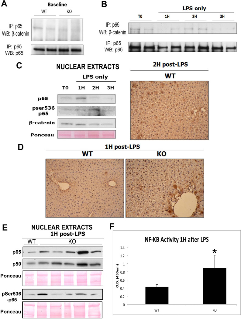
Figure 6. The decreased association of p65 and β-catenin in KOs contributes to protection from injury after LPS treatment.
(A) IP shows that p65 and β-catenin associate strongly in WT livers at baseline but less so in KO livers.
(B) Association of p65/β-catenin decreases after administration of LPS in the WT especially at 1H and 3H, as assessed by IP.
(C) Representative WB of WT liver nuclear lysates shows p65 translocation to the nucleus at 1H while its peak activation (ser536-phospho-p65) occurs at 2H after LPS administration. Ponceau represents loading control. The right panel shows representative nuclear expression of ser536-phosho-p65 in around 50% of hepatocytes in WT at 2H after LPS by IHC.
(D) NF-κB is active in the nucleus 1H after LPS treatment in the KO but not in WT, as shown by IHC for ser536-phospho-p65.
(E) Representative WB shows increased p65 and p50 along with ser536-phospho-p65 in the KO livers over the WT at 1H after LPS.
(F) Transcriptional activity of NF-κB is increased in KOs as compared to WTs 1H after LPS treatment alone, as measured by colorimetric assay (*P<0.05).

