Dear Editor,
The recent controversial studies of man-made avian flu viruses caused a media storm, and brought new concerns to the potential of an avian influenza H5N1 virus pandemic, which has been pending since 19971,2. Although the estimated mortality rate of avian influenza A H5N1 virus infection in humans could be as high as 60%, the World Health Organization (WHO) phase of pandemic alert is currently set at 3, due to that there has not been human-to-human or community-level transmission (http://www.who.int/influenza/preparedness/pandemic/h5n1phase/en/index.html). However, the newly created H5N1 virus strains, which are genetically altered, are transmissible among ferrets, and thus may trigger a real pandemic that could potentially result in millions of deaths according to Science Insider3. While it is arguably a bit too late to debate whether regulations or mandatory reviews should be applied to these dual-use studies, in the matter of fact, these viruses that are probably among the most dangerous infectious agents known already exist. Therefore, a top priority at present is to find effective prophylactic or therapeutic agents that would help to control a pandemic of avian influenza A H5N1 viruses.
Previous reports have demonstrated that the high mortality in humans infected with avian influenza A H5N1 is partly due to acute lung injury or the resulting severe condition, acute respiratory distress syndrome (ARDS)4,5. There are few treatment choices for ARDS, aside from mechanical supporting equipment and empirical treatments. The use of cortisones is controversial.
We have recently discovered that avian influenza A H5N1 virus infection causes acute lung injury by inducing autophagic alveolar epithelial cell death6. Importantly, we found that autophagy inhibitors are effective in ameliorating murine acute lung injury induced by live avian influenza A H5N1 virus infections6. We thus hypothesize that if a drug that is currently in clinical use can act to inhibit autophagy, then such a drug might be a good candidate for treating H5N1 infections.
To test this, we have focused our efforts on chloroquine (CQ), as CQ is the only oral clinical drug that is known to be an autophagy inhibitor7. CQ, or N′-(7-chloroquinolin-4-yl)-N,N-diethyl-pentane-1,4-diamine, was discovered in 1934 by Hans Andersag and his co-workers at Bayer Laboratories and was introduced into clinical practice in 1947 as a prophylactic treatment for malaria8. Currently, CQ and its hydroxyl form, HCQ, are used as anti-inflammatory agents for the treatment of rheumatoid arthritis, lupus erythematosus and amoebic hepatitis. More recently, CQ has been studied for its potential use as an enhancing agent in cancer therapies as well as novel antagonists to chemokine receptor CXCR4 in pancreatic cancer8,9.
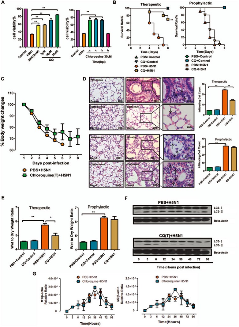
We first tested whether CQ could inhibit cell death in the human lung carcinoma A549 cells infected with live avian influenza A H5N1 virus. The cell viability was improved both prophylactically and therapeutically in a dose-dependent manner with CQ treatments, and the efficacy of CQ was much higher than that of the potent autophagy inhibitor 3-methyladenine (3-MA) (Figure 1A). Next, we tested the potential therapeutic effect of CQ in a mouse model of live H5N1 infections. We found that when CQ was administered therapeutically at a dose equivalent to that for human clinical use, the survival rate of H5N1 virus-infected mice was improved dramatically (from 0% to 70% at day 8 post infection), and the body weight changes also showed a trend of improvement, but prophylactic application of CQ showed no protective effect (Figure 1B and 1C). We analyzed the lung histopathology in these mice and found fewer infiltrating leukocytes in the CQ therapeutic group, but not in the CQ prophylactic group (Figure 1D). Lung edema, as determined by the increased wet/dry weight ratio of lung tissue, was also significantly reduced by therapeutic CQ treatment, but not by prophylactic CQ treatment (Figure 1E). Taken together, our results demonstrate that CQ, a known autophagy inhibitor that is in clinical use, could efficiently ameliorate acute lung injury and dramatically improve the survival rate in mice infected with live avian influenza A H5N1 virus.
Figure 1.
Chloroquine (CQ) is a highly effective therapeutic but not prophylactic agent against avian influenza A H5N1 virus infection in mice. (A) MTS assay of A549 cells treated with 3-MA (3 mM) or chloroquine (3, 10 or 30 μM) 1 h before or treated with chloroquine (30 μM) 1, 3, 6 h after infection with control or H5N1 virus (4 MOI) for 48 h. (B) Survival rates of BALB/c mice receiving therapeutic treatment of CQ (i.p. 50 mg/kg) or vehicle control for 6 h and then once per day for 1 week after the intratracheal instillation of vehicle control or H5N1 virus (106 TCID50) and survival rates for the prophylactic treatment of CQ (i.p. 50 mg/kg) 2 h and 0.5 h before the intratracheal instillation of vehicle control or H5N1 virus (106 TCID50) (n = 10 mice per group). (C) Changes in body weights of BALB/c mice receiving therapeutic treatment of CQ as described above. The values are means ± SEM from ten mice. (D) Representative lung histopathology of BALB/c mice with therapeutic or prophylactic treatment of CQ as described above. Lung tissues were obtained on day 4 or 5 after virus infection. The black arrows pointed at the monocytes and neutrophils of the infiltrating cells. The bar graph shows the mean number of lung infiltrating cells ± SEM from 100 microscopic fields of each group (Original magnification was 200× microscopic fields from the lungs of BALB/c mice infected with H5N1 virus with or withour therapeutic or prophylactic treatment of CQ were partially magnified to 400× 100 fields were analyzed. n = 3 mice per group). (E) Wet/dry weight ratios of the lungs of BALB/c mice with therapeutic or prophylactic treatment of CQ as described above. Lung tissues were obtained on day 4 or 5 after virus instillation (n = 4-6 mice per group). (F) Western blot analysis of LC3-I and LC3-II in mouse lung tissue receiving therapeutic treatment of CQ as described above. Blots were analyzed with antibodies against the indicated proteins. (G) Real-time quantitative PCR analysis of M1's and M2's relative ratios to β-actin in mouse lung tissue receiving therapeutic treatment of CQ as described above (n = 3 mice per group).
CQ acts to raise the lysosomal pH, leading to the inhibition of both the fusion of autophagosomes with lysosomes and lysosomal protein degradation7. As a lysosomotropic agent, CQ could prevent endosomal acidification, which might inhibit influenza virus endocytotic cell entry10,11. CQ was therefore proposed to be a candidate prophylactic agent for influenza virus. However, government-sponsored clinical trials have shown insufficient prophylactic effects against influenza infection12, which is consistent with our mouse results. CQ could also inhibit the innate immune responses through TLR signaling pathways and act as novel antagonists to chemokine receptor CXCR4 in pancreatic cancer9,13. We have shown that CQ treatment clearly inhibited the autophagy in mouse lung induced by avian influenza A H5N1 virus while the virus loads and proinflammatory cytokines were not significantly affected (Figure 1F, 1G, Supplementary information, Figure S1). Further studies are necessary to elucidate the precise molecular mechanisms by which CQ ameliorates murine acute lung injury induced by avian influenza A H5N1 virus.
This notwithstanding, our study strongly suggests that CQ (and potentially its derivatives) should be evaluated as a candidate drug for clinical treatment of H5N1-infected patients. Moreover, a systematic screening of common clinical drugs for potential autophagy inhibitors may lead to the identification of other novel treatments against avian flu.
Acknowledgments
This work is supported by the Ministry of Science and Technology of China (2009CB522105), the National Natural Science Foundation of China (81230002), and the 111 Project (B08007). CJ is a Hsien Wu professor of Biochemistry.
Footnotes
(Supplementary information is linked to the online version of the paper on the Cell Research website.)
Supplementary Information
(A, B, C) Real time quantitativePCR analysis of IL-1β's, IL-6's and TNF-α's relative ratio to beta-actin in mice lung tissue receiving therapeutic treatment of CQ as described above.
Materials and Methods
References
- Herfst S, Schrauwen EJ, Linster M, et al. Science. 2012. pp. 1534–1541. [DOI] [PMC free article] [PubMed]
- Imai M, Watanabe T, Hatta M, et al. Nature. 2012. pp. 420–428. [DOI] [PMC free article] [PubMed]
- Enserink M. Science. 2011. pp. 1192–1193. [DOI] [PubMed]
- Bauer TT, Ewig S, Rodloff AC, et al. Clin Infect Dis. 2006. pp. 748–756. [DOI] [PMC free article] [PubMed]
- Wang H, Jiang C. Sci China C Life Sci. 2009. pp. 459–463. [DOI] [PMC free article] [PubMed]
- Sun Y, Li C, Shu Y, et al. Sci Signal. 2012. p. ra16. [DOI] [PubMed]
- Carew JS, Espitia CM, Esquivel JA, 2nd, et al. J Biol Chem. 2011. pp. 6602–6613. [DOI] [PMC free article] [PubMed]
- Solomon VR, Lee H. Eur J Pharmacol. 2009. pp. 220–233. [DOI] [PubMed]
- Kim J, Yip ML, Shen X, et al. PLoS One. 2012. p. e 31004. [DOI] [PMC free article] [PubMed]
- Savarino A. Lancet Infect Dis. 2011. pp. 653–654. [DOI] [PMC free article] [PubMed]
- Wang H, Jiang C. Sci China C Life Sci. 2009. pp. 464–469. [DOI] [PMC free article] [PubMed]
- Paton NI, Lee L, Xu Y, et al. Lancet Infect Dis. 2011. pp. 677–683. [DOI] [PubMed]
- Lund J, Sato A, Akira S, et al. J Exp Med. 2003. pp. 513–520. [DOI] [PMC free article] [PubMed]
Associated Data
This section collects any data citations, data availability statements, or supplementary materials included in this article.
Supplementary Materials
(A, B, C) Real time quantitativePCR analysis of IL-1β's, IL-6's and TNF-α's relative ratio to beta-actin in mice lung tissue receiving therapeutic treatment of CQ as described above.
Materials and Methods


