FIGURE 3.
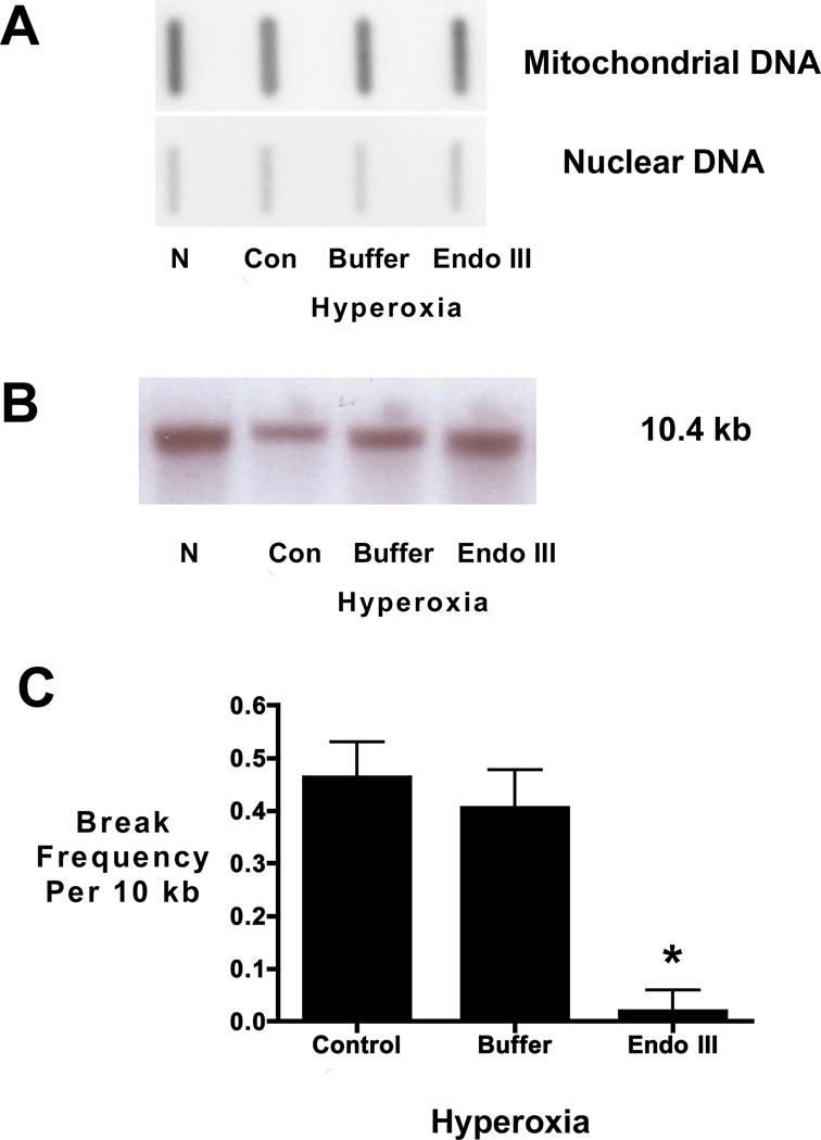
Hyperoxia causes mtDNA damage in fetal rat lung explants that is prevented by the Endo III fusion protein construct. A) Representative slot blot analysis of mtDNA content in fetal rat lung explants cultured for 24 hours in normoxic or hyperoxic conditions in the absence (control and buffer) or presence of Endo III fusion protein construct (EndoIII). No change in mitochondrial DNA content was detected in fetal lung explants as a function of treatment. Representative of 3 experiments. B) Representative Southern blot of alkali-labile mtDNA damage in fetal rat lung explants. Note decreased hybridization intensity in hyperoxia compared to normoxia indicating increased mtDNA damage in hyperoxic fetal lung explants. Note, too, restoration of band intensity when hyperoxic explants were pretreated with the Endo III-fusion protein. C) Histogram depicting the changes in equilibrium lesion density calculated using the Poisson equation Break frequency= -ln (Po) where Po is the quotient of the band intensity of the hyperoxic cultures (control, buffer, or EndoIII) relative to the band intensity of the normoxic explant culture. Calculated changes in equilibrium lesion density normalized to normoxic explants also indicate that hyperoxia increases mtDNA damage which is prevented when mtDNA repair is enhanced *P <0.05 N=4.

