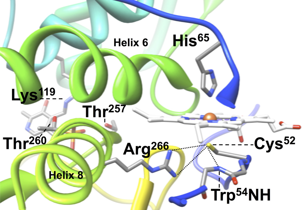
Figure 1.
Location of key residues that interact with the heme and PLP in hCBS. Labeled are: the Fe(III) heme ligands Cys52 and His65; the cysteine(thiolate) hydrogen bonding partners Arg266 and the amide backbone of Trp54; the PLP phosphate hydrogen bonding partners Thr257 and Thr260; and the PLP internal aldimine forming Lys119. Data taken from PDB file 1JBQ (14). The polypeptide backbone is colored using a rainbow scheme from N-terminus (blue) to C-terminus (red).

