Abstract
Neurotransmitter release is a multistep process that is coordinated by a large number of synaptic proteins and depends on proper protein–protein interactions. Using morphological, capacitance, and amperometric measurements, we investigated the effect of tomosyn, a Syntaxin-binding protein, on the different kinetic components of exocytosis in adrenal chromaffin cells. Overexpression of tomosyn decreased the release probability and led to a 50% reduction in the number of fusion-competent vesicles. The number of docked vesicles and the fusion kinetics of single vesicles were not altered suggesting that tomosyn inhibits the priming step. Interestingly, this inhibition is partially relieved at elevated calcium concentration. Calcium ramp experiments supported the latter finding and indicated that the reduction in secretion is caused by a shift in the calcium-dependence of release. These results indicate that secretion is not entirely blocked but occurs at higher calcium concentrations. We suggest that tomosyn inhibits the priming step and impairs the efficiency of vesicle pool refilling in a calcium-dependent manner.
Communication between neurons occurs by the release of neurotransmitter into the synaptic cleft and the subsequent activation of postsynaptic receptors. Neurotransmitter release is mediated by the fusion of synaptic vesicles and is restricted to presynaptic active zones (1). The vesicles in the synapse undergo a multistep cycle that includes vesicles docking at the active zone, priming (the formation of fusion-competent vesicles), fusion, and recycling (2–4). To achieve rapid and efficient synaptic transmission, synapses contain specific protein machinery that mediates these processes. The modulation of these steps is believed to account for several forms of short-term synaptic plasticity (5, 6).
Although numerous proteins have been implicated in the synaptic vesicle cycle, the molecular mechanisms underlying defined steps are only beginning to emerge (7–10). Among the well characterized proteins associated with the synaptic vesicle cycle are Syntaxin, Synaptobrevin (also known as VAMP), and synaptosome-associated protein of 25 kDa (SNAP-25). These proteins form the soluble N-ethylmaleimide-sensitive factor attachment protein receptor (SNARE) complex, which plays an essential role in priming and/or in the fusion reaction itself (11–14). Changes in the quantity or availability of SNARE complexes directly affect the number of fusion-competent vesicles and neurotransmitter release (15–19).
Under resting conditions, the major restriction for SNARE complex formation is the availability of its different components. For example, high affinity binding of Munc-18 to Syntaxin can prevent assembly of the core complex (10, 20). Recent studies provide accumulating evidence regarding the involvement of a previously uncharacterized protein family in regulation of the SNARE complex (21–25). Tomosyn, a brain-specific member of this family, was identified as a binding partner for Syntaxin. Tomosyn can displace Munc-18 from Syntaxin and has been shown to form a four-protein complex composed of tomosyn, SNAP-25, Syntaxin, and Synaptotagmin (21). The C-terminal domain of tomosyn is homologous to the SNARE motif of Synaptobrevin and was suggested to mediate the interaction with Syntaxin (21, 26). Based on these data, it was suggested that tomosyn plays a positive role in neurotransmission by enhancing the availability of Syntaxin to form SNARE complexes (21, 24). In this scenario, formation of the four-protein complex is an intermediate step in a sequence of events leading to SNARE complex formation through the replacement of tomosyn with Synaptobrevin (21, 24). However, there are several caveats to this hypothesis: Synaptobrevin does not dissociate tomosyn from the four protein complex in vitro (21). Furthermore, the Synaptobrevin homology region of tomosyn binds to Syntaxin with relatively low affinity in vitro (24). In addition, Sro7p and Sro77p, two tomosyn yeast homologues, do not possess a Synaptobrevin-like domain but still bind to the yeast Syntaxin homologue Sso1/2p and to the yeast SNAP-25 homologue Sec9p (27). These findings suggest that other domains of tomosyn are involved in the interaction with Syntaxin and imply a function for tomosyn that differs from that proposed earlier (21, 24). Recent studies demonstrated that tomosyn inhibits secretion from PC12 cells. However, the molecular mechanism of this inhibition was not determined and it is not known which step in the process of secretion is affected by tomosyn (21, 22).
In an attempt to assess the role of tomosyn, we overexpressed tomosyn in adrenal chromaffin cells and quantified the effect on catecholamine secretion. We demonstrate that, in contrast to the current view, tomosyn down-regulates secretion and inhibits the calcium-dependent priming step. Interestingly, the inhibitory effect of tomosyn could be partially relieved by elevated calcium levels, suggesting that the regulatory interactions of tomosyn are calcium-sensitive.
Materials and Methods
Plasmid Construction. The gene coding for m-tomosyn was amplified from a rat brain cDNA library (kindly provided by N. Brose, Göttingen, Germany) by PCR. The resulting fragments were subcloned into pSP73 (Promega). The viral vector pSFV1 (Invitrogen) was modified by insertion of an oligonucleotide cassette into its XmaI site to generate singular ClaI and BssHII sites. The gene coding for GFP was modified by PCR to generate a 5′ BglII and 3′ BamHI site and inserted into the BamHI site of the modified pSFV1 to yield pSFV1-GFP. The coding sequence of m-tomosyn was inserted into pSFV1-GFP by using the BamHI and ClaI sites. For the expression of m-tomosyn without fused GFP, the gene was modified by PCR to generate a 5′ Kozak consensus sequence and inserted into pSFV1-IRES-EGFP (28) by using the BamHI and BssHII sites. The sequence of each construct was verified by DNA sequencing. For details on immunocytochemistry and Western, see Supporting Text, which is published as supporting information on the PNAS web site.
Chromaffin Cell Preparation, Infection, and Electron Microscopy. Isolated bovine adrenal chromaffin cells were prepared and cultured as described before (29). Cells were used 2–3 days after preparation. Infection was performed on cultured cells 5–48 h after plating (29). For electron microscopy, chromaffin cells were plated on collagen-coated coverslips (Cellocate, Eppendorf, Germany) and infected with GFP-Tomosyn. 12–18 h after infection, cells were observed under a fluorescence microscope and the location of the infected/control cells were mapped. Coverslips were then fixed and embedded as described before (30). Ultrathin (70–90 nm) sections were cut by using an ultramicrotome (Leica Ultracut UCT), contrasted with lead citrate and viewed under a Philips Technai electron microscope at 120 KV. Pictures were prepared by using a charge coupled device camera Megaview III and analyzed with analysis software. Measurements were done up to a distance of 1,000 nm from the plasma membrane.
Membrane Capacitance and Amperometric Measurements. Conventional whole-cell recordings and capacitance measurements were performed as described (29, 30) and analyzed by using Igor Pro (Wavemetrics, Lake Oswego, OR). Experiments were performed 12–24 h after infection at 30–32°C. Analysis and comparison were always performed from pairs of control and tomosyn-overexpressing cells from the same batch of cells. Statistical analysis was done by using Student's t test or Mann–Whitney nonparametric test. Given values represent mean ± SEM (29). For analysis of single amperometric spikes, chromaffin cells were perfused intracellularly with solution containing 16 μM free calcium. Amperometric activity was monitored by using 5-μm carbon fibers as described (22). Data analysis was performed with igor pro by using a macro procedure that analyzed individual current transients >10 pA higher than the local baseline current. To prevent overrepresentation of cells with higher release probability, the data from each cell were binned into a cumulative probability histogram. Bins were averaged across cells and the data were presented in average cumulative histograms as mean ± SEM.
Photolysis of Caged Ca2+, Ca2+ Ramp Experiments, and [Ca2+]i Measurements. Photolysis experiments were done as described (15, 30). Flashes of UV light were generated by a flash lamp (T.I.L.L. Photonics) and fluorescent excitation light was generated by a monochromator (T.I.L.L. Photonics) that were coupled by using a Dual Port condenser (T.I.L.L. Photonics) into the epifluorescence port of an IX-50 Olympus microscope equipped with a ×40 objective (UAPO/340; Olympus, Tokyo). Fura2FF (TefLabs, Austin, TX) was excited at 350/380 nm and detected through a 500-nm long-pass filter (T.I.L.L. Photonics). Calcium ramps were elicited by the fluorescence excitation light alternating between 350 and 380 nm such that photolysis of NPEGTA (G. Ellis-Davis; MCP, Hahnemann University, Philadelphia) could be combined with simultaneous measurement of [Ca2+]i. Rate constants were calculated as described (28, 31) and binned according to the corresponding calcium values to create the plot of rate constants versus [Ca2+]i on a double logarithmic scale (see Fig. 5). Given values represent mean ± SEM.
Fig. 5.
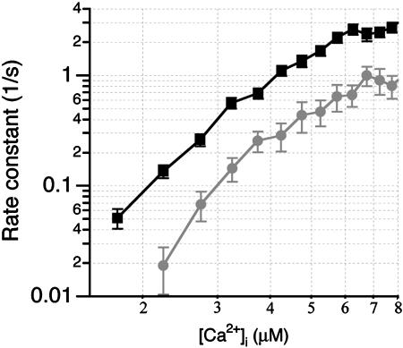
Tomosyn increases the requirement for calcium in exocytosis. Calcium ramp experiments were performed by slow release of Ca2+ from NP-EGTA by using weak UV-illumination. Membrane capacitance and Ca2+ concentration were monitored simultaneously during the 4 s of stimulation, and rate constants of exocytosis were analyzed and plotted on a double-logarithmic scale versus the respective [Ca2+]i. Cells overexpressing tomosyn (gray circles, n = 12) secreted with slower rate constants than control cells (black squares, n = 18) for equivalent [Ca2+]i. The calcium-dependence curve for tomosyn cells was shifted toward higher calcium concentrations, but its slope remained unchanged.
Results
Overexpression of Full-Length Tomosyn Reduces the Vesicular Release Probability in Adrenal Chromaffin Cells. Assuming that displacement of Munc18 from Syntaxin by tomosyn has a positive effect on secretion, overexpression of tomosyn may lead to a higher concentration of free Syntaxin molecules in the synapse and to a higher priming rate and enhanced secretion. To test this hypothesis, we overexpressed tomosyn as an internal ribosome entry site (IRES)-GFP construct in adrenal chromaffin cells. The expression level of exogenous, overexpressed tomosyn was on average 13 times higher than the expression level of endogenous tomosyn, as determined by immunofluorescence and Western analysis (see Fig. 6, which is published as supporting information on the PNAS web site). In addition, we verified that the expression level of Syntaxin, a tomosyn interacting partner, was similar in control cells and cells overexpressing tomosyn (see Fig. 6). Thus, the observed effects could be attributed directly to the overexpression of tomosyn in these cells.
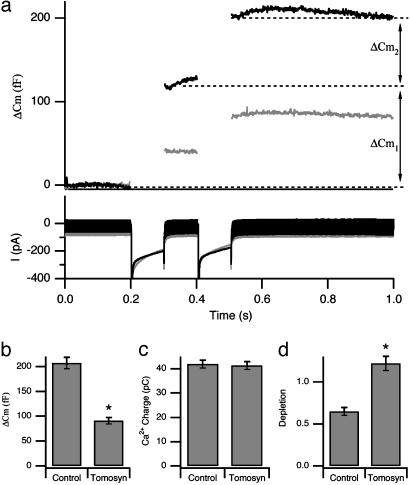
Our initial goal was to investigate the effect of tomosyn overexpression on depolarization-induced secretion. Chromaffin cells were stimulated with a dual-pulse depolarization protocol (Fig. 1a), and calcium current and membrane capacitance were assayed simultaneously before and after stimulation. Control (uninfected) chromaffin cells responded to the dual-pulse depolarization protocol with a robust increase in membrane capacitance. The secretory response was significantly reduced in chromaffin cells overexpressing tomosyn (Fig. 1 a and b), whereas the calcium currents were similar in both groups (Fig. 1c). These results suggest that tomosyn attenuates the response of the exocytotic machinery to calcium. We next measured the size of the readily releasable pool (RRP) by using a dual pulse protocol (32, 33). The RRP is composed of vesicles that are docked to the plasma membrane and are in a fusion-competent state. The degree of RRP depletion is measured as the ratio R = ΔCm2/ΔCm1 (32) and gives a good indication for the probability of release. In control cells, the RRP was effectively depleted by this protocol, whereas in tomosyn cells, no depletion was observed and the second response was slightly larger than the first (Fig. 1a). This secretory facilitation (Fig. 1d) implies that tomosyn overexpression reduces the probability of release.
Fig. 1.
Tomosyn reduces the probability of release in chromaffin cells. (a) Changes in capacitance (Cm, upper trace) and current (I, lower trace) were simultaneously measured in response to a dual-depolarization protocol. (b) The average capacitance increase in tomosyn-overexpressing cells was ≈50% of that in control cells (207 ± 11.8 fF control; black, 91 ± 6.8 fF tomosyn; gray), whereas calcium influx (c) was identical in control and tomosyn cells (41.9 ± 1.7 pC control, 41.4 ± 1.6 pC tomosyn). (d) In control cells, the RRP was effectively depleted by this protocol, as indicated by the ratio between the second and first capacitance increase (ΔCm2/ΔCm1 = 0.65 ± 0.05), whereas tomosyn cells exhibited facilitation (ΔCm2/ΔCm1 = 1.22 ± 0.08), indicating a decrease in the probability of release in these cells. Results were averaged from 41 traces from 12 tomosyn and 49 traces from 12 control cells. ΔCm1 and ΔCm2 are shown for control data for clarification. Asterisks indicate a significant change (P < 0.0001).
Because a reliable calculation of pool size depends on significant pool depletion (R < 0.7; refs. 32 and 33), we could not reliably quantify the size of the RRP in these cells. Therefore, we used only the sum of the capacitance increases to estimate the effect. Accordingly, secretion was reduced in tomosyn cells by >50% as compared to control cells (Fig. 1). This is consistent with the reduced secretion that was observed recently when tomosyn was expressed in PC12 cells (21, 22).
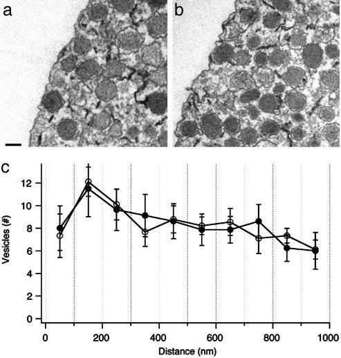
Tomosyn Overexpression Has No Detectable Effect on Vesicle Docking or Fusion Kinetics. The decrease in secretion reported in the previous section might be caused by (i) a decrease in the number of docked vesicles, (ii) attenuation of vesicle priming, or (iii) modification of the calcium-dependent fusion reaction. To test the first possibility, we performed electron microscopy experiments and compared the number of vesicles close to the plasma membrane in tomosyn cells to that in control cells. Quantitative analysis revealed that the number of vesicles located within 100 nm of the plasma membrane, the total number of vesicles, and the overall morphology were similar in tomosyn and control cells (Fig. 2). These results rule out the possibility that the decrease in secretion in tomosyn cells was mediated by a reduced number of “morphologically docked” vesicles.
Fig. 2.
The number of morphologically docked vesicles is unchanged in tomosyn cells. Representative electron micrographs of two sections from control (a) and tomosyn-overexpressing (b) bovine chromaffin cells. The overall distribution of organelles within a tomosyn-expressing cell is similar to that of control cell. (Bar represents 200 nm.) (c) Quantitative analysis revealed that the number of “morphologically docked” vesicles (within a distance of 100 nm from the plasma membrane) and the overall relative distribution and density of vesicles (within a distance of 1,000 nm from the plasma membrane) are similar in control cells (open circles, 750 vesicles from nine cells) and tomosyn cells (filled circles, 668 vesicles from eight cells).
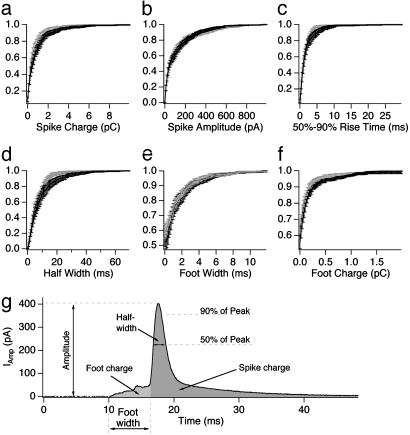
To examine a possible effect of tomosyn on the fusion kinetics of a single vesicle we next conducted a detailed analysis of amperometric spikes. As expected from the capacitance experiments, the frequency of the amperometric spikes in tomosyn cells was smaller than in control cells (not shown). However, the kinetic properties of single spikes were not significantly different in tomosyn cells (Fig. 3). This suggests that, in agreement with recent data (22), tomosyn does not alter the fusion step. We conclude from these experiments that tomosyn is not directly involved in vesicle docking nor in the fusion reaction itself. The effects of tomosyn on secretion are therefore best explained by modulation of vesicle priming. Further evidence in support of this hypothesis is presented below.
Fig. 3.
Individual spike characteristics are similar in control and tomosyn cells. (a–f) Amperometric spike properties from control cells (black, 806 spikes from eight cells) and tomosyn-overexpressing cells (gray, 511 spikes from six cells). Data are presented in normalized cumulative histograms to which each cell contributes equally, irrespective of the number of spikes it produced. (g) An example of a typical spike showing the relevant parameters for analysis.
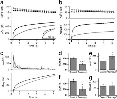
Overexpression of Tomosyn Inhibits Secretion by Attenuating the Priming Process. To measure the effect of tomosyn overexpression on the different kinetic components of exocytosis, we conducted a series of flash photolysis experiments. Capacitance was measured in the whole-cell configuration and secretion was triggered by photorelease of calcium from the caged compound NPEGTA by a flash of UV light (15, 30). Control chromaffin cells responded to the flash stimulation with a typical biphasic capacitance increase, in which an exocytotic burst was followed by a sustained phase of secretion (Fig. 4a Lower). The exocytotic burst occurs during the first second after the flash and represents the fusion of release-competent vesicles. The sustained component, measured after the burst, represents vesicle recruitment and subsequent fusion (15, 34). Cells overexpressing tomosyn showed a markedly different secretion pattern: whereas the exocytotic burst was reduced, the sustained component was enhanced (Fig. 4 a, d, and e). Average [Ca2+]i during the flash stimulation in tomosyn cells was slightly higher than in control cells, excluding the possibility that the reduced secretion resulted from lower [Ca2+]i (Fig. 4a Upper). A second flash stimulation, applied to the same cells after a 2-min recovery period, was also smaller in tomosyn cells compared with control cells, indicating that tomosyn inhibits vesicle pools refilling after stimulation (Fig. 4 b and f and g). The flash experiments discussed above were performed separately with cells expressing either a GFP-tomosyn fusion protein or with cells expressing tomosyn separated from GFP by an internal ribosome entry site element. The results obtained with both constructs were indistinguishable, allowing us to pool the two data sets.
Fig. 4.
Impaired exocytotic response to elevated [Ca2+]i in cells overexpressing tomosyn. (a) Averaged calcium ([Ca2+]i, Upper) and capacitance change (Cm, Lower) in control cells (black, n = 29) and cells overexpressing tomosyn (gray, n = 23) in response to flash photolysis of caged Ca2+. Arrow indicates the time of flash stimulation. The size of the exocytotic burst in tomosyn and control cells was measured as the average capacitance increase during the first second after the flash. The exocytotic burst in tomosyn cells was significantly reduced (d; 206 ± 29 fF tomosyn, 418 ± 36 fF control). The sustained component, represented by the capacitance increase during the remaining time of stimulation, was significantly enhanced (e; 200 ± 31 fF tomosyn, 128 ± 31 fF control). The capacitance trace in tomosyn cells was normalized to control for the first second after the flash (a Inset). A significant reduction is observed in the fast component of the burst. (b) Response to a second flash stimulation in tomosyn cells was attenuated as well, with a similar reduction in the exocytotic burst (f; 226 ± 71 fF tomosyn, gray; 488 ± 64 fF control, black), but no significant change in the sustained component (g; 143 ± 51 fF tomosyn, 130 ± 27 fF control). (c) Averaged recordings of amperometric current (Upper) and the integral of the amperometric current (Lower) in response to flash photolysis of caged calcium (indicated by an arrow) from control (black, n = 12) and tomosyn (gray, n = 14) cells. [Ca2+]i was kept between 10 and 20 μM for 5 s in all experiments. Compared with control cells, tomosyn cells display a reduced exocytotic burst and an enhanced sustained component. Note that the integral of the amperometric current agrees with capacitance data shown in a. *, P < 0.05; ***, P < 0.0001.
Previous work has shown that the exocytotic burst can be further resolved into a fast component with a time constant of ≈30 ms and a slow component with a time constant of ≈200 msec (15, 34). The fast burst component represents the fusion of vesicles from the RRP, and the slow component represents fusion of vesicles from the slowly releasable pool (SRP; ref. 34). We performed a similar analysis to determine which of the distinct phases of the exocytotic burst was affected by overexpression of tomosyn. Our results demonstrate that the amplitudes of the fast and slow burst component in tomosyn cells were reduced by 87% and 54%, respectively, as compared to control cells (Table 1 and Fig. 4a Inset). The time constant of the fast burst component was slightly attenuated. However, because in most of tomosyn cells (16 of 22) the fast component was absent, the latter observation is based on very few data and thus not statistically significant. The time constant of the slow burst component was not significantly affected by overexpression of tomosyn. These results suggest that tomosyn overexpression substantially inhibits the priming of vesicles into the RRP.
Table 1. Effect of tomosyn on the different kinetic components of the exocytotic burst.
| Fast component, fF | Fast τ, ms | Slow component, fF | Slow τ, ms | |
|---|---|---|---|---|
| First flash | ||||
| Control | 175.2 ± 26.3 (n = 32) | 22.8 ± 1.7 (n = 30) | 184.5 ± 18.8 (n = 32) | 253.9 ± 27.4 (n = 32) |
| Tomosyn | 23.8 ± 10.2 (n = 22)** | 43.3 ± 7.9 (n = 6)† | 90.6 ± 28 (n = 21)* | 322.0 ± 74.2 (n = 11) |
| Second flash | ||||
| Control | 238.6 ± 33.4 (n = 22) | 31.8 ± 3.8 (n = 21) | 304.9 ± 44 (n = 22) | 280.0 ± 35.5 (n = 21) |
| Tomosyn | 42.3 ± 71.4 (n = 16)** | 35.4 ± 8.9 (n = 7)† | 87.2 ± 33.3 (n = 16)* | 296.3 ± 49 (n = 10) |
, P < 0.0005
, P < 0.0001
The fast component was absent from most tomosyn cells
As previously noted, tomosyn-infected cells showed a significant increase in the sustained component during the first flash stimulation. This finding is intriguing because it suggests that tomosyn inhibits priming under resting conditions but that this inhibition is relieved under high [Ca2+]i. Because membrane capacitance measurements do not distinguish between exocytotic and endocytotic activity, we could not rule out the possibility that the enhanced sustained component in tomosyn cells reflects inhibition of endocytosis rather than enhanced exocytosis. To explore this possibility, we used carbon fiber amperometry in combination with flash photolysis experiments. The amperometric current provides a direct measure of catecholamine release and is not influenced by endocytosis. Tomosyn overexpression caused a reduction in secretion during the exocytotic burst and enhanced secretion during the sustained component phase. This can be seen in both the level of amperometric activity (Fig. 4c Upper) and the integral of the amperometric current (Fig. 4c Lower). These results are consistent with the findings described in the previous section, indicating that the observed effects of tomosyn can be accounted for by modulation of exocytosis and not of endocytosis.
Tomosyn Raises the Requirement for Ca2+ in Exocytosis and Shifts the Calcium-Dependence of Release. In the previous section we showed that tomosyn causes a reduction in exocytosis, possibly through a decline in the probability of release. Furthermore, the inhibition caused by tomosyn overexpression is relieved upon elevation of [Ca2+]i. Further support for these findings came from two sets of amperometric measurements that we performed: control cells responded to application of 70 mM KCl with a spike frequency of 22 spikes per min, whereas tomosyn cells showed a markedly attenuated response (5.8 spikes per min; data not shown). However, when we intracellularly dialyzed the cells with 16 μM calcium, the spike number in tomosyn cells was elevated >3-fold (20 spikes per min), whereas in control cells only a slight elevation was observed (31 spikes per min). Thus, at low intracellular calcium levels, fewer vesicles are primed in tomosyn cells and the size of the burst component is smaller. However, elevation of calcium concentration above 10 μM for several seconds overcomes the inhibition imposed by tomosyn.
To further characterize the Ca2+-dependence of secretion, we performed a set of “calcium-ramp” experiments in which the intracellular Ca2+ concentration is raised slowly, rather than abruptly as in the flash experiments. This is achieved by weak UV-illumination that causes controlled photolysis of the NPEGTA (28, 31). During the period of stimulation, [Ca2+]i increases, leading to an increased rate of secretion. This allowed us to determine the dependence of the rate constant of secretion on [Ca2+]i. As shown in Fig. 5, a shift in the rate constants toward higher [Ca2+]i without a significant change in the slope was observed in tomosyn-overexpressing cells. This indicates that overexpression of tomosyn raises the requirement for Ca2+ in exocytosis without changing the degree of calcium cooperativity. Because we did not observe a change in spike kinetics (Fig. 3), we suggest that tomosyn does not alter the calcium-dependent fusion step or the fusion reaction itself, but rather attenuates vesicle priming.
Discussion
In the present study we show that tomosyn, a Syntaxin binding protein, inhibits vesicle priming and that this inhibition is relieved at high calcium concentration. The involvement of calcium in the regulatory action of tomosyn may reflect an interaction with calcium-responsive proteins associated with the SNARE complex. Our experiments suggest that the modulation exerted by tomosyn is activity-dependent, a phenomenon known to be associated with synaptic plasticity.
Previous studies reported that tomosyn dissociates Munc18 from Syntaxin1 and may enhance SNARE complex formation and secretion (21, 24). We demonstrate that this prediction is not fulfilled. Rather, our results support recent findings that show that tomosyn inhibits secretion from PC12 cells (21, 22). We further characterized the effect of tomosyn and suggest that tomosyn inhibits priming and reduces the number of fusion-competent vesicles.
Although we demonstrate that tomosyn overexpression inhibits secretion, this inhibition is relieved following elevation of [Ca2+]i. This unique phenomenon is illustrated by several experiments. During the 5 s of flash stimulation, when calcium concentration is >10 μM, tomosyn-overexpressing cells exhibit enhanced late secretion. The amperometric spike frequency was very low during subtle increases in calcium (application of 70 mM KCl) but increased significantly after dialysis of high calcium into the cell. Finally, the calcium ramp experiment shows a shift in the calcium-dependence curve, indicating that secretion is not completely blocked but occurs at higher calcium concentrations than in control cells.
According to the current model for secretion from chromaffin cells, the inhibition of priming, the reduced fast exocytotic burst, and the shift in the calcium-secretion relationship could indicate that tomosyn (i) interferes with priming of vesicles into the RRP (see ref. 35), (ii) reduces the release rate, or (iii) alters the calcium-dependent fusion step. A reduced rate or a change in the calcium-dependence of fusion should lead to a change in the time constants of the burst components and to a change in the slope of the calcium-secretion relationship, which we do not observe. Because, in addition, we demonstrate that tomosyn reduces mainly the size of the RRP, we conclude that tomosyn inhibits the maturation of vesicles into the RRP (35). A putative molecular explanation is discussed below.
According to our findings we suggest the following model: under control conditions, calcium elevation allows the final association of Synaptotagmin with preassembled SNARE complexes and with phospholipids and thus drives vesicle fusion (36–40). Overexpression of tomosyn might affect the number of preassembled SNARE complexes associated with a single vesicle (41, 42), causing a reduced release probability and a higher requirement for calcium. Such a mechanism might underlie both the inhibition of priming and the shift in the calcium-dependence curve. Although this hypothesis fits most of the data presented, it seems that the effect of tomosyn is more complex than being a simple inhibition of a single step. Further experiments will be needed to elucidate its precise molecular mechanism.
Inhibition of exocytosis and a similar shift in the calcium-dependence curve was observed after cleavage of SNAP-25 or overexpression of SNAP-25 mutants, whereas high calcium levels rescued secretion under these conditions (18, 37, 40, 43, 44). These findings suggest a general mechanism in which high calcium levels overcome inhibitory effects on SNARE complex formation.
Tomosyn possesses two domains that might contribute to its function. Its C-terminal end contains a coil-coiled, VAMP-homology domain, and its N-terminal half contains a WD-repeat motif, homologous to several β propeller-like proteins (27). It was postulated that tomosyn utilizes an R-SNARE motif to interact with Syntaxin and to substitute for one of the components of the SNARE complex (26). Indeed, tomosyn coiled-coil domain was shown to bind Syntaxin and to compete with Synaptobrevin for binding of Syntaxin and SNAP25 (22). However, the binding kinetics are very slow, and binding occurs at very high concentrations of the coiled coil domain. This binding causes inhibition of secretion in a cell-free preparation derived from PC12 cells. However, expression of the C-terminal part containing the coiled coil domain in PC12 cells did not inhibit secretion (21). It is possible that, under physiological conditions, when trans-SNARE complexes exist, the coiled-coil domain is not sufficient to achieve inhibition. Alternatively, other domains of the protein may participate in the inhibition.
The WD-repeats consist of highly conserved repetitive units that create a β-sheet propeller-like structure (45). This structure creates a stable platform that can reversibly form complexes with several proteins, thus coordinating sequential and/or simultaneous interactions with several partners. Therefore, the WD40 domain of tomosyn might be involved in the sequential binding/unbinding of Munc-18/Syntaxin/Synaptobrevin and thus regulate SNARE complex formation. Thus, it still remains to be determined which domains mediate the observed effect of tomosyn.
The most puzzling finding in this study was that high calcium relieves the inhibition of tomosyn. Although tomosyn does not possess a calcium-binding motif, it interacts with two calcium-responsive proteins (21): the calcium sensor protein Synaptotagmin (46) and the SNARE component SNAP-25 (28, 40). Interaction with either of these proteins might alter secretion in a manner demonstrated in this study. One can speculate that high calcium causes detachment of tomosyn from the four-protein intermediate complex. Tomosyn can then dislocate to the cytosol or bind once more to new sites when calcium is lowered, causing further inhibition.
The levels of tomosyn expression in the brain are very low, estimated to be only 3% of Syntaxin levels (21). Changes in the level of tomosyn might constitute a mechanism that modulates release probability and synaptic strength, as was suggested recently for Dunc13 (47). The regulation of SNARE complex formation by different mechanisms can affect the extent of secretion and modulate synaptic efficacy (23, 48). Interestingly, tomosyn and Munc13-1 are the only proteins known to displace Syntaxin from Munc18 in vitro, yet their effects on priming seem to be opposed (21, 49). Whereas overexpression of Munc13-1 induces a substantial increase in vesicle priming (30), tomosyn overexpression reduces the release probability and blocks priming. In addition, tomosyn is unique in that it changes the calcium-dependence of secretion. The exact molecular events involving the action of tomosyn are unknown. Nonetheless, our findings suggest that tomosyn belongs to a previously uncharacterized group of SNARE modulators that regulate vesicle priming in a calcium-dependent manner.
Supplementary Material
Acknowledgments
We thank Yoel Kloog, Uri Nili, and David Stevens for comments on the manuscript, Frederique Varoqueaux and Heidi de Wit for suggestions for the electron microscopy, Vera Shinder and Shira Tzadikario for excellent technical assistance with electron microscopy, and Detlef Hof for help with amperometry. This research was supported by Israel Science Foundation Grant 424/02-16.6 and by grants from the Minerva Junior research group and the Adams supercenter for brain studies (to U.A.) and from the Deutsche Forschungsgemeinschaft (to J.R.).
Abbreviations: SNARE, soluble N-ethylmaleimide-sensitive factor attachment protein receptor; SNAP-25, synaptosome-associated protein of 25 kDa; RRP, readily releasable pool.
References
- 1.Garner, C. C., Kindler, S. & Gundelfinger, E. D. (2000) Curr. Opin. Neurobiol. 10, 321-327. [DOI] [PubMed] [Google Scholar]
- 2.Richmond, J. & Broadie, K. (2002) Curr. Opin. Neurobiol. 12, 499. [DOI] [PubMed] [Google Scholar]
- 3.Sudhof, T. C. (2000) Neuron 28, 317-320. [DOI] [PubMed] [Google Scholar]
- 4.Rosenmund, C., Rettig, J. & Brose, N. (2003) Curr. Opin. Neurobiol. 13, 509-519. [DOI] [PubMed] [Google Scholar]
- 5.Rettig, J. & Neher, E. (2002) Science 298, 781-785. [DOI] [PubMed] [Google Scholar]
- 6.Zucker, R. S. (1999) Curr. Opin. Neurobiol. 9, 305-313. [DOI] [PubMed] [Google Scholar]
- 7.Augustine, G. J., Burns, M. E., DeBello, W. M., Hilfiker, S., Morgan, J. R., Schweizer, F. E., Tokumaru, H. & Umayahara, K. (1999) J. Physiol. (London) 520, 33-41. [DOI] [PMC free article] [PubMed] [Google Scholar]
- 8.Fernandez-Chacon, R. & Sudhof, T. C. (1999) Annu. Rev. Physiol. 61, 753-776. [DOI] [PubMed] [Google Scholar]
- 9.Brunger, A. T. (2000) Curr. Opin. Neurobiol. 10, 293-302. [DOI] [PubMed] [Google Scholar]
- 10.Rizo, J. & Sudhof, T. C. (2002) Nat. Rev. Neurosci. 3, 641-653. [DOI] [PubMed] [Google Scholar]
- 11.Jahn, R. & Sudhof, T. C. (1999) Annu. Rev. Biochem. 68, 863-911. [DOI] [PubMed] [Google Scholar]
- 12.Sutton, R. B., Fasshauer, D., Jahn, R. & Brunger, A. T. (1998) Nature 395, 347-353. [DOI] [PubMed] [Google Scholar]
- 13.Weber, T., Zemelman, B. V., McNew, J. A., Westermann, B., Gmachl, M., Parlati, F., Sollner, T. H. & Rothman, J. E. (1998) Cell 92, 759-772. [DOI] [PubMed] [Google Scholar]
- 14.Weis, W. I. & Scheller, R. H. (1998) Nature 395, 328-329. [DOI] [PubMed] [Google Scholar]
- 15.Xu, T., Binz, T., Niemann, H. & Neher, E. (1998) Nat. Neurosci. 1, 192-200. [DOI] [PubMed] [Google Scholar]
- 16.Xu, T., Rammner, B., Margittai, M., Artalejo, A. R., Neher, E. & Jahn, R. (1999) Cell 99, 713-722. [DOI] [PubMed] [Google Scholar]
- 17.Lonart, G. & Sudhof, T. C. (2000) J. Biol. Chem. 275, 27703-27707. [PubMed] [Google Scholar]
- 18.Trudeau, L. E., Fang, Y. & Haydon, P. G. (1998) Proc. Natl. Acad. Sci. USA 95, 7163-7168. [DOI] [PMC free article] [PubMed] [Google Scholar]
- 19.Fergestad, T., Wu, M. N., Schulze, K. L., Lloyd, T. E., Bellen, H. J. & Broadie, K. (2001) J. Neurosci. 21, 9142-9150. [DOI] [PMC free article] [PubMed] [Google Scholar]
- 20.Misura, K. M., Scheller, R. H. & Weis, W. I. (2000) Nature 404, 355-362. [DOI] [PubMed] [Google Scholar]
- 21.Fujita, Y., Shirataki, H., Sakisaka, T., Asakura, T., Ohya, T., Kotani, H., Yokoyama, S., Nishioka, H., Matsuura, Y., Mizoguchi, A., et al. (1998) Neuron 20, 905-915. [DOI] [PubMed] [Google Scholar]
- 22.Hatsuzawa, K., Lang, T., Fasshauer, D., Bruns, D. & Jahn, R. (2003) J. Biol. Chem. 278, 31159-31166. [DOI] [PubMed] [Google Scholar]
- 23.Scales, S. J., Hesser, B. A., Masuda, E. S. & Scheller, R. H. (2002) J. Biol. Chem. 277, 28271-28279. [DOI] [PubMed] [Google Scholar]
- 24.Yokoyama, S., Shirataki, H., Sakisaka, T. & Takai, Y. (1999) Biochem. Biophys. Res. Commun. 256, 218-222. [DOI] [PubMed] [Google Scholar]
- 25.Widberg, C. H., Bryant, N. J., Girotti, M., Rea, S. & James, D. E. (2003) J. Biol. Chem. 278, 35093-35101. [DOI] [PubMed] [Google Scholar]
- 26.Masuda, E. S., Huang, B. C., Fisher, J. M., Luo, Y. & Scheller, R. H. (1998) Neuron 21, 479-480. [DOI] [PubMed] [Google Scholar]
- 27.Lehman, K., Rossi, G., Adamo, J. E. & Brennwald, P. (1999) J. Cell Biol. 146, 125-140. [DOI] [PMC free article] [PubMed] [Google Scholar]
- 28.Sorensen, J. B., Matti, U., Wei, S. H., Nehring, R. B., Voets, T., Ashery, U., Binz, T., Neher, E. & Rettig, J. (2002) Proc. Natl. Acad. Sci. USA 99, 1627-1632. [DOI] [PMC free article] [PubMed] [Google Scholar]
- 29.Ashery, U., Betz, A., Xu, T., Brose, N. & Rettig, J. (1999) Eur. J. Cell Biol. 78, 525-532. [DOI] [PubMed] [Google Scholar]
- 30.Ashery, U., Varoqueaux, F., Voets, T., Betz, A., Thakur, P., Koch, H., Neher, E., Brose, N. & Rettig, J. (2000) EMBO J. 19, 3586-3596. [DOI] [PMC free article] [PubMed] [Google Scholar]
- 31.Xu, T. & Bajjalieh, S. M. (2001) Nat. Cell Biol. 3, 691-698. [DOI] [PubMed] [Google Scholar]
- 32.Gillis, K. D., Mossner, R. & Neher, E. (1996) Neuron 16, 1209-1220. [DOI] [PubMed] [Google Scholar]
- 33.Smith, C., Moser, T., Xu, T. & Neher, E. (1998) Neuron 20, 1243-1253. [DOI] [PubMed] [Google Scholar]
- 34.Voets, T., Neher, E. & Moser, T. (1999) Neuron 23, 607-615. [DOI] [PubMed] [Google Scholar]
- 35.Sorensen, J. B., Fernandez-Chacon, R., Sudhof, T. C. & Neher, E. (2003) J. Gen. Physiol. 122, 265-276. [DOI] [PMC free article] [PubMed] [Google Scholar]
- 36.Davis, A. F., Bai, J., Fasshauer, D., Wolowick, M. J., Lewis, J. L. & Chapman, E. R. (1999) Neuron 24, 363-376. [DOI] [PubMed] [Google Scholar]
- 37.Gerona, R. R., Larsen, E. C., Kowalchyk, J. A. & Martin, T. F. (2000) J. Biol. Chem. 275, 6328-6336. [DOI] [PubMed] [Google Scholar]
- 38.Shin, O. H., Rhee, J. S., Tang, J., Sugita, S., Rosenmund, C. & Sudhof, T. C. (2003) Neuron 37, 99-108. [DOI] [PubMed] [Google Scholar]
- 39.Yoshihara, M. & Littleton, J. T. (2002) Neuron 36, 897-908. [DOI] [PubMed] [Google Scholar]
- 40.Zhang, X., Kim-Miller, M. J., Fukuda, M., Kowalchyk, J. A. & Martin, T. F. (2002) Neuron 34, 599-611. [DOI] [PubMed] [Google Scholar]
- 41.Lang, T., Bruns, D., Wenzel, D., Riedel, D., Holroyd, P., Thiele, C. & Jahn, R. (2001) EMBO J. 20, 2202-2213. [DOI] [PMC free article] [PubMed] [Google Scholar]
- 42.Tokumaru, H., Umayahara, K., Pellegrini, L. L., Ishizuka, T., Saisu, H., Betz, H., Augustine, G. J. & Abe, T. (2001) Cell 104, 421-432. [DOI] [PubMed] [Google Scholar]
- 43.Capogna, M., McKinney, R. A., O'Connor, V., H., G. B. & Thompson, S. M. (1997) J. Neurosci. 17, 7190-7202. [DOI] [PMC free article] [PubMed] [Google Scholar]
- 44.Wei, S., Xu, T., Ashery, U., Kollewe, A., Matti, U., Antonin, W., Rettig, J. & Neher, E. (2000) EMBO J. 19, 1279-1289. [DOI] [PMC free article] [PubMed] [Google Scholar]
- 45.Smith, T. F., Gaitatzes, C., Saxena, K. & Neer, E. J. (1999) Trends Biochem. Sci. 24, 181-185. [DOI] [PubMed] [Google Scholar]
- 46.Geppert, M., Goda, Y., Hammer, R. E., Li, C., Rosahl, T. W., Stevens, C. F. & Sudhof, T. C. (1994) Cell 79, 717-727. [DOI] [PubMed] [Google Scholar]
- 47.Aravamudan, B. & Broadie, K. (2003) J. Neurobiol. 54, 417-438. [DOI] [PubMed] [Google Scholar]
- 48.Lao, G., Scheuss, V., Gerwin, C. M., Su, Q., Mochida, S., Rettig, J. & Sheng, Z. H. (2000) Neuron 25, 191-201. [DOI] [PubMed] [Google Scholar]
- 49.Sassa, T., Harada, S., Ogawa, H., Rand, J. B., Maruyama, I. N. & Hosono, R. (1999) J. Neurosci. 19, 4772-4777. [DOI] [PMC free article] [PubMed] [Google Scholar]
Associated Data
This section collects any data citations, data availability statements, or supplementary materials included in this article.


