Abstract
Most living organisms, including humans, age. Over time the ability to do physical and intellectual work deteriorates, and susceptibility to infectious, metabolic, and neurodegenerative diseases increases, which leads to general fitness decline and ultimately to death. Work in model organisms has demonstrated that genetic and environmental manipulations can prevent numerous age-associated diseases, improve health at advanced age, and increase life span. Calorie restriction (CR) (consumption of a diet with fewer calories but containing all the essential nutrients) is the most robust manipulation, genetic or environmental, to extend longevity and improve health parameters in laboratory animals. However, outside of the protected laboratory environment, the effects of CR are much less certain. Understanding the molecular mechanisms of CR may lead to the development of novel therapies to combat diseases of aging and to improve the quality of life. Sirtuins, a family of NAD+-dependent enzymes, mediate a number of metabolic and behavioral responses to CR and are intriguing targets for pharmaceutical interventions. We review the molecular understanding of CR; the role of sirtuins in CR; and the effects of sirtuins on physiology, mood, and behavior.
Keywords: SIRT1, sirtuins, longevity
INTRODUCTION
The desire of people to have longer and healthier lives is as ancient as humanity. It has been almost 80 years since the counterintuitive and unexpected demonstration that retarding the growth of rats or mice by restricting calorie consumption increases their life span (1). Subsequently, researchers demonstrated that calorie restriction (CR), also termed dietary restriction (DR), extends longevity in numerous model organisms, such as yeast (2), worms (3), flies (4), spiders (5, 6), mice (7), and possibly rhesus monkeys (8).
CR is a rather severe reduction in the amount of food (calories) consumed by the organism compared with normal or ad libitum food intake. The response to CR is thought to have evolved naturally as animals dealt with food scarcity throughout their lives. From an evolutionary point of view, the pressure of natural selection is strong before sexual maturity and decreases dramatically after reproduction. However, attempts to reproduce during times of food scarcity are often futile, as progeny are unlikely to survive. Therefore, the ability to extend reproductive fitness and longevity in order to postpone reproduction until more prosperous times would be selected for (9).
Not only does CR extend the length of life, but it also prevents or delays the severity of numerous age-associated diseases (10, 11). However, maintaining such a strict diet is not practical for most humans. Additionally, some effects of CR may result in more harm than good when practiced outside of laboratory, an environment rife with infectious diseases. Therapies based on molecular mechanisms of CR may be safer alternatives to prevent age-associated diseases and increase the health span of humans. The detailed mechanisms by which CR extends longevity and health span of model organisms are still being elaborated, but the research in this area is exciting and advancing quickly.
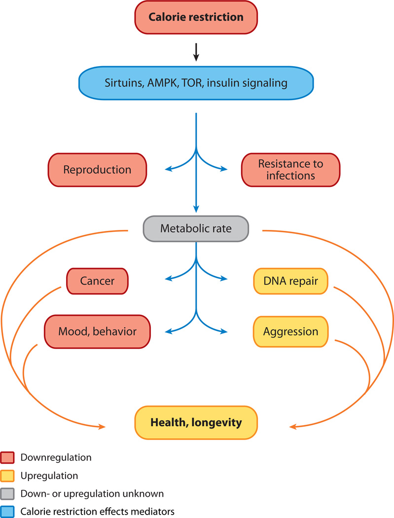
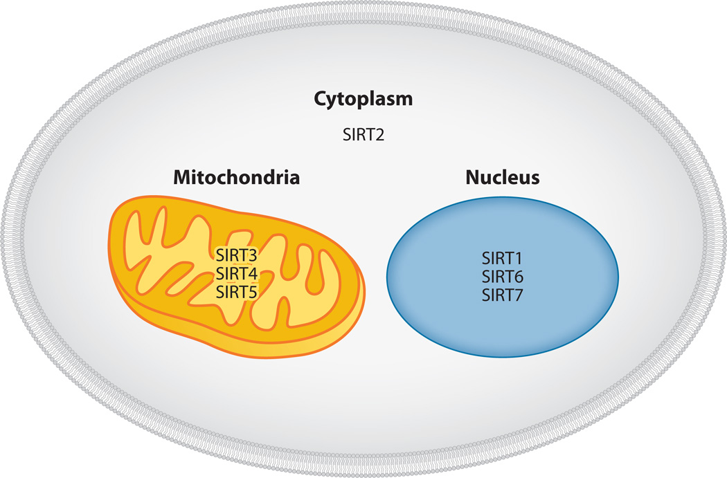
There is ample evidence that the mechanisms involve significant alterations in energy metabolism; response to oxidative damage; insulin sensitivity; inflammation; and changes in the neuroendocrine, paracrine, and reproductive systems (12). These changes are likely triggered by metabolic response regulators in cells, which can sense caloric intake and signal to metabolic pathways affecting aging and diseases (Figure 1). Among these regulators are the sirtuins, a family of enzymes that were initially shown to slow aging in yeast. Sirtuins are NAD+-dependent enzymes, which makes them an excellent link between the nutritional status of the organism and cellular genetic programs. These enzymes contain homologous domains that bind NAD+ (13), and they can be found in organisms ranging from bacteria to humans (14). Mammals have seven sirtuin homologs (SIRT1–7). Three sirtuins (SIRT1, -6, and -7) are found mostly in the nucleus, three (SIRT3, -4, and -5) are in mitochondria, and one (SIRT2) is in the cytoplasm (Figure 2). SIRT1, -2, -3, -6, and -7 can catalyze deacetylation of protein substrates. SIRT4 has not yet been shown to have detectable deacetylase activity but has ADP-ribosyltransferase activity (15). SIRT5 was recently demonstrated to have desuccinylase and demalonylase activities that are more robust than its weak deacetylase activity (16, 17).
Figure 1.
Calorie restriction impacts numerous physiological functions and parameters, which may ultimately affect health and longevity. The most heavily influenced physiological characteristics are presented in this flowchart. A few genes may mediate the impact of calorie restriction; the most fundamental ones are genes encoding nutritional sensors such as sirtuins (SIRT1–7), adenosine monophosphate–activated protein kinase (AMPK), and target of rapamycin (TOR) and genes involved in insulin signaling. Downstream effects of calorie restriction include decreases in cancer incidence, the suppression of reproduction, alterations of metabolic functions with increased fat oxidation, alterations of mood (higher degrees of anxiety and susceptibility to depression), increased aggression, and increased DNA repair. Calorie restriction robustly increases the longevity and health of laboratory animals; however, its applicability to humans is unknown.
Figure 2.
Sirtuins are a family of proteins ranging from bacteria to humans and contain conserved sirtuin motifs. Mammals have seven homologs of sir2 enzymes (SIRT1–7). SIRT1, -6, and -7 are found primarily in the nucleus; SIRT3, -4, and -5 are mitochondrial; and SIRT2 is cytoplasmic. Sirtuins have NAD+-dependent protein deacetylase activity and ADP-ribosyltransferase activity. In addition, SIRT5 has desuccinylase and demalonylase activity.
Consistent with evolutionary theory, CR in Drosophila melanogaster reduces fecundity (18) and downregulates the expression of genes necessary for gametogenesis and eggshell formation (19). Similarly, Caenorhabditis elegans reproductive output is reduced by various regimes of dietary restriction (20). Rodents subjected to a diet of 30% or fewer calories relative to fully fed control rodents also have reduced reproductive output (21). Women who practice weight control are likelier to experience reproductive failure (22). Interestingly, both SIRT1 knockout male and female mice, which can survive in outbred backgrounds, are sterile (23). These observations are consistent with the disposable soma theory, according to which, during times of plentiful nutrition, resources are directed toward reproduction (24). However, during famine, resources are diverted from reproduction to the maintenance and repair of other organs necessary for increased durability and longevity.
Changes in energy metabolism often accompany CR (25). CR lowers body temperature in mice (26), rats, rhesus monkeys (27), and humans (28). The life span of poikilothermic invertebrates can be extended by reducing ambient temperature (29). Therefore, a link between basal metabolic rate, temperature, and longevity has been proposed. Intriguingly, transgenic mice with a reduced core body temperature have an increased life span (30). The sirtuin SIRT3 regulates global lysine acetylation in mitochondria (31) and is responsible for mitochondrial metabolic strategy and thermogenesis (32). For example, SIRT3 mediates fatty-acid oxidation by deacetylating enzymes in the β-oxidation pathway (33). SIRT3 is closely linked to CR: CR triggers an increase in SIRT3 abundance (34), and the lack of SIRT3 negates the strong protective effect of CR [e.g., against hearing loss in aged rodents (35)].
CR also reduces the incidence of cancers, such as virally induced leukemia (36), chemically induced mammary and colon tumors (37), and spontaneous aging-associated tumors (38). CR also activates SIRT1 (39). Because SIRT1 can suppress the activity of p53 by deacetylating it (40), the role of SIRT1 in cancer protection must be due to effects on additional pathways. For example, genetic activation of SIRT1 protects against tumors in the gut (41) and the pancreas (42), likely by suppressing β-catenin activity.
SIRTUINS’ ROLE IN PHYSIOLOGY AND METABOLISM
SIRT1 is the most-studied sirtuin in the context of metabolism. This sirtuin influences the differentiation of preadipocytes (43) by repressing peroxisome proliferator–activated receptor γ (PPAR-γ). Additionally, SIRT1 deacetylates and thus activates PPAR-γ coactivator PGC-1α (44), which in turn can activate mitochondrial biogenesis as well as induce endothelial nitric oxide synthase expression (45). Activation of SIRT1 also causes deacetylation and activation of peroxisome proliferator–activated receptor α (PPAR-α) to turn on genes required for the increase in fatty-acid oxidation (46). In summary, these changes activate oxidative metabolism in muscle, fat cells, and the liver to improve insulin sensitivity and prevent progression of metabolic age-associated disorders.
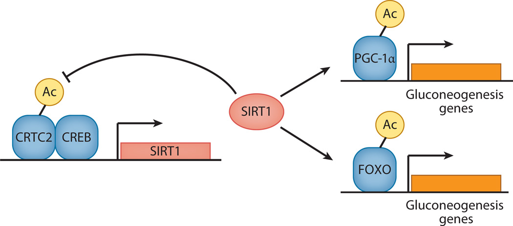
In the fasting liver, SIRT1 controls pathways that are responsible for upregulation of gluconeogenesis (47). In the early stages of fasting, gluconeogenesis is turned on by CREB/CRTC2, which also activate SIRT1 transcription (48). However, SIRT1 activation triggers deacetylation and subsequent ubiquitination and degradation of CRTC2, which lowers the expression of CREB targets. At the same time, SIRT1 deacetylates and thus activates PGC-1α and FOXO transcriptional factors to turn on genes necessary for response to long-term fasting (Figure 3). The formation of ketone bodies is also triggered at this time.
Figure 3.
SIRT1 mediates the liver’s response to fasting. In the early stages of fasting, CREB/CRTC2 turn on gluconeogenesis and increase SIRT1 production. Via negative feedback, SIRT1 deacetylates CRTC2, which causes SIRT1 degradation. At the same time, SIRT1 deacetylates and activates PGC-1α and FOXO transcriptional factors to turn on genes necessary for response to long-term fasting.
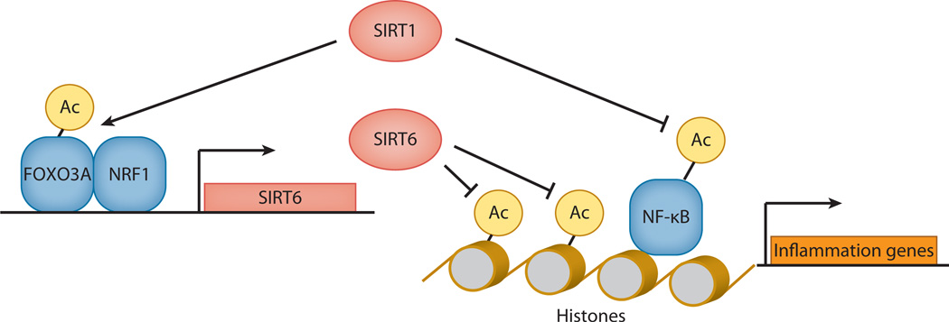
Sirtuins also play an important role in controlling inflammation (Figure 4), which contributes to numerous age-associated diseases. Both SIRT1 and SIRT6 suppress the activity of a key activator of inflammation: nuclear factor κB (NF-κB). SIRT1 can directly deacetylate the p65 subunit of NF-κB and thus reduce the ability of NF-κB to activate the transcription of proinflammatory genes (49). SIRT6 deacetylates histones at the promoters of NF-κB-regulated genes to repress them and thus suppress inflammation (50). SIRT1 and SIRT6 may work in concert, as SIRT1 can activate SIRT6 transcription by deacetylating and activating the FOXO3a/NRF1 complex on the SIRT6 promoter (51).
Figure 4.
SIRT1 and SIRT6 may work in concert to suppress inflammation. SIRT1 directly deacetylates the p65 subunit of NF-κB and thus reduces the ability of NF-κB to activate the transcription of proinflammatory genes. Additionally, SIRT1 activates the production of SIRT6, which deacetylates histones at the promoters of NF-κB-regulated genes to suppress inflammation.
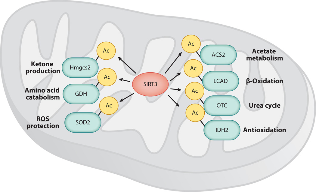
Mitochondrial sirtuins govern key aspects of energy metabolism, especially during prolonged fasting or CR. In addition to its role in fat oxidation, SIRT3 promotes the catabolism of acetate and amino acids (Figure 5). For example, it deacetylates glutamate dehydrogenase (GDH), which converts glutamine into α-ketoglutaric acid (aKG); aKG can be used in the Krebs cycle for energy production. Another, recently identified target of SIRT3 is lysine 88 of ornithine transcarbamoylase (52). Deacetylation of this enzyme by SIRT3 activates the urea cycle to dispose of ammonia, which is essential when energy is produced by amino acid digestion. Another mitochondrial sirtuin, SIRT5, can also activate the urea cycle by deacetylating (or more likely desuccinylating) carbamoyl phosphate synthetase 1 (53). SIRT3 also deacetylates Hmgcs2 (3-hydroxy-3-methylglutaryl CoA synthase 2), which is essential for ketone synthesis (54). Acetyl-CoA synthetase 2 (ACS2) is tightly controlled by acetylation, and SIRT3-mediated deacetylation on lysine 635 stimulates ACS2 activity (55). One target for SIRT3 control of fatty-acid oxidation is lysine 42 of long-chain acyl-CoA dehydrogenase (56). Interestingly, SIRT3 activates mitochondrial pathways that mitigate oxidative damage. For example, SIRT3 stimulates the activity of isocitrate dehydrogenase 2 to drive the glutathione detoxification system (52) and the activity of the superoxide dismutase SOD2, a critical antioxidant, by deacetylating lysines 53, 68, and 122 of this enzyme (57, 58). Because SIRT3 also deacetylates components of the electron transport chain (59, 60), it may reduce reactive oxygen species production.
Figure 5.
SIRT3 controls key aspects of mitochondrial function. SIRT3 deacetylates and activates glutamate dehydrogenase (GDH), ornithine transcarbamoylase (OTC), 3-hydroxy-3-methylglutaryl CoA synthase 2 (Hmgcs2), acetyl-CoA synthetase 2 (ACS2), and long-chain acyl-CoA dehydrogenase (LCAD). Such activation of mitochondrial metabolism is conducted in unison with the activation of systems that detoxify reactive oxygen species (ROS). SIRT3 activates isocitrate dehydrogenase 2 (IDH2) and superoxide dismutase (SOD2) by deacetylating lysines 53, 68, and 122 of SOD2 to stimulate ROS detoxification.
Another mitochondrial sirtuin, SIRT4, regulates fatty-acid oxidation and mitochondrial gene expression in liver and muscle cells. However, the direction of this regulation appears to be opposite of that of SIRT3. To wit, SIRT4 inhibition increases fat oxidative capacity and mitochondrial function (61). SIRT4 can directly downregulate GDH activity by ADP-ribosylation to regulate amino acid catabolism (15). Additionally, SIRT4 regulates insulin secretion by β cells. Depletion of SIRT4 from insulin-producing INS-1E cells results in increased insulin secretion in response to glucose (62).
These data suggest that sirtuins evolved to control and fine-tune a complex network of metabolic enzymes in response to energy availability. Thus, they are excellent targets to provide therapeutic benefits for age-associated metabolic diseases, such as type 2 diabetes (see below).
SIRTUINS’ ROLE IN AGING AND AGE-ASSOCIATED DISEASES
Age is the biggest risk factor for a number of diseases, such as cardiovascular diseases, diabetes, neurodegenerative disorders, and cancer. Sirtuins mediate the effects of CR (63). Indeed, SIRT1 is necessary for increased activity and longevity of animals subjected to CR (64). Levels of SIRT1 and SIRT3 go up in many tissues of animals subjected to CR (39, 65). SIRT1 can upregulate its own transcription (66), so its protein levels reflect its activity. CR activates SIRT1 via an increase in the available NAD+ due to glycolysis slowing down during food limitation. These effects have been directly demonstrated in muscle and white adipose tissue (67). SIRT3 transcription is also likely under SIRT1 control (34). SIRT1-overexpressing animals recapitulate many characteristics of CR (68).
Thus, (a) genetic or pharmacological manipulation of SIRT1 or (b) diet may protect against diseases. Increased SIRT1 can protect against diabetes (69–71), Alzheimer’s disease (72), Huntington’s disease (73), Parkinson’s disease, (74) and amyotrophic lateral sclerosis (75).
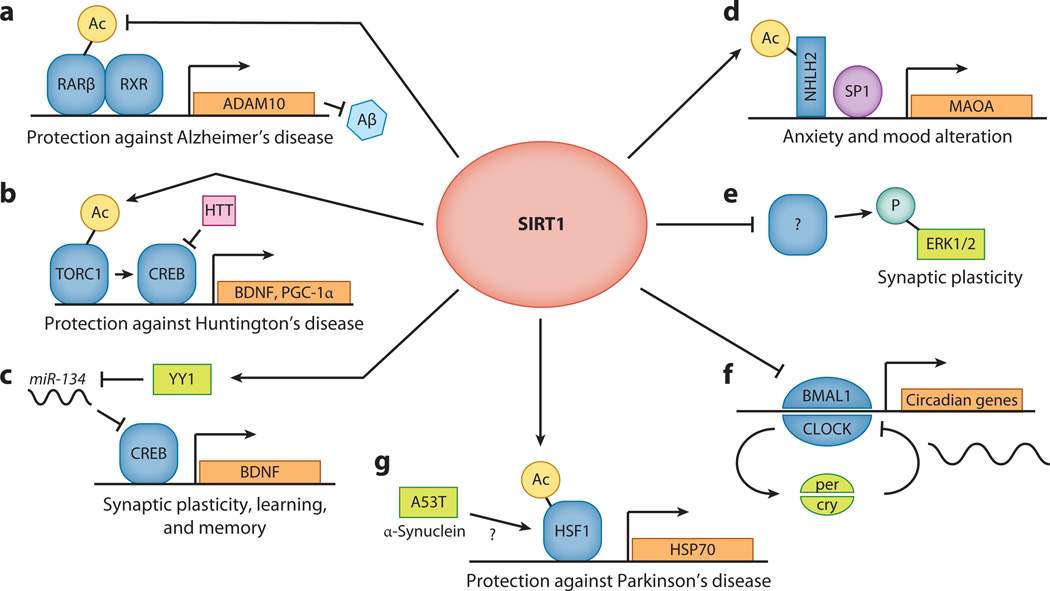
There are several mechanisms by which SIRT1 appears to protect against neurodegenerative diseases (Figure 6). In Alzheimer’s disease, SIRT1 can deacetylate retinoic acid receptor β (RARβ). This action results in the heterodimerization of RARβ with retinoid X receptor on the promoter of ADAM10, which encodes α-secretase. Increases in the abundance and activity of α-secretase direct amyloid precursor peptide (APP) processing away from production of the toxic Aβ amyloid peptide, resulting in less Aβ production (72). In Huntington’s disease, SIRT1 can deacetylate and activate TORC1, which promotes TORC1 dephosphorylation and the interaction of TORC1 with CREB. The TORC1-CREB complex activates the production of brain-derived neurotrophic factor and PGC-1α, both of which are protective in Huntington’s disease (73). SIRT1 also limits miR-134 expression, thereby limiting the expression of CREB and its targets (76). In Parkinson’s disease, SIRT1 deacetylates heat shock factor 1 (HSF1) to increase the activation of its targets, such as heat shock protein 70 (77), and thus protects mice expressing the α-synuclein Parkinson’s disease mutant A53T/A53T (74). Interestingly, induction of HSF1 activity requires the overexpression of both SIRT1 and the A53T disease gene.
Figure 6.
SIRT1 in the brain is involved in a number of processes that affect neuronal health, neurogenesis, and behavior. (a) SIRT1 can deacetylate retinoic acid receptor β (RARβ), which causes the heterodimerization of RARβ with retinoid X receptor (RXR) on the promoter of ADAM10, which encodes α-secretase. Increases in the abundance and activity of ADAM10 direct amyloid precursor peptide (APP) processing toward the α-secretase pathway. This in turn leads to less β-secretase cleavage of APP and thus less Aβ production, which is protective against Alzheimer’s disease. (b) SIRT1 deacetylates and activates TORC1 by promoting its dephosphorylation and its interaction with CREB. Brain-derived neurotrophic factor (BDNF) and PGC-1α are key targets of SIRT1 and TORC1 transcriptional activity. Both targets counteract the toxic effects of the Huntingtin protein and protect against Huntington’s disease. (c) SIRT1 limits miR-134 expression via a repressor complex containing the transcription factor YY1. If SIRT1 is downregulated and miR-134 expression is increased, CREB and BDNF are downregulated, thereby impairing synaptic plasticity. (d) SIRT1 deacetylates and thus activates the neuronal helix-loop-helix 2 (NHLH2) transcription factor, which in conjunction with SP1 controls the transcription of monoamine oxidase A (MAOA). MAOA activity alters the abundance of neurotransmitters, such as serotonin and noradrenaline, which in turn influence an animal’s mood and behavior. (e) SIRT1 knockout results in decreased extracellular signal–regulated kinase 1/2 (ERK1/2) phosphorylation and in altered expression of hippocampal genes involved in synaptic function. SIRT1 knockout animals have compromised cognitive functions, reinforcing the notion that SIRT1 is indispensable for normal learning, memory, and synaptic plasticity. (f) SIRT1 binds the CLOCK-BMAL1 complex in a circadian manner and promotes the deacetylation and degradation of cytochrome PER2. The PER2/CRY complex suppresses the activity of CLOCK/BMAL1, forming a negative feedback loop that results in periodic oscillation of the abundance of these molecules. SIRT1 is required for normal circadian transcription of core clock genes in the liver and connects cellular metabolism to the circadian rhythm. (g) SIRT1 protects against Parkinson’s disease by deacetylating heat shock factor 1 (HSF1) to increase activation of its targets, such as heat shock protein 70 (HSP70). Interestingly, the induction of HSF1 activity requires the overexpression of both SIRT1 and the A53T disease gene.
Deletion of the mitochondrial sirtuin SIRT3 globally increases acetylation of mitochondrial proteins and, as mentioned above, completely abolishes the protective effects of CR on inner-ear neurons (35). In this context, activation of SIRT1 and/or SIRT3 in certain tissues may be a powerful tool for combating diseases of aging without the need for extreme dieting.
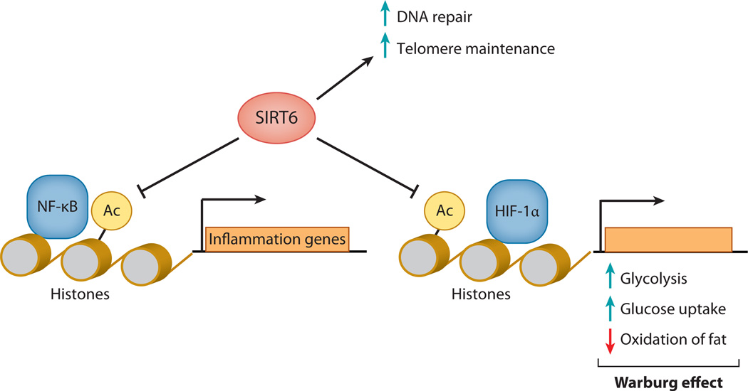
Another important member of the sirtuin family is SIRT6, which has anti-inflammatory actions, as described above. SIRT6 is a nuclear protein and has both NAD+-dependent ADP-ribosyltransferase (78) and NAD+-dependent deacetylase (79) activities. SIRT6 knockout is postnatally lethal due to severe hypoglycemia (80), but these mice can be kept alive if their diet is supplemented with high levels of glucose. Hypoxia-inducible factor 1α (HIF-1α) may be an important target of SIRT6 and the main link between this sirtuin and glucose homeostasis (Figure 7). Indeed, phenotypes of SIRT6 knockouts resemble phenotypes of HIF-1α hyperactivation (81). Other functions attributed to SIRT6 are DNA repair (82) and telomere maintenance (83). Intriguingly, ubiquitous overexpression of SIRT6 extends the longevity of male mice (84). The mechanisms behind this longevity extension and the sexual dimorphism of its prolongevity properties are not known and are an intriguing topic of research.
Figure 7.
SIRT6 inhibits carcinogenesis by inhibiting the Warburg effect, contributes to DNA repair activation, and suppresses inflammation. Hypoxia-inducible factor 1α (HIF-1α) may be the major target of SIRT6 and the main link between this sirtuin and glucose homeostasis. DNA repair and telomere maintenance also depend on SIRT6 activity.
SIRT1 INFLUENCES NEUROLOGICAL PROCESSES: MEMORY, MOOD, AND BEHAVIOR
SIRT1 plays a role in learning and memory. SIRT1 knockout results in decreased extracellular signal–regulated kinase 1/2 (ERK1/2) phosphorylation and in altered expression of hippocampal genes involved in synaptic function (85). Moreover, SIRT1 brain-specific knockouts show reduced long-term potentiation in tissue slices compared with the wild type (76). Consequently, SIRT1 knockout animals have compromised cognitive functions, reinforcing the notion that that SIRT1 is important for normal learning, memory, and synaptic plasticity. SIRT1 also regulates the circadian clock system in peripheral tissues such as the liver, although a role in central circadian control in the brain has not yet been shown. SIRT1 binds the CLOCK-BMAL1 complex in a circadian manner and promotes the deacetylation and degradation of cytochrome PER2 (86, 87) (Figure 6f). Finally, SIRT1 is involved in the formation of addictions, for example, addiction to cocaine (88).
Equally important but less well studied is the impact of CR on behavior. CR in rats alters behavior so that animals become more aggressive, with diminished sexual activity (89). Food-restricted monkeys display heightened aggression during feeding times (90). Research in model organisms has provided ample evidence for the link between metabolism and behavior. Although the connection between CR, psychopathology, and physical activity is evident, the mechanisms are still poorly understood (91). Interestingly, the nutritional status of parents may have lasting effects on the behavior of progeny. For example, animals perinatally exposed to maternal CR demonstrated heightened anxiety-like behaviors (92) later in life. Furthermore, chronic food restriction in young rats results in depression and anxiety-like behaviors (93).
The link between mood and nutritional status of the organism may be mediated by metabolic genes (e.g., Figure 1), which sense energy status and affect neurological processes. One of the genes responsible for this link is SIRT1 (94). In humans, variants of the SIRT1 gene are associated with prevalence of major depressive disorder (95) and anxiety (96). The molecular mechanism behind the impact of SIRT1 on anxiety and mood involves serotonin signaling (Figure 6). SIRT1 deacetylates the helix-loop-helix transcriptional factor NHLH2 on lysine 49, which leads to its activation on the monoamine oxidase A (MAOA) promoter. MAOAis the major brain enzyme that degrades serotonin and noradrenalin, and its overactivation leads to decreased levels of serotonin. This reduction promotes anxiety and makes animals more prone to depression-like behaviors (96). Indeed, transgenic mice that overexpress SIRT1 in the brain have elevated levels of MAOA, reduced levels of serotonin, and a higher prevalence of anxiety-like and depression-like behaviors. Importantly, treatment of these transgenic animals with antidepression drugs, such as phenelzine and fluoxetine, normalizes their behavior (96). Evolutionarily, it is reasonable that CR would also affect mood and behavior. For example, during food scarcity an animal would have to scour a larger area in the face of predators. CR may enhance the survival of such an animal to exhibit heightened anxiety and vigilance in such an environment (96).
The connection between nutritional status of the organism and psychiatric disorders has far-reaching implications. One of the most severe disorders reminiscent of CR is anorexia nervosa (AN), which has the highest rate of mortality among all mental disorders (97) and is the most prevalent in young women (98). From the physiological and psychological points of view, there are common features between people suffering from AN and those practicing CR (28, 99). The manipulation of genes suspected to mediate the effect of CR could be a therapeutic approach to treat psychiatric disorders such as AN.
Another interesting disorder related to the intersection of metabolic state and neuropsychiatric processes is autism spectrum disorder (ASD). Major comorbid features of autism include mental retardation (30–60%) (100), anxiety, and mood disorders (101). Maternal metabolic conditions increase the risk for autism and other neurodevelopmental disorders (102). Once again, the link may be mediated by genes that relay metabolic state information and connect it with the expression of genes such as SIRT1. Copy number variations in the 10q21.3 region (immediately upstream of the SIRT1 coding sequence), which may impact SIRT1 transcription, are associated with the risk of autism in humans (103). That SIRT1 plays a role in learning, memory formation (76, 85), and mood (96) raises the possibility that SIRT1 may be a pharmacological target to alleviate at least some of the ASD symptoms. As we deepen our understanding of neuropsychiatric processes, we come to a realization that metabolic response genes play a far greater and more important role than previously imagined and that such genes may lead to new targets to heal our minds and bodies simultaneously.
THE LIMITATIONS AND DANGERS OF CALORIE RESTRICTION
Although CR extends longevity in numerous species, the mechanism of its action differs from species to species. The main cause of aging of the single-cell organism Saccharomyces cerevisiae is the accumulation of extrachromosomal rDNA circles (104). A likely cause of aging and advanced age mortality in D. melanogaster is systemic infection (19). Short-lived mice usually suffer from cancer, whereas the biggest risk factor for death in aging humans is cardiovascular disease (105). CR is expected to have a positive impact on these and other diseases of aging, but it is still unproven whether CR has long-term benefits for nonobese human subjects. We cannot rule out possible negative effects of CR that would outweigh its positive effects. To wit, the vast majority of mammalian CR experiments are conducted in mice housed in pathogen-free and environmentally protected facilities. However, when challenged with polymicrobial infection, calorie-restricted animals performed significantly worse (106). Similarly, CR significantly worsened the outcome of animals infected with intestinal parasites (107). Indeed, some scientists are skeptical whether CR will work in humans (108).
In addition, CR has a number of negative effects on quality of life, which could outweigh the positive effects CR might have on human health. For example, sex hormone (both serum total and free testosterone) is reduced in men practicing CR compared with equally lean endurance runners or age-matched controls eating Western diets (109). Decreases in the circulating levels of this hormone not only may negatively impact quality of life due to decreased sex drive (110) but also may result in more severe problems such as loss of muscle mass and loss of bone density. Indeed, bone mineral density is significantly reduced in humans practicing CR (111), and osteoporosis is one of the most severe problems that humans of advanced age face. In the view of these data, individuals practicing CR should be cautious, as it may be more sensible and beneficial to utilize therapies based on specific genetic mechanisms of CR action.
BENEFICIAL EFFECTS OF SMALL-MOLECULE SIRT1 ACTIVATORS IN ANIMAL MODELS AND HUMANS
Great interest exists in developing small molecules that act as CR mimetics by activating sirtuins, triggering protection from age-associated diseases and perhaps contributing to a longer life span. One of the first natural compounds discovered to activate SIRT1 is resveratrol (112). Following its discovery, novel synthetic and more potent compounds were developed (69). Resveratrol has many effects that are promising from a drug development point of view. For example, it improves insulin sensitivity (113), inhibits tumor growth (114), suppresses inflammation, promotes cardiovascular health (115), and protects against neurodegenerative diseases (116). In the murine model of obesity (ob/ob mice), resveratrol significantly increased the health and longevity of treated animals. There is debate on whether resveratrol has a direct effect (117) or an indirect effect (118) on SIRT1. Other compounds, such as oxazolo[4,5-b]pyridines (119) and imidazo[1,2-b]thiazole (120), activate SIRT1. Moreover, the NAD+ precursors NMN and nicotinamide riboside increase NAD+ levels in tissues, drive SIRT1 activation, and enhance oxidative metabolism to protect against obesity induced by a high-fat diet (121, 122). Private companies such as Sirtris Pharmaceuticals (which has been acquired by GlaxoSmithKline) identified many of the synthetic compounds that activate SIRT1 (69), some of which are in clinical trials (see http://ClinicalTrials.gov).
CONCLUSION
To date there is no known intervention that can extend human longevity. Even though CR results in impressive health improvement in rodents, its applicability to higher organisms is uncertain, and CR may even be dangerous. However, the elucidation of the molecular mechanisms of CR encourages the development of pharmacological approaches that can mimic the beneficial effects of CR. Such approaches may lead to healthier lives and perhaps longer lives.
Because sirtuins mediate many of the metabolic responses of organisms to diet, these proteins offer one set of targets for pharmacological approaches. In addition, research aimed at understanding the role of sirtuins in brain function has begun to uncover a long-suspected coordination between metabolism and behavior. Studies of sirtuins have generated insights into diet and aging that may improve human health. These findings imply that sirtuin drugs might be used to improve not only metabolic health but also neuropsychiatric health.
ACKNOWLEDGMENTS
We apologize to authors who were not cited due to space limitation. Work in L.G.’s lab was supported by the Glenn Medical Foundation and the National Institutes of Health.
Footnotes
DISCLOSURE STATEMENT
L.G. consults for Sirtris/GSK Company. S.L. has no affiliations, memberships, funding, or financial holdings that might be perceived as affecting the objectivity of this review.
LITERATURE CITED
- 1.McCay CM, Crowell MF, Maynard LA. The effect of retarded growth upon the length of life span and upon the ultimate body size. Nutrition. 1935;5:155–171. discussion 72. [PubMed] [Google Scholar]
- 2.Guarente L. Calorie restriction and SIR2 genes—towards a mechanism. Mech. Ageing Dev. 2005;126:923–928. doi: 10.1016/j.mad.2005.03.013. [DOI] [PubMed] [Google Scholar]
- 3.Houthoofd K, Vanfleteren JR. The longevity effect of dietary restriction in Caenorhabditis elegans. Exp. Gerontol. 2006;41:1026–1031. doi: 10.1016/j.exger.2006.05.007. [DOI] [PubMed] [Google Scholar]
- 4.Partridge L, Piper MD, Mair W. Dietary restriction in Drosophila. Mech. Ageing Dev. 2005;126:938–950. doi: 10.1016/j.mad.2005.03.023. [DOI] [PubMed] [Google Scholar]
- 5.Jones W. Longevity in a fasting spider. Science. 1884;3:4. doi: 10.1126/science.ns-3.48.4-c. [DOI] [PubMed] [Google Scholar]
- 6.Austad SN. Life extension by dietary restriction in the bowl and doily spider, Frontinella pyramitela. Exp. Gerontol. 1989;24:83–92. doi: 10.1016/0531-5565(89)90037-5. [DOI] [PubMed] [Google Scholar]
- 7.Piper MD, Bartke A. Diet and aging. Cell Metab. 2008;8:99–104. doi: 10.1016/j.cmet.2008.06.012. [DOI] [PubMed] [Google Scholar]
- 8.Colman RJ, Anderson RM, Johnson SC, Kastman EK, Kosmatka KJ, et al. Caloric restriction delays disease onset and mortality in rhesus monkeys. Science. 2009;325:201–204. doi: 10.1126/science.1173635. [DOI] [PMC free article] [PubMed] [Google Scholar]
- 9.Libert S, Pletcher SD. Modulation of longevity by environmental sensing. Cell. 2007;131:1231–1234. doi: 10.1016/j.cell.2007.12.002. [DOI] [PubMed] [Google Scholar]
- 10.Wu P, Shen Q, Dong S, Xu Z, Tsien JZ, Hu Y. Calorie restriction ameliorates neurodegenerative phenotypes in forebrain-specific presenilin-1 and presenilin-2 double knockout mice. Neurobiol. Aging. 2008;29:1502–1511. doi: 10.1016/j.neurobiolaging.2007.03.028. [DOI] [PubMed] [Google Scholar]
- 11.Weiss EP, Fontana L. Caloric restriction: powerful protection for the aging heart and vasculature. AmJPhysiol. Heart Circ. Physiol. 2011;301:H1205–H1219. doi: 10.1152/ajpheart.00685.2011. [DOI] [PMC free article] [PubMed] [Google Scholar]
- 12.Redman LM, Ravussin E. Caloric restriction in humans: impact on physiological, psychological, and behavioral outcomes. Antioxid. Redox Signal. 2011;14:275–287. doi: 10.1089/ars.2010.3253. [DOI] [PMC free article] [PubMed] [Google Scholar]
- 13.Sanders BD, Jackson B, Marmorstein R. Structural basis for sirtuin function: what we know and what we don’t. Biochim. Biophys. Acta. 2010;1804:1604–1616. doi: 10.1016/j.bbapap.2009.09.009. [DOI] [PMC free article] [PubMed] [Google Scholar]
- 14.North BJ, Schwer B, Ahuja N, Marshall B, Verdin E. Preparation of enzymatically active recombinant class III protein deacetylases. Methods. 2005;36:338–345. doi: 10.1016/j.ymeth.2005.03.004. [DOI] [PubMed] [Google Scholar]
- 15.Haigis MC, Mostoslavsky R, Haigis KM, Fahie K, Christodoulou DC, et al. SIRT4 inhibits glutamate dehydrogenase and opposes the effects of calorie restriction in pancreatic β cells. Cell. 2006;126:941–954. doi: 10.1016/j.cell.2006.06.057. [DOI] [PubMed] [Google Scholar]
- 16.Du J, Zhou Y, Su X, Yu JJ, Khan S, et al. Sirt5 is a NAD-dependent protein lysine demalonylase and desuccinylase. Science. 2011;334:806–809. doi: 10.1126/science.1207861. [DOI] [PMC free article] [PubMed] [Google Scholar]
- 17.Peng C, Lu Z, Xie Z, Cheng Z, Chen Y, et al. The first identification of lysine malonylation substrates and its regulatory enzyme. Mol. Cell. Proteomics. 2011;10 doi: 10.1074/mcp.M111.012658. M111 012658. [DOI] [PMC free article] [PubMed] [Google Scholar]
- 18.Bross TG, Rogina B, Helfand SL. Behavioral, physical, and demographic changes in Drosophila populations through dietary restriction. Aging Cell. 2005;4:309–317. doi: 10.1111/j.1474-9726.2005.00181.x. [DOI] [PubMed] [Google Scholar]
- 19.Pletcher SD, Libert S, Skorupa D. Flies and their golden apples: the effect of dietary restriction on Drosophila aging and age-dependent gene expression. Ageing Res. Rev. 2005;4:451–480. doi: 10.1016/j.arr.2005.06.007. [DOI] [PubMed] [Google Scholar]
- 20.Walker G, Houthoofd K, Vanfleteren JR, Gems D. Dietary restriction in C. elegans : from rate-ofliving effects to nutrient sensing pathways. Mech. Ageing Dev. 2005;126:929–937. doi: 10.1016/j.mad.2005.03.014. [DOI] [PubMed] [Google Scholar]
- 21.Rocha JS, Bonkowski MS, de Franca LR, Bartke A. Effects of mild calorie restriction on reproduction, plasma parameters and hepatic gene expression in mice with altered GH/IGF-I axis. Mech. Ageing Dev. 2007;128:317–331. doi: 10.1016/j.mad.2007.02.001. [DOI] [PubMed] [Google Scholar]
- 22.Bates GW, Bates SR, Whitworth NS. Reproductive failure in women who practice weight control. Fertil. Steril. 1982;37:373–378. [PubMed] [Google Scholar]
- 23.McBurney MW, Yang X, Jardine K, Hixon M, Boekelheide K, et al. The mammalian SIR2α protein has a role in embryogenesis and gametogenesis. Mol. Cell. Biol. 2003;23:38–54. doi: 10.1128/MCB.23.1.38-54.2003. [DOI] [PMC free article] [PubMed] [Google Scholar]
- 24.Drenos F, Kirkwood TB. Modelling the disposable soma theory of ageing. Mech. Ageing Dev. 2005;126:99–103. doi: 10.1016/j.mad.2004.09.026. [DOI] [PubMed] [Google Scholar]
- 25.Walford RL, Spindler SR. The response to calorie restriction in mammals shows features also common to hibernation: a cross-adaptation hypothesis. J. Gerontol. A. 1997;52:B179–B183. doi: 10.1093/gerona/52a.4.b179. [DOI] [PubMed] [Google Scholar]
- 26.Weindruch R, Walford RL, Fligiel S, Guthrie D. The retardation of aging in mice by dietary restriction: longevity, cancer, immunity and lifetime energy intake. J. Nutr. 1986;116:641–654. doi: 10.1093/jn/116.4.641. [DOI] [PubMed] [Google Scholar]
- 27.Lane MA, Baer DJ, Rumpler WV, Weindruch R, Ingram DK, et al. Calorie restriction lowers body temperature in rhesus monkeys, consistent with a postulated anti-aging mechanism in rodents. Proc. Natl. Acad. Sci. USA. 1996;93:4159–4164. doi: 10.1073/pnas.93.9.4159. [DOI] [PMC free article] [PubMed] [Google Scholar]
- 28.Soare A, Cangemi R, Omodei D, Holloszy JO, Fontana L. Long-term calorie restriction, but not endurance exercise, lowers core body temperature in humans. Aging. 2011;3:374–379. doi: 10.18632/aging.100280. [DOI] [PMC free article] [PubMed] [Google Scholar]
- 29.Lamb MJ. Temperature and lifespan in Drosophila . Nature. 1968;220:808–809. doi: 10.1038/220808a0. [DOI] [PubMed] [Google Scholar]
- 30.Conti B, Sanchez-Alavez M, Winsky-Sommerer R, Morale MC, Lucero J, et al. Transgenic mice with a reduced core body temperature have an increased life span. Science. 2006;314:825–828. doi: 10.1126/science.1132191. [DOI] [PubMed] [Google Scholar]
- 31.Lombard DB, Alt FW, Cheng HL, Bunkenborg J, Streeper RS, et al. Mammalian Sir2 homolog SIRT3 regulates global mitochondrial lysine acetylation. Mol. Cell. Biol. 2007;27:8807–8814. doi: 10.1128/MCB.01636-07. [DOI] [PMC free article] [PubMed] [Google Scholar]
- 32.Shi T, Wang F, Stieren E, Tong Q. SIRT3, a mitochondrial sirtuin deacetylase, regulates mitochondrial function and thermogenesis in brown adipocytes. J. Biol. Chem. 2005;280:13560–13567. doi: 10.1074/jbc.M414670200. [DOI] [PubMed] [Google Scholar]
- 33.Hirschey MD, Shimazu T, Huang JY, Schwer B, Verdin E. SIRT3 regulates mitochondrial protein acetylation and intermediary metabolism. Cold Spring Harb. Symp. Quant. Biol. 2011;76:267–277. doi: 10.1101/sqb.2011.76.010850. [DOI] [PubMed] [Google Scholar]
- 34.Bell EL, Guarente L. The SirT3 divining rod points to oxidative stress. Mol. Cell. 2011;42:561–568. doi: 10.1016/j.molcel.2011.05.008. [DOI] [PMC free article] [PubMed] [Google Scholar]
- 35.Someya S, Yu W, Hallows WC, Xu J, Vann JM, et al. Sirt3 mediates reduction of oxidative damage and prevention of age-related hearing loss under caloric restriction. Cell. 2010;143:802–812. doi: 10.1016/j.cell.2010.10.002. [DOI] [PMC free article] [PubMed] [Google Scholar]
- 36.Sarkar RN, Das S. Effect of calorie restriction on the development of virus induced leukaemia in mice. Proc. Soc. Exp. Biol. Med. 1990;193:164–166. doi: 10.3181/00379727-193-2-rc1. [DOI] [PubMed] [Google Scholar]
- 37.Klurfeld DM, Weber MM, Kritchevsky D. Inhibition of chemically induced mammary and colon tumor promotion by caloric restriction in rats fed increased dietary fat. Cancer Res. 1987;47:2759–2762. [PubMed] [Google Scholar]
- 38.Kritchevsky D. Caloric restriction and cancer. J. Nutr. Sci. Vitaminol. 2001;47:13–19. doi: 10.3177/jnsv.47.13. [DOI] [PubMed] [Google Scholar]
- 39.Cohen HY, Miller C, Bitterman KJ, Wall NR, Hekking B, et al. Calorie restriction promotes mammalian cell survival by inducing the SIRT1 deacetylase. Science. 2004;305:390–392. doi: 10.1126/science.1099196. [DOI] [PubMed] [Google Scholar]
- 40.Vaziri H, Dessain SK, Ng Eaton E, Imai SI, Frye RA, et al. hSIR2SIRT1 functions as an NAD-dependent p53 deacetylase. Cell. 2001;107:149–159. doi: 10.1016/s0092-8674(01)00527-x. [DOI] [PubMed] [Google Scholar]
- 41.Firestein R, Blander G, Michan S, Oberdoerffer P, Ogino S, et al. The SIRT1 deacetylase suppresses intestinal tumorigenesis and colon cancer growth. PLoS ONE. 2008;3:e2020. doi: 10.1371/journal.pone.0002020. [DOI] [PMC free article] [PubMed] [Google Scholar]
- 42.Cho IR, Koh SS, Malilas W, Srisuttee R, Moon J, et al. SIRT1 inhibits proliferation of pancreatic cancer cells expressing pancreatic adenocarcinoma up-regulated factor (PAUF), a novel oncogene, by suppression of β-catenin. Biochem. Biophys. Res. Commun. 2012 doi: 10.1016/j.bbrc.2012.05.107. In press. [DOI] [PubMed] [Google Scholar]
- 43.Picard F, Kurtev M, Chung N, Topark-Ngarm A, Senawong T, et al. Sirt1 promotes fat mobilization in white adipocytes by repressing PPAR-γ. Nature. 2004;429:771–776. doi: 10.1038/nature02583. [DOI] [PMC free article] [PubMed] [Google Scholar]
- 44.Rodgers JT, Lerin C, Haas W, Gygi SP, Spiegelman BM, Puigserver P. Nutrient control of glucose homeostasis through a complex of PGC-1α and SIRT1. Nature. 2005;434:113–118. doi: 10.1038/nature03354. [DOI] [PubMed] [Google Scholar]
- 45.Nisoli E, Tonello C, Cardile A, Cozzi V, Bracale R, et al. Calorie restriction promotes mitochondrial biogenesis by inducing the expression of eNOS. Science. 2005;310:314–317. doi: 10.1126/science.1117728. [DOI] [PubMed] [Google Scholar]
- 46.Purushotham A, Schug TT, Xu Q, Surapureddi S, Guo X, Li X. Hepatocyte-specific deletion of SIRT1 alters fatty acid metabolism and results in hepatic steatosis and inflammation. Cell Metab. 2009;9:327–338. doi: 10.1016/j.cmet.2009.02.006. [DOI] [PMC free article] [PubMed] [Google Scholar]
- 47.Liu Y, Dentin R, Chen D, Hedrick S, Ravnskjaer K, et al. A fasting inducible switch modulates gluconeogenesis via activator/coactivator exchange. Nature. 2008;456:269–273. doi: 10.1038/nature07349. [DOI] [PMC free article] [PubMed] [Google Scholar]
- 48.Noriega LG, Feige JN, Canto C, Yamamoto H, Yu J, et al. CREBand ChREBP oppositely regulate SIRT1 expression in response to energy availability. EMBO Rep. 2011;12:1069–1076. doi: 10.1038/embor.2011.151. [DOI] [PMC free article] [PubMed] [Google Scholar]
- 49.Yeung F, Hoberg JE, Ramsey CS, Keller MD, Jones DR, et al. Modulation of NF-κB-dependent transcription and cell survival by the SIRT1 deacetylase. EMBO J. 2004;23:2369–2380. doi: 10.1038/sj.emboj.7600244. [DOI] [PMC free article] [PubMed] [Google Scholar]
- 50.Kawahara TL, Michishita E, Adler AS, Damian M, Berber E, et al. SIRT6 links histone H3 lysine-9 deacetylation to NF-κB-dependent gene expression and organismal life span. Cell. 2009;136:62–74. doi: 10.1016/j.cell.2008.10.052. [DOI] [PMC free article] [PubMed] [Google Scholar]
- 51.Kim HS, Xiao C, Wang RH, Lahusen T, Xu X, et al. Hepatic-specific disruption of SIRT6 in mice results in fatty liver formation due to enhanced glycolysis and triglyceride synthesis. Cell Metab. 2010;12:224–236. doi: 10.1016/j.cmet.2010.06.009. [DOI] [PMC free article] [PubMed] [Google Scholar]
- 52.Hallows WC, Yu W, Smith BC, Devries MK, Ellinger JJ, et al. Sirt3 promotes the urea cycle and fatty acid oxidation during dietary restriction. Mol. Cell. 2011;41:139–149. doi: 10.1016/j.molcel.2011.01.002. [DOI] [PMC free article] [PubMed] [Google Scholar]
- 53.Nakagawa T, Lomb DJ, Haigis MC, Guarente L. SIRT5 deacetylates carbamoyl phosphate synthetase-1 and regulates the urea cycle. Cell. 2009;137:560–570. doi: 10.1016/j.cell.2009.02.026. [DOI] [PMC free article] [PubMed] [Google Scholar]
- 54.Shimazu T, Hirschey MD, Hua L, Dittenhafer-Reed KE, Schwer B, et al. SIRT3 deacetylates mitochondrial 3-hydroxy-3-methylglutaryl CoA synthase 2 and regulates ketone body production. Cell Metab. 2010;12:654–661. doi: 10.1016/j.cmet.2010.11.003. [DOI] [PMC free article] [PubMed] [Google Scholar]
- 55.Hallows WC, Lee S, Denu JM. Sirtuins deacetylate and activate mammalian acetyl-CoA synthetases. Proc. Natl. Acad. Sci. USA. 2006;103:10230–10235. doi: 10.1073/pnas.0604392103. [DOI] [PMC free article] [PubMed] [Google Scholar]
- 56.Hirschey MD, Shimazu T, Goetzman E, Jing E, Schwer B, et al. SIRT3 regulates mitochondrial fatty-acid oxidation by reversible enzyme deacetylation. Nature. 2010;464:121–125. doi: 10.1038/nature08778. [DOI] [PMC free article] [PubMed] [Google Scholar]
- 57.Qiu X, Brown K, Hirschey MD, Verdin E, Chen D. Calorie restriction reduces oxidative stress by SIRT3-mediated SOD2 activation. Cell Metab. 2010;12:662–667. doi: 10.1016/j.cmet.2010.11.015. [DOI] [PubMed] [Google Scholar]
- 58.Tao R, Coleman MC, Pennington JD, Ozden O, Park SH, et al. Sirt3-mediated deacetylation of evolutionarily conserved lysine 122 regulates MnSOD activity in response to stress. Mol. Cell. 2010;40:893–904. doi: 10.1016/j.molcel.2010.12.013. [DOI] [PMC free article] [PubMed] [Google Scholar]
- 59.Ahn BH, Kim HS, Song S, Lee IH, Liu J, et al. A role for the mitochondrial deacetylase Sirt3 in regulating energy homeostasis. Proc. Natl. Acad. Sci. USA. 2008;105:14447–14452. doi: 10.1073/pnas.0803790105. [DOI] [PMC free article] [PubMed] [Google Scholar]
- 60.Jing E, Emanuelli B, Hirschey MD, Boucher J, Lee KY, et al. Sirtuin-3 (Sirt3) regulates skeletal muscle metabolism and insulin signaling via altered mitochondrial oxidation and reactive oxygen species production. Proc. Natl. Acad. Sci. USA. 2011;108:14608–14613. doi: 10.1073/pnas.1111308108. [DOI] [PMC free article] [PubMed] [Google Scholar]
- 61.Nasrin N, Wu X, Fortier E, Feng Y, Bare OC, et al. SIRT4 regulates fatty acid oxidation and mitochondrial gene expression in liver and muscle cells. J. Biol. Chem. 2010;285:31995–32002. doi: 10.1074/jbc.M110.124164. [DOI] [PMC free article] [PubMed] [Google Scholar]
- 62.Ahuja N, Schwer B, Carobbio S, Waltregny D, North BJ, et al. Regulation of insulin secretion by SIRT4, a mitochondrial ADP-ribosyltransferase. J. Biol. Chem. 2007;282:33583–33592. doi: 10.1074/jbc.M705488200. [DOI] [PubMed] [Google Scholar]
- 63.Guarente L. Sirtuins, aging, and metabolism. Cold Spring Harb. Symp. Quant. Biol. 2011;76:81–90. doi: 10.1101/sqb.2011.76.010629. [DOI] [PubMed] [Google Scholar]
- 64.Chen D, Steele AD, Lindquist S, Guarente L. Increase in activity during calorie restriction requires Sirt1. Science. 2005;310:1641. doi: 10.1126/science.1118357. [DOI] [PubMed] [Google Scholar]
- 65.Haigis MC, Sinclair DA. Mammalian sirtuins: biological insights and disease relevance. Annu. Rev. Pathol. 2010;5:253–295. doi: 10.1146/annurev.pathol.4.110807.092250. [DOI] [PMC free article] [PubMed] [Google Scholar]
- 66.Xiong S, Salazar G, Patrushev N, Alexander RW. FoxO1 mediates an autofeedback loop regulating SIRT1 expression. J. Biol. Chem. 2011;286:5289–5299. doi: 10.1074/jbc.M110.163667. [DOI] [PMC free article] [PubMed] [Google Scholar]
- 67.Chen D, Bruno J, Easlon E, Lin SJ, Cheng HL, et al. Tissue-specific regulation of SIRT1 by calorie restriction. Genes Dev. 2008;22:1753–1757. doi: 10.1101/gad.1650608. [DOI] [PMC free article] [PubMed] [Google Scholar]
- 68.Bordone L, Cohen D, Robinson A, Motta MC, van Veen E, et al. SIRT1 transgenic mice show phenotypes resembling calorie restriction. Aging Cell. 2007;6:759–767. doi: 10.1111/j.1474-9726.2007.00335.x. [DOI] [PubMed] [Google Scholar]
- 69.Milne JC, Lambert PD, Schenk S, Carney DP, Smith JJ, et al. Small molecule activators of SIRT1 as therapeutics for the treatment of type 2 diabetes. Nature. 2007;450:712–716. doi: 10.1038/nature06261. [DOI] [PMC free article] [PubMed] [Google Scholar]
- 70.Lagouge M, Argmann C, Gerhart-Hines Z, Meziane H, Lerin C, et al. Resveratrol improves mitochondrial function and protects against metabolic disease by activating SIRT1 and PGC-1α. Cell. 2006;127:1109–1122. doi: 10.1016/j.cell.2006.11.013. [DOI] [PubMed] [Google Scholar]
- 71.Banks AS, Kon N, Knight C, Matsumoto M, Gutierrez-Juarez R, et al. SirT1 gain of function increases energy efficiency and prevents diabetes in mice. Cell Metab. 2008;8:333–341. doi: 10.1016/j.cmet.2008.08.014. [DOI] [PMC free article] [PubMed] [Google Scholar]
- 72.Donmez G, Wang D, Cohen DE, Guarente L. SIRT1 suppresses β-amyloid production by activating the α-secretase gene ADAM10. Cell. 2010;142:320–332. doi: 10.1016/j.cell.2010.06.020. [DOI] [PMC free article] [PubMed] [Google Scholar] [Retracted]
- 73.Jeong H, Cohen DE, Cui L, Supinski A, Savas JN, et al. Sirt1 mediates neuroprotection from mutant huntingtin by activation of the TORC1 and CREB transcriptional pathway. Nat. Med. 2012;18:159–165. doi: 10.1038/nm.2559. [DOI] [PMC free article] [PubMed] [Google Scholar]
- 74.Donmez G, Arun A, Chung CY, McLean PJ, Lindquist S, Guarente L. SIRT1 protects against α-synuclein aggregation by activating molecular chaperones. J. Neurosci. 2012;32:124–132. doi: 10.1523/JNEUROSCI.3442-11.2012. [DOI] [PMC free article] [PubMed] [Google Scholar] [Retracted]
- 75.Kim D, Nguyen MD, Dobbin MM, Fischer A, Sananbenesi F, et al. SIRT1 deacetylase protects against neurodegeneration in models for Alzheimer’s disease and amyotrophic lateral sclerosis. EMBO J. 2007;26:3169–3179. doi: 10.1038/sj.emboj.7601758. [DOI] [PMC free article] [PubMed] [Google Scholar]
- 76.Gao J, Wang WY, Mao YW, Graff J, Guan JS, et al. A novel pathway regulates memory and plasticity via SIRT1 and miR-134. Nature. 2010;466:1105–1109. doi: 10.1038/nature09271. [DOI] [PMC free article] [PubMed] [Google Scholar]
- 77.Westerheide SD, Anckar J, Stevens SM, Jr, Sistonen L, Morimoto RI. Stress-inducible regulation of heat shock factor 1 by the deacetylase SIRT1. Science. 2009;323:1063–1066. doi: 10.1126/science.1165946. [DOI] [PMC free article] [PubMed] [Google Scholar]
- 78.Liszt G, Ford E, Kurtev M, Guarente L. Mouse Sir2 homolog SIRT6 is a nuclear ADP-ribosyltransferase. J. Biol. Chem. 2005;280:21313–21320. doi: 10.1074/jbc.M413296200. [DOI] [PubMed] [Google Scholar]
- 79.Michishita E, McCord RA, Berber E, Kioi M, Padilla-Nash H, et al. SIRT6 is a histone H3 lysine-9 deacetylase that modulates telomeric chromatin. Nature. 2008;452:492–496. doi: 10.1038/nature06736. [DOI] [PMC free article] [PubMed] [Google Scholar]
- 80.Mostoslavsky R, Chua KF, Lombard DB, Pang WW, Fischer MR, et al. Genomic instability and aging-like phenotype in the absence of mammalian SIRT6. Cell. 2006;124:315–329. doi: 10.1016/j.cell.2005.11.044. [DOI] [PubMed] [Google Scholar]
- 81.Zhong L, D’Urso A, Toiber D, Sebastian C, Henry RE, et al. The histone deacetylase Sirt6 regulates glucose homeostasis via Hif1 α. Cell. 2010;140:280–293. doi: 10.1016/j.cell.2009.12.041. [DOI] [PMC free article] [PubMed] [Google Scholar]
- 82.Kaidi A, Weinert BT, Choudhary C, Jackson SP. Human SIRT6 promotes DNA end resection through CtIP deacetylation. Science. 2010;329:1348–1353. doi: 10.1126/science.1192049. [DOI] [PMC free article] [PubMed] [Google Scholar] [Retracted]
- 83.Tennen RI, Bua DJ, Wright WE, Chua KF. SIRT6 is required for maintenance of telomere position effect in human cells. Nat. Commun. 2011;2:433. doi: 10.1038/ncomms1443. [DOI] [PMC free article] [PubMed] [Google Scholar]
- 84.Kanfi Y, Naiman S, Amir G, Peshti V, Zinman G, et al. The sirtuin SIRT6 regulates lifespan in male mice. Nature. 2012;483:218–221. doi: 10.1038/nature10815. [DOI] [PubMed] [Google Scholar]
- 85.Michan S, Li Y, Chou MM, Parrella E, Ge H, et al. SIRT1 is essential for normal cognitive function and synaptic plasticity. J. Neurosci. 2010;30:9695–9707. doi: 10.1523/JNEUROSCI.0027-10.2010. [DOI] [PMC free article] [PubMed] [Google Scholar]
- 86.Nakahata Y, Kaluzova M, Grimaldi B, Sahar S, Hirayama J, et al. TheNAD+-dependent deacetylase SIRT1 modulates CLOCK-mediated chromatin remodeling and circadian control. Cell. 2008;134:329–340. doi: 10.1016/j.cell.2008.07.002. [DOI] [PMC free article] [PubMed] [Google Scholar]
- 87.Asher G, Gatfield D, Stratmann M, Reinke H, Dibner C, et al. SIRT1 regulates circadian clock gene expression through PER2 deacetylation. Cell. 2008;134:317–328. doi: 10.1016/j.cell.2008.06.050. [DOI] [PubMed] [Google Scholar]
- 88.Renthal W, Kumar A, Xiao G, Wilkinson M, Covington HE, 3rd, et al. Genome-wide analysis of chromatin regulation by cocaine reveals a role for sirtuins. Neuron. 2009;62:335–348. doi: 10.1016/j.neuron.2009.03.026. [DOI] [PMC free article] [PubMed] [Google Scholar]
- 89.Vitousek KM, Manke FP, Gray JA, Vitousek MN. Caloric restriction for longevity. II. The systematic neglect of behavioural and psychological outcomes in animal research. Eur. Eat. Disord. Rev. 2004;12:338–360. [Google Scholar]
- 90.Ramsey JJ, Colman RJ, Binkley NC, Christensen JD, Gresl TA, et al. Dietary restriction and aging in rhesus monkeys: the University of Wisconsin study. Exp. Gerontol. 2000;35:1131–1149. doi: 10.1016/s0531-5565(00)00166-2. [DOI] [PubMed] [Google Scholar]
- 91.Holtkamp K, Hebebrand J, Herpertz-Dahlmann B. The contribution of anxiety and food restriction on physical activity levels in acute anorexia nervosa. Int. J. Eat. Disord. 2004;36:163–171. doi: 10.1002/eat.20035. [DOI] [PubMed] [Google Scholar]
- 92.Levay EA, Paolini AG, Govic A, Hazi A, Penman J, Kent S. Anxiety-like behaviour in adult rats perinatally exposed to maternal calorie restriction. Behav. Brain Res. 2008;191:164–172. doi: 10.1016/j.bbr.2008.03.021. [DOI] [PubMed] [Google Scholar]
- 93.Jahng JW, Kim JG, Kim HJ, Kim BT, Kang DW, Lee JH. Chronic food restriction in young rats results in depression- and anxiety-like behaviors with decreased expression of serotonin reuptake transporter. Brain Res. 2007;1150:100–107. doi: 10.1016/j.brainres.2007.02.080. [DOI] [PubMed] [Google Scholar]
- 94.Jeninga EH, Schoonjans K, Auwerx J. Reversible acetylation of PGC-1: connecting energy sensors and effectors to guarantee metabolic flexibility. Oncogene. 2010;29:4617–4624. doi: 10.1038/onc.2010.206. [DOI] [PMC free article] [PubMed] [Google Scholar]
- 95.Kishi T, Yoshimura R, Kitajima T, Okochi T, Okumura T, et al. SIRT1 gene is associated with major depressive disorder in the Japanese population. J. Affect. Disord. 2010;126:167–173. doi: 10.1016/j.jad.2010.04.003. [DOI] [PubMed] [Google Scholar]
- 96.Libert S, Pointer K, Bell EL, Das A, Cohen DE, et al. SIRT1 activates MAO-A in the brain to mediate anxiety and exploratory drive. Cell. 2011;147:1459–1472. doi: 10.1016/j.cell.2011.10.054. [DOI] [PMC free article] [PubMed] [Google Scholar]
- 97.Smink FR, van Hoeken D, Hoek HW. Epidemiology of eating disorders: incidence, prevalence and mortality rates. Curr. Psychiatry Rep. 2012 doi: 10.1007/s11920-012-0282-y. In press. [DOI] [PMC free article] [PubMed] [Google Scholar]
- 98.D’Andrea G, Ostuzzi R, Bolner A, Colavito D, Leon A. Is migraine a risk factor for the occurrence of eating disorders? Prevalence and biochemical evidences. Neurol. Sci. 2012;33(Suppl. 1):71–76. doi: 10.1007/s10072-012-1045-6. [DOI] [PubMed] [Google Scholar]
- 99.Misra M, Aggarwal A, Miller KK, Almazan C, Worley M, et al. Effects of anorexia nervosa on clinical, hematologic, biochemical, and bone density parameters in community-dwelling adolescent girls. Pediatrics. 2004;114:1574–1583. doi: 10.1542/peds.2004-0540. [DOI] [PubMed] [Google Scholar]
- 100.Kumar RA, Marshall CR, Badner JA, Babatz TD, Mukamel Z, et al. Association and mutation analyses of 16p11.2 autism candidate genes. PLoS ONE. 2009;4:e4582. doi: 10.1371/journal.pone.0004582. [DOI] [PMC free article] [PubMed] [Google Scholar]
- 101.Lecavalier L. Behavioral and emotional problems in young people with pervasive developmental disorders: relative prevalence, effects of subject characteristics, and empirical classification. J. Autism Dev. Disord. 2006;36:1101–1114. doi: 10.1007/s10803-006-0147-5. [DOI] [PubMed] [Google Scholar]
- 102.Krakowiak P, Walker CK, Bremer AA, Baker AS, Ozonoff S, et al. Maternal metabolic conditions and risk for autism and other neurodevelopmental disorders. Pediatrics. 2012;129:e1121–e1128. doi: 10.1542/peds.2011-2583. [DOI] [PMC free article] [PubMed] [Google Scholar]
- 103.Szatmari P, Paterson AD, Zwaigenbaum L, Roberts W, Brian J, et al. Mapping autism risk loci using genetic linkage and chromosomal rearrangements. Nat. Genet. 2007;39:319–328. doi: 10.1038/ng1985. [DOI] [PMC free article] [PubMed] [Google Scholar]
- 104.Sinclair DA, Guarente L. Extrachromosomal rDNAcircles—a cause of aging in yeast. Cell. 1997;91:1033–1042. doi: 10.1016/s0092-8674(00)80493-6. [DOI] [PubMed] [Google Scholar]
- 105.Paffenbarger RS, Jr, Hyde RT, Hsieh CC, Wing AL. Physical activity, other life-style patterns, cardiovascular disease and longevity. Acta Med. Scand. Suppl. 1986;711:85–91. doi: 10.1111/j.0954-6820.1986.tb08936.x. [DOI] [PubMed] [Google Scholar]
- 106.Sun D, Muthukumar AR, Lawrence RA, Fernandes G. Effects of calorie restriction on polymicrobial peritonitis induced by cecum ligation and puncture in young C57BL/6 mice. Clin. Diagn. Lab. Immunol. 2001;8:1003–1111. doi: 10.1128/CDLI.8.5.1003-1011.2001. [DOI] [PMC free article] [PubMed] [Google Scholar]
- 107.Kristan DM. Chronic calorie restriction increases susceptibility of laboratory mice (Mus musculus) to a primary intestinal parasite infection. Aging Cell. 2007;6:817–825. doi: 10.1111/j.1474-9726.2007.00345.x. [DOI] [PubMed] [Google Scholar]
- 108.Shanley DP, Kirkwood TB. Caloric restriction does not enhance longevity in all species and is unlikely to do so in humans. Biogerontology. 2006;7:165–168. doi: 10.1007/s10522-006-9006-1. [DOI] [PubMed] [Google Scholar]
- 109.Fontana L, Weiss EP, Villareal DT, Klein S, Holloszy JO. Long-term effects of calorie or protein restriction on serum IGF-1 and IGFBP-3 concentration in humans. Aging Cell. 2008;7:681–687. doi: 10.1111/j.1474-9726.2008.00417.x. [DOI] [PMC free article] [PubMed] [Google Scholar]
- 110.Speakman JR, Mitchell SE. Caloric restriction. Mol. Aspects Med. 2011;32:159–221. doi: 10.1016/j.mam.2011.07.001. [DOI] [PubMed] [Google Scholar]
- 111.Villareal DT, Kotyk JJ, Armamento-Villareal RC, Kenguva V, Seaman P, et al. Reduced bone mineral density is not associated with significantly reduced bone quality in men and women practicing long-term calorie restriction with adequate nutrition. Aging Cell. 2011;10:96–102. doi: 10.1111/j.1474-9726.2010.00643.x. [DOI] [PMC free article] [PubMed] [Google Scholar]
- 112.Howitz KT, Bitterman KJ, Cohen HY, Lamming DW, Lavu S, et al. Small molecule activators of sirtuins extend Saccharomyces cerevisiae lifespan. Nature. 2003;425:191–196. doi: 10.1038/nature01960. [DOI] [PubMed] [Google Scholar]
- 113.Baur JA, Pearson KJ, Price NL, Jamieson HA, Lerin C, et al. Resveratrol improves health and survival of mice on a high-calorie diet. Nature. 2006;444:337–342. doi: 10.1038/nature05354. [DOI] [PMC free article] [PubMed] [Google Scholar]
- 114.Jang M, Cai L, Udeani GO, Slowing KV, Thomas CF, et al. Cancer chemopreventive activity of resveratrol, a natural product derived from grapes. Science. 1997;275:218–220. doi: 10.1126/science.275.5297.218. [DOI] [PubMed] [Google Scholar]
- 115.Baur JA, Sinclair DA. Therapeutic potential of resveratrol: the in vivo evidence. Nat. Rev. Drug Discov. 2006;5:493–506. doi: 10.1038/nrd2060. [DOI] [PubMed] [Google Scholar]
- 116.Richard T, Pawlus AD, Iglesias ML, Pedrot E, Waffo-Teguo P, et al. Neuroprotective properties of resveratrol and derivatives. Ann. NY Acad. Sci. 2011;1215:103–108. doi: 10.1111/j.1749-6632.2010.05865.x. [DOI] [PubMed] [Google Scholar]
- 117.Dai H, Kustigian L, Carney D, Case A, Considine T, et al. SIRT1 activation by small molecules: kinetic and biophysical evidence for direct interaction of enzyme and activator. J. Biol. Chem. 2010;285:32695–32703. doi: 10.1074/jbc.M110.133892. [DOI] [PMC free article] [PubMed] [Google Scholar]
- 118.Park SJ, Ahmad F, Philp A, Baar K, Williams T, et al. Resveratrol ameliorates aging-related metabolic phenotypes by inhibiting cAMP phosphodiesterases. Cell. 2012;148:421–433. doi: 10.1016/j.cell.2012.01.017. [DOI] [PMC free article] [PubMed] [Google Scholar]
- 119.Bemis JE, Vu CB, Xie R, Nunes JJ, Ng PY, et al. Discovery of oxazolo[4,5-b]pyridines and related heterocyclic analogs as novel SIRT1 activators. Bioorg. Med. Chem. Lett. 2009;19:2350–2353. doi: 10.1016/j.bmcl.2008.11.106. [DOI] [PubMed] [Google Scholar]
- 120.Vu CB, Bemis JE, Disch JS, Ng PY, Nunes JJ, et al. Discovery of imidazo[1,2-b]thiazole derivatives as novel SIRT1 activators. J. Med. Chem. 2009;52:1275–1283. doi: 10.1021/jm8012954. [DOI] [PubMed] [Google Scholar]
- 121.Canto C, Houtkooper RH, Pirinen E, Youn DY, Oosterveer MH, et al. The NAD+ precursor nicotinamide riboside enhances oxidative metabolism and protects against high-fat diet-induced obesity. Cell Metab. 2012;15:838–847. doi: 10.1016/j.cmet.2012.04.022. [DOI] [PMC free article] [PubMed] [Google Scholar]
- 122.Revollo JR, Korner A, Mills KF, Satoh A, Wang T, et al. Nampt/PBEF/visfatin regulates insulin secretion in β cells as a systemic NAD biosynthetic enzyme. Cell Metab. 2007;6:363–375. doi: 10.1016/j.cmet.2007.09.003. [DOI] [PMC free article] [PubMed] [Google Scholar]


