Abstract
A novel bacterium-free approach for rapid assembly of flavivirus infectious cDNAs using circular polymerase extension reaction was applied to generate infectious cDNA for the virulent New South Wales isolate of the Kunjin strain of West Nile virus (KUNV) that recently emerged in Australia. Recovered virus recapitulated the genetic heterogeneity present in the original isolate. The approach was utilized to generate viral mutants with designed phenotypic properties and to identify E protein glycosylation as one of the virulence determinants.
TEXT
Generation of flavivirus infectious clones has been traditionally hindered by the toxicity of their full-length cDNAs in bacteria. Various approaches have been employed to overcome this problem, including the use of very-low-copy-number plasmids (1, 2), bacterial artificial chromosomes (3), cosmid vectors (4), specific Escherichia coli strains (5, 6), mutation of cryptic bacterial promoter sites in the flavivirus genome (7), separation of the genome into two plasmids followed by in vitro ligation and RNA transcription (8–12), and use of a yeast recombination system to assemble full-length clones (13, 14). All of these approaches require substantial efforts, are time-consuming, and are prone to introducing undesired mutations during amplification of plasmid DNA in bacteria and during in vitro RNA transcription by bacteriophage DNA-dependent RNA polymerases (T7 or SP6). A bacterium-free approach involving either in vitro ligation of two large overlapping cDNA fragments generated by reverse transcription and PCR (RT-PCR) or linking these two large cDNA fragments by fusion PCR was developed for tick-borne encephalitis virus (TBEV) (15). The resulting product containing the SP6 RNA polymerase promoter upstream of TBEV sequence was then used to produce RNA by in vitro transcription and virus was recovered by injecting in vitro-transcribed RNA into the brain of suckling mice. This approach allows rapid generation of infectious cDNA without a need for plasmid DNA amplification in bacteria; however, it still requires an in vitro RNA transcription step as well as the highly sensitive suckling mouse model to recover infectious virus. The former is prone to introduction of undesired mutations, and the latter is not applicable for flaviviruses that do not replicate well in mice (e.g., dengue viruses). We and others have previously developed infectious cDNA clones for West Nile virus (WNV) and Japanese encephalitis virus (JEV) in which the in vitro RNA transcription step is eliminated by replacing SP6 or T7 polymerase promoters with the cytomegalovirus (CMV) promoter, thus allowing transcription of viral RNA in cells directly from plasmid DNAs by the cellular RNA polymerase II (16–18). Although an infectious cDNA clone for the Kunjin strain of West Nile virus (KUNV) has been demonstrated to be relatively stable, further manipulations, including mutagenesis could render it less stable (A. Khromykh, unpublished results). To solve the problem of stability for CMV-based JEV and WNV infectious DNAs, the authors had to introduce introns into the viral genome (17, 18). An additional modification of the infectious cDNA approach to eliminate potential instability was recently developed for rapid generation of WNV mutants in which DNA fragments harboring mutated structural genes were ligated in vitro with the genome in which structural genes had been deleted under the control of a CMV promoter, and the in vitro ligation reaction mixture was then directly transfected into mammalian cells to recover infectious viruses (19). Despite bringing a substantial advance in rapid generation of flavivirus infectious clones, these CMV-based approaches still require cloning and amplification of at least one plasmid DNA containing a large portion of the flavivirus genome in bacteria.
Herein we describe a novel approach for rapid generation of flavivirus infectious cDNAs that completely eliminates the need for plasmid DNA propagation in bacteria and for in vitro RNA transcription; it also allows generation of viruses that accurately represent native viral heterogeneity. The approach is based on using a circular polymerase extension cloning (CPEC) reaction with Phusion high-fidelity DNA polymerase, which was successfully employed previously for assembly of complex gene libraries and pathways (20). The goal of CPEC is to assemble multiple RT-PCR amplicons in the right order in a vector. To this end, the RT-PCR fragments are designed to have overlapping ends and the first and the last RT-PCR amplicons also have overlapping ends with the vector fragment. When the fragments and the vector are mixed and denatured, the overlapping ends anneal to each other. DNA polymerase then extends the fragments, using the neighboring fragment as a primer. If the overlapping fragments are designed to have similar annealing temperatures, this reaction generates a circular end product. Depending on the number of RT-PCR fragments to be assembled, the reaction requires anywhere from 2 to 20 cycles to complete the assembly. The generated circular product can then be transformed directly into competent cells without any additional manipulations. The use of high-fidelity Phusion DNA polymerase for both RT-PCR amplification of fragments and for CPEC reaction ensures accurate amplification of required sequences.
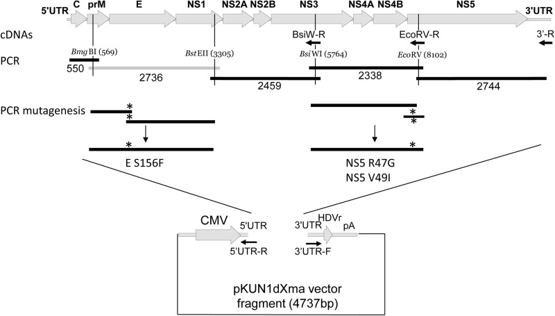
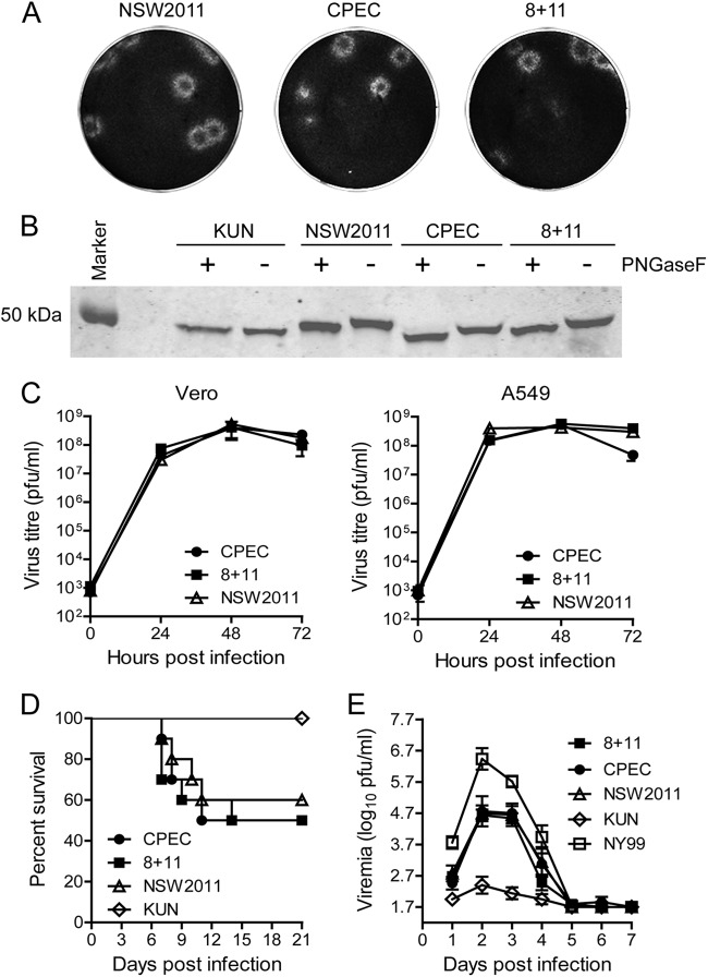
To adapt this approach for generation of infectious DNA for the NSW2011 isolate of KUNV, we used the truncated pKUN1dXma vector containing the CMV promoter immediately upstream of the first nucleotide of KUNV sequence and the hepatitis delta virus ribozyme sequence followed by the simian virus 40 (SV40) poly(A) signal placed directly downstream of the last nucleotide of the KUNV sequence (Fig. 1) (16). The vector fragment was amplified using primers complementary to the 5′- and 3′-terminal KUNV sequences to contain the first 87 and the last 127 nucleotides of the KUNV genome, which are virtually identical to the corresponding NSW2011 sequences (21). To generate overlapping RT-PCR fragments, viral RNA isolated from a low-passage-number stock of the NSW2011 isolate was first converted to three cDNA fragments using the 3′-R, EcoRV-R, and BsiW-R primers and Moloney murine leukemia virus (MMLV) reverse transcriptase (Promega) followed by PCR amplification of five DNA fragments using Phusion high-fidelity DNA polymerase (Thermo Scientific) and corresponding primers (Fig. 1). The primers were designed to contain overlapping ends for two neighboring fragments, similar annealing temperatures, and unique restriction sites in the NSW2011 genome to allow any future cloning if required. Two strategies were then employed to assemble infectious cDNA. The first strategy involved assembly of infectious full-length cDNA from all the PCR-amplified viral cDNA fragments and the vector fragment entirely by CPEC reaction without any intermediate steps in bacteria. The second (back-up) strategy involved production of two plasmid cDNAs, one (pNSW2011-1) containing most of the viral genome, except for the ∼2.7-kb BmgBI-BstEII fragment in the pKUN1dXma vector assembled by CPEC, and the second plasmid (pUC-BmgB-BstE) containing the BmgBI-BstEII fragment in the pUC19 vector obtained by conventional cloning. CPEC reactions for both strategies were performed using 200 ng of vector fragment and equimolar amounts of each of the other PCR fragments. The total amount of DNA used in each reaction was approximately 700 ng. The CPEC reactions were initiated by denaturation for 45 s at 98°C, followed by addition of Phusion high-fidelity DNA polymerase and 20 cycles of denaturing for 15 s at 98°C, annealing for 30 s at 60°C, and extension for 7 min (strategy 1) or 6.5 min (strategy 2) at 72°C. The CPEC was completed by 15 min of extension at 72°C. The CPEC reaction mixture containing full-length cDNA was directly transfected into HEK293T cells using Lipofectamine LTX reagent (Invitrogen) as described by the manufacturer. In strategy 2, the CPEC reaction mixture for generation of the pNSW2011-1 plasmid was transformed into DH5α cells. Ten out of 10 bacterial colonies contained plasmid DNA of the expected size, and restriction digestion with HindIII resulted in appearance of DNA fragments of the expected molecular weights (not shown). A blunt-ended PCR-amplified fragment containing BmgBI and BstEII restriction sites was cloned into the SmaI-digested pUC19 vector to obtain a pUC-BmgB-BstE plasmid. To obtain the full-length infectious DNA, the BmgBI-BstEII fragment was excised from the pUC-BmgB-BstE plasmid (clone 11) and ligated with BmgBI- and BstEII-digested CPEC-generated vector plasmid pNSW2011-1 (clone 8). The ligation reaction mixture (8 + 11) was then transfected directly into HEK293T cells using Lipofectamine LTX reagent. Culture fluid was harvested at 4 days after transfection with CPEC reaction mixture or ligation reaction mixture, at which point viruses reached titers of 8 × 10⁷ PFU/ml for the virus generated by strategy 1 (CPEC virus) and 2 × 10⁷ PFU/ml for the virus generated by strategy 2 (8 + 11 virus). These culture fluids were then amplified once in Vero cells to prepare high-titer stocks for further experiments; both viruses accumulated to the titers exceeding 108 PFU/ml by 3 days after infection (8 + 11, 8.8 × 10⁸ PFU/ml; CPEC, 5.5 × 10⁸ PFU/ml). Comparison of the plaque morphologies on Vero cells showed that viruses obtained using both strategy 1 (CPEC) and strategy 2 (8 + 11) formed plaques very similar in size to each other and to those of the parental NSW2011 isolate (Fig. 2A). The E proteins of both recovered viruses and the original NSW2011 isolates were glycosylated as expected (21), as shown by faster migration of E protein posttreatment with peptide:N-glycosidase F (PNGaseF) (Fig. 2B). Both recovered viruses also demonstrated indistinguishable growth kinetics in Vero and A549 cells (Fig. 2C) and virulence in 4 week-old mice (Fig. 2D) and 4-day-old chickens (Fig. 2E) compared to each other and to the original NSW2011 isolate. Notably, recovered viruses and the original NSW2011 isolate were more virulent in chickens than the prototype KUNV strain MRM61C and less virulent than the NY99 strain of WNV, which correlates with our previous findings in mice (21).
Fig 1.
Assembly of infectious cDNA for the wild-type and mutant NSW2011 viruses by CPEC reaction. A schematic representation of the WNV genome is shown at the top of the figure, with individual genes and untranslated regions (UTR) indicated. Shown are locations of the unique restriction sites with numbers indicating nucleotide positions in the NSW2011 genome (GenBank accession no. JN887352). The primers used to generate cDNA fragments are shown by arrows and labeled by the designations of unique restriction sites. Lines show PCR fragments, with numbers representing their size in base pairs (bp). Asterisks show approximate locations of introduced mutations. The lightly shaded box for the BmgBI-BstEII fragment indicates that this fragment was cloned separately from other fragments in strategy 2. E S156F, PCR fragment contacting the S156F mutation in E protein; NS5 R47G and NS5 V49I, PCR fragments containing the corresponding mutations in the NS5 protein; CMV, cytomegalovirus promoter; HDVr, hepatitis delta virus ribosome; pA, SV40 poly(A) signal.
Fig 2.
In vitro and in vivo properties of the recovered wild-type NSW2011 viruses. (A) Plaque morphology in Vero cells at 4 days after infection. Cells were fixed with 4% formaldehyde and stained with 0.2% crystal violet. (B) Western blot with anti-E antibodies. Lysates from viral culture supernatants were treated with PNGaseF (+) or mock treated (−). Samples were separated on 10% SDS-PAGE gels, transferred to nitrocellulose membrane, and probed with MAb 4G2. KUN, Kunjin virus. (C) Viral growth kinetics in Vero and A549 cells. Cells were infected at a multiplicity of infection (MOI) of 1, and culture supernatant was collected at the indicated time points. The viral titer was determined by plaque assay on Vero cells. Error bars represent the standard errors of the means of 2 samples. (D) Survival of 28-day-old CD-1 mice (n = 10) challenged intraperitoneally with 1,000 PFU of virus. Mice were monitored for 21 days and sacrificed when signs of encephalitis were evident. (E) Virulence in 4-day-old chickens (n = 8) following subcutaneous inoculation with ∼1,000 PFU of the indicated viruses. Infected chickens were bled daily for the first 7 days after infection, and virus titers in the serum were determined by plaque assays on Vero cells.
Sequencing of RT-PCR products generated from the RNA of recovered viruses identified two sites in the NS5 gene showing mixed bases in the sequencing chromatograms (Table 1) indicating the presence of mixed virus populations. These were present in both viruses obtained by two different strategies (Table 1), suggesting that they may be present in the original viral RNA used to generate infectious DNAs. Retrospective sequencing of the original virus isolate indeed confirmed the presence of mixed bases at the same positions (Table 1). The result indicates that the CPEC-based strategy for generating infectious DNA may be used to accurately recapitulate natural viral heterogeneity. In addition to the mixed bases at the same positions in the NS5 gene, 8 + 11 virus obtained by strategy 2 contained two mutations, a codon change from leucine to valine at position 287 of the NS1 gene and a silent mutation in the NS3 gene, that were not present in the original viral isolate (Table 1). The results show that propagation in bacteria of the plasmids used to generate infectious DNA for 8 + 11 virus led to the appearance of mutations that can be avoided if infectious DNA is assembled entirely from PCR fragments by CPEC reaction without any bacterial propagation steps.
Table 1.
Sequence analysis of generated viruses
| Virus | Mixed nucleotides or mutations in RNA | Corresponding amino acid position(s) in viral proteins | Polymorphism in original NSW2011 isolate |
|---|---|---|---|
| CPEC | 8295 U/A | NS5 201 L/H | Yes |
| 9935 U/A | NS5 757 C/A | Yes | |
| 8 + 11 | C3311G | NS1 L287V | No |
| U5041C | NS3 L149 silent | No | |
| 8295 U/A | NS5 201 L/H | Yes | |
| 9935 U/A | NS5 757 C/A | Yes |
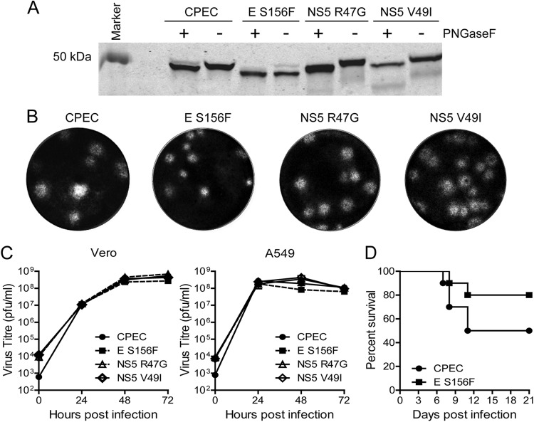
To demonstrate the utility of the CPEC method, three viral mutants were generated by site-directed PCR mutagenesis of corresponding cDNA fragments followed by CPEC. The locations of mutations were chosen so that the phenotype of mutants could be easily distinguished from that of the wild type: i.e., removal of glycosylation motif in E (E S156F) and change in the reactivity of the NS5 protein to the 5H1 monoclonal antibody (NS5 R47G and NS5 V49I) (22–25). To generate the E S156F mutant, two overlapping PCR fragments were generated using the BmgBI-BstEII fragment as the template (Fig. 1). To generate NS5 mutants, two sets of overlapping fragments were generated using the BsiWI-EcoRV fragment as the template (Fig. 1). These sets of overlapping fragments were then used to generate new mutated BmgBI-BstEII (E S156F) and BsiWI-EcoRV (NS5 R47G and NS5 V49I) fragments, respectively (Fig. 1). These mutated fragments were then used in combination with other original PCR fragments covering the rest of the genome for the CPEC reaction and recovery of mutant viruses as per the method described above. Secreted viruses were first harvested 3 days after transfection, when they reached titers of 8.8 × 10⁵ PFU/ml for E S156F virus, 1.8 × 10⁶ PFU/ml for NS5 R47G virus, and 9.8 × 10⁵ PFU/ml for NS5 V49I virus. The viruses were then amplified once in Vero cells to prepare virus stocks for further studies: the titers were 8.3 × 10⁷ PFU/ml for E S156F virus, 4 × 10⁸ PFU/ml for NS5 R47G virus, and 3.8 × 10⁸ PFU/ml for NS5 V49I virus. Sequencing of the corresponding RT-PCR products generated from RNA of recovered viruses confirmed the presence of introduced mutations. E S156F mutant virus lacked E glycosylation (Fig. 3A) and formed smaller plaques (Fig. 3B), as expected. A minor proportion of E protein appeared to be glycosylated (Fig. 3B), a phenomenon that may occur during virus passaging in cells (26). All mutant viruses replicated with similar efficiencies to each other and to the parental CPEC virus in Vero and A549 cells (Fig. 3C). As expected from previous studies, E S156F mutant virus showed decreased virulence in 4-week-old mice compared to the parent CPEC virus (Fig. 3D). The results demonstrate that glycosylation of E protein contributes to the virulence of the NSW2011 isolate.
Fig 3.
In vitro and in vivo properties of NSW2011 mutant viruses generated by CPEC. (A) Western blot with anti-E antibodies. Lysates from viral culture supernatants were treated with PNGaseF (+) or mock treated (−). Samples were separated on 10% SDS-PAGE gels, transferred to nitrocellulose membrane, and probed with anti-E MAb 4G2. (B) Plaque morphology in Vero cells at 4 days after infection. Cells were fixed with 4% formaldehyde and stained with 0.2% crystal violet. (C) Viral growth kinetics in Vero and A549 cells. Cells were infected at an MOI of 1, and culture supernatant was collected at the indicated time points. Viral titers were determined by plaque assay on Vero cells. Error bars represent the standard errors of the means of 3 independent experiments. (D) Survival of 28-day-old CD-1 mice (n = 10) challenged intraperitoneally with 1,000 PFU of the indicated viruses. Mice were monitored for 21 days and sacrificed when signs of encephalitis were evident.
Both NS5 mutants were glycosylated (Fig. 3A), formed similar plaques to those formed by CPEC virus (Fig. 3A), and had similar growth kinetics to the CPEC virus (Fig. 3C). Monoclonal antibody (MAb)-binding assays confirmed the lack of glycosylation for E S156F mutant, i.e., unglycosylated KUNV and the E S156F mutant bound to MAb 3.101C, which specifically recognizes unglycosylated E protein (J. Hobson-Peters and R. A. Hall, unpublished data), while the NSW2011, CPEC, and 8 + 11 viruses did not (Table 2). Reactivity to the 5H1 MAb, which recognizes an epitope containing residues 47 and 49 in KUNV NS5 protein (R47 and I49) but not in NY99 NS5 protein (G47 and V49) (24) showed that NSW2011 NS5 (R47 and V49) and the NS5 R47G mutant didn't bind 5H1 MAb, while KUNV and the NS5 V49I mutant did (Table 2). The results confirm our previous data distinguishing NSW2011 NS5 protein from KUNV NS5 protein in binding to 5H1 MAb (21) and showing the important role for residue 49 in this binding (24). Thus, all CPEC-generated mutant viruses behaved as expected in in vitro assays, while the E S156F mutant also behaved as expected in vivo. The results demonstrate the utility of the CPEC method for rapid generation of viral mutants. The ability to quickly exchange desired cDNA fragments makes the CPEC approach also very useful for generating chimeric viruses between different viral strains as well as for replacing regions containing an undesired polymorphism by well-characterized and stable sequences from cloned cDNA fragments.
Table 2.
Reactivity of parental and generated viruses with monoclonal antibodies
| Virus | Virus reactivity with MAb: |
||
|---|---|---|---|
| 4G2 (glycosylated and nonglycosylated E) | 5H1 (residue 49 in NS5) | 3.101C (nonglycosylated E) | |
| KUN | + | + | + |
| NSW2011 | + | − | − |
| 8 + 11 | + | − | − |
| CPEC | + | − | − |
| E S156F | + | − | + |
| NS5 R47G | + | − | − |
| NS5 V49I | + | + | − |
Although our finding that the CPEC method allows accurate recapitulation of heterogeneous viral population is limited to only two loci in the genome that were identified to contain a mixed virus population in our NSW2011 virus preparation, the robustness, high fidelity, and the lack of bacterial amplification and in vitro RNA transcription steps afforded by the CPEC method should allow accurate recreation of more heterogeneous flavivirus populations. This in turn should facilitate studies investigating the potential role of viral heterogeneity in the adaptation of flaviviruses to different hosts.
ACKNOWLEDGMENTS
We thank Jody Hobson-Peters for assistance with ELISA and the PNGase F assay, Peter Kirkland for providing the NSW2011 isolate and its sequence, and Theodore Pierson for useful suggestions. We thank Barbara Arnts and Rebecca West for assistance with the animal experiments.
The animal experiments were conducted with approval from the University of Queensland Experimentation Ethics Committee in accordance with the guidelines for animal experimentation as set by the National Health and Medical Research Council, Australia.
The work was supported by grants to A.A.K. from the National Health and Medical Research Council.
Footnotes
Published ahead of print 12 December 2012
REFERENCES
- 1. Bredenbeek PJ, Kooi EA, Lindenbach B, Huijkman N, Rice CM, Spaan WJ. 2003. A stable full-length yellow fever virus cDNA clone and the role of conserved RNA elements in flavivirus replication. J. Gen. Virol. 84:1261–1268 [DOI] [PubMed] [Google Scholar]
- 2. Zhao Z, Date T, Li Y, Kato T, Miyamoto M, Yasui K, Wakita T. 2005. Characterization of the E-138 (Glu/Lys) mutation in Japanese encephalitis virus by using a stable, full-length, infectious cDNA clone. J. Gen. Virol. 86:2209–2220 [DOI] [PubMed] [Google Scholar]
- 3. Yun SI, Kim SY, Rice CM, Lee YM. 2003. Development and application of a reverse genetics system for Japanese encephalitis virus. J. Virol. 77:6450–6465 [DOI] [PMC free article] [PubMed] [Google Scholar]
- 4. Zhang F, Huang Q, Ma W, Jiang S, Fan Y, Zhang H. 2001. Amplification and cloning of the full-length genome of Japanese encephalitis virus by a novel long RT-PCR protocol in a cosmid vector. J. Virol. Methods 96:171–182 [DOI] [PubMed] [Google Scholar]
- 5. Lai CJ, Zhao BT, Hori H, Bray M. 1991. Infectious RNA transcribed from stably cloned full-length cDNA of dengue type 4 virus. Proc. Natl. Acad. Sci. U. S. A. 88:5139–5143 [DOI] [PMC free article] [PubMed] [Google Scholar]
- 6. Schoggins JW, Dorner M, Feulner M, Imanaka N, Murphy MY, Ploss A, Rice CM. 2012. Dengue reporter viruses reveal viral dynamics in interferon receptor-deficient mice and sensitivity to interferon effectors in vitro. Proc. Natl. Acad. Sci. U. S. A. 109:14610–14615 [DOI] [PMC free article] [PubMed] [Google Scholar]
- 7. Pu SY, Wu RH, Yang CC, Jao TM, Tsai MH, Wang JC, Lin HM, Chao YS, Yueh A. 2011. Successful propagation of flavivirus infectious cDNAs by a novel method to reduce the cryptic bacterial promoter activity of virus genomes. J. Virol. 85:2927–2941 [DOI] [PMC free article] [PubMed] [Google Scholar]
- 8. Audsley M, Edmonds J, Liu W, Mokhonov V, Mokhonova E, Melian EB, Prow N, Hall RA, Khromykh AA. 2011. Virulence determinants between New York 99 and Kunjin strains of West Nile virus. Virology 414:63–73 [DOI] [PMC free article] [PubMed] [Google Scholar]
- 9. Beasley DW, Whiteman MC, Zhang S, Huang CY, Schneider BS, Smith DR, Gromowski GD, Higgs S, Kinney RM, Barrett AD. 2005. Envelope protein glycosylation status influences mouse neuroinvasion phenotype of genetic lineage 1 West Nile virus strains. J. Virol. 79:8339–8347 [DOI] [PMC free article] [PubMed] [Google Scholar]
- 10. Mandl CW, Ecker M, Holzmann H, Kunz C, Heinz FX. 1997. Infectious cDNA clones of tick-borne encephalitis virus European subtype prototypic strain Neudoerfl and high virulence strain Hypr. J. Gen. Virol. 78:1049–1057 [DOI] [PubMed] [Google Scholar]
- 11. Shi PY, Tilgner M, Lo MK, Kent KA, Bernard KA. 2002. Infectious cDNA clone of the epidemic West Nile virus from New York City. J. Virol. 76:5847–5856 [DOI] [PMC free article] [PubMed] [Google Scholar]
- 12. Sumiyoshi H, Hoke CH, Trent DW. 1992. Infectious Japanese encephalitis virus RNA can be synthesized from in vitro-ligated cDNA templates. J. Virol. 66:5425–5431 [DOI] [PMC free article] [PubMed] [Google Scholar]
- 13. Kelly EP, Puri B, Sun W, Falgout B. 2010. Identification of mutations in a candidate dengue 4 vaccine strain 341750 PDK20 and construction of a full-length cDNA clone of the PDK20 vaccine candidate. Vaccine 28:3030–3037 [DOI] [PubMed] [Google Scholar]
- 14. Polo S, Ketner G, Levis R, Falgout B. 1997. Infectious RNA transcripts from full-length dengue virus type 2 cDNA clones made in yeast. J. Virol. 71:5366–5374 [DOI] [PMC free article] [PubMed] [Google Scholar]
- 15. Gritsun TS, Gould EA. 1995. Infectious transcripts of tick-borne encephalitis virus, generated in days by RT-PCR. Virology 214:611–618 [DOI] [PubMed] [Google Scholar]
- 16. Hall RA, Nisbet DJ, Pham KB, Pyke AT, Smith GA, Khromykh AA. 2003. DNA vaccine coding for the full-length infectious Kunjin virus RNA protects mice against the New York strain of West Nile virus. Proc. Natl. Acad. Sci. U. S. A. 100:10460–10464 [DOI] [PMC free article] [PubMed] [Google Scholar]
- 17. Seregin A, Nistler R, Borisevich V, Yamshchikov G, Chaporgina E, Kwok CW, Yamshchikov V. 2006. Immunogenicity of West Nile virus infectious DNA and its noninfectious derivatives. Virology 356:115–125 [DOI] [PubMed] [Google Scholar]
- 18. Yamshchikov V, Mishin V, Cominelli F. 2001. A new strategy in design of +RNA virus infectious clones enabling their stable propagation in E. coli. Virology 281:272–280 [DOI] [PubMed] [Google Scholar]
- 19. Lin TY, Dowd KA, Manhart CJ, Nelson S, Whitehead SS, Pierson TC. 2012. A novel approach for the rapid mutagenesis and directed evolution of the structural genes of West Nile virus. J. Virol. 86:3501–3512 [DOI] [PMC free article] [PubMed] [Google Scholar]
- 20. Quan J, Tian J. 2009. Circular polymerase extension cloning of complex gene libraries and pathways. PLoS One 4:e6441 doi:10.1371/journal.pone.0006441 [DOI] [PMC free article] [PubMed] [Google Scholar]
- 21. Frost MJ, Zhang J, Edmonds JH, Prow NA, Gu X, Davis R, Hornitzky C, Arzey KE, Finlaison D, Hick P, Read A, Hobson-Peters J, May FJ, Doggett SL, Haniotis J, Russell RC, Hall RA, Khromykh AA, Kirkland PD. 2012. Characterization of virulent West Nile virus Kunjin strain, Australia, 2011. Emerg. Infect. Dis. 18:792–800 [DOI] [PMC free article] [PubMed] [Google Scholar]
- 22. Hall RA, Burgess GW, Kay BH, Clancy P. 1991. Monoclonal antibodies to Kunjin and Kokobera viruses. Immunol. Cell Biol. 69:47–49 [DOI] [PubMed] [Google Scholar]
- 23. Hall RA, Kay BH, Burgess GW, Clancy P, Fanning ID. 1990. Epitope analysis of the envelope and non-structural glycoproteins of Murray Valley encephalitis virus. J. Gen. Virol. 71:2923–2930 [DOI] [PubMed] [Google Scholar]
- 24. Hall RA, Tan SE, Selisko B, Slade R, Hobson-Peters J, Canard B, Hughes M, Leung JY, Balmori-Melian E, Hall-Mendelin S, Pham KB, Clark DC, Prow NA, Khromykh AA. 2009. Monoclonal antibodies to the West Nile virus NS5 protein map to linear and conformational epitopes in the methyltransferase and polymerase domains. J. Gen. Virol. 90:2912–2922 [DOI] [PubMed] [Google Scholar]
- 25. Hobson-Peters J, Toye P, Sanchez MD, Bossart KN, Wang LF, Clark DC, Cheah WY, Hall RA. 2008. A glycosylated peptide in the West Nile virus envelope protein is immunogenic during equine infection. J. Gen. Virol. 89:3063–3072 [DOI] [PubMed] [Google Scholar]
- 26. Scherret JH, Mackenzie JS, Khromykh AA, Hall RA. 2001. Biological significance of glycosylation of the envelope protein of Kunjin virus. Ann. N. Y. Acad. Sci. 951:361–363 [DOI] [PubMed] [Google Scholar]


