Inverted hyperplastic polyps (IHPs) of the colon are unusual variant of exophytic hyperplastic polyps that are characterized by epithelial displacement or inversion of the epithelium into the submucosa.1 To date, only a few cases of IHPs associated with adenoma or adenocarcinoma have been reported.2-4 However, due to the rarity of these polyps, their malignant potential is poorly understood.
A 56-year-old man was referred to Uijeongbu St. Mary's Hospital due to a positive fecal occult-blood test during a routine health check-up. The patient had a medical history of surgery for intestinal perforation and hypertension. On admission, physical examination revealed no abnormal findings and all laboratory examination were within normal range.
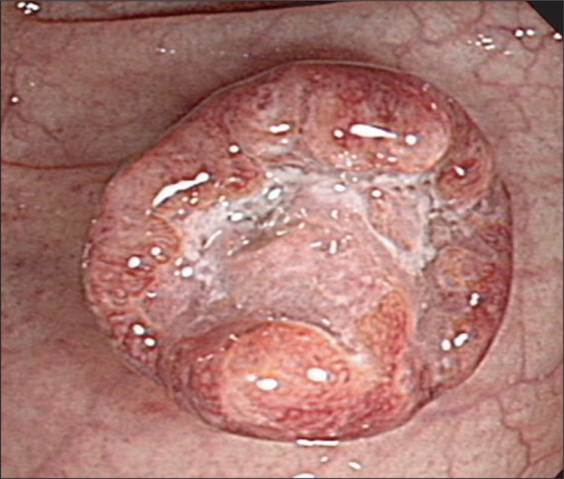
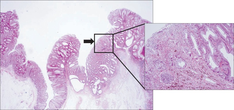
We performed colonoscopy for further evaluation. The colonoscopy showed a subpedunculated polyp with a large dimple, 20 mm in diameter, in the sigmoid colon (Fig. 1). An endoscopic mucosal resection (EMR) using the inject and cut technique was performed and the polyp was resected en bloc. The pathology examination showed an intramucosal cancer in an IHP (Fig. 2). The intramucosal cancer consisted mainly of well-differentiated adenocarcinoma (Fig. 2). Abdominal computed tomography showed no discernible malignant lesion. Follow-up colonoscopy revealed no remarkable findings 1 year after EMR.
Fig. 1.

Colonoscopy shows a subpeduculated polyp with a large dimple located in the sigmoid colon.
Fig. 2.
Microscopic image of the resected specimen showing an inverted hyperplastic polyp associated with intramucosal cancer, as indicated by the arrowhead in the low-magnified image (H&E stain, ×100). The intramucosal cancer consisted mainly of well-differentiated adenocarcinoma in the high-magnified image (H&E stain, ×400).
IHPs of the colon were initially described by Sobin5 in 1985. They represent an unusual morphological variant of hyperplastic polyps that shows epithelial displacement into the submucosa.5 IHPs of the colon are most commonly sessile lesions and are predominantly located in the left colon.1 Although the pathogenesis and natural history remain unknown, IHPs are thought to result from local trauma.1
The characteristic endoscopic features of IHPs include sessile or superficial elevated lesions with a pitted surface or central depression.6-8 Because distinctive surface pattern of IHPs is similar to that of invasive carcinoma or depressed type adenoma, IHPs are difficult to diagnose endoscopically.2,6 Observation of the pit pattern using magnifying colonoscope may be useful to precisely diagnose IHPs because it has the same surface and cellular features as ordinary hyperplastic polyps.6 The current case showed atypical endoscopic findings with large depressed portion. In addition, the histology showed the presence of well-differentiated carcinoma in the depressed portion.
To date, only a small number of cases associated with adenomas or adenocarcinoma have been described. Among them, three cases with an IHP were associated with adenomas,3 and one case with invasive cancer.4 For several years, hyperplastic polyps have been considered as non-neoplastic lesions without malignant potential. However, some hyperplastic polyps show molecular features similar to those of colorectal carcinomas. Several subtypes of serrated polyps, such as sessile serrated adenoma (traditional serrated adenoma), and mixed polyp, have been proposed to be the precursors of colorectal carcinomas.9 However, due to the rarity of IHPs, their malignant potential is poorly understood.
IHPs of the colon are usually asymptomatic and are often detected incidentally on endoscopy.3 In some cases, the main clinical feature of IHPs of the colon is gastrointestinal bleeding or abdominal pain.2 Usually, an endoscopic biopsy is not sufficient to make an accurate diagnosis of an IHP of the colon. The final diagnosis of an IHP of the colon depends on the pathological examinations of EMR and endoscopic polypectomy specimens.8 Treatment for IHPs continues to be debated. Since IHPs have benign cellular characteristics of exophytic hyperplastic polyps, overtreatment should be avoided. However, endoscopic treatment is necessary in cases with associated adenomatous findings, due to their malignant potential.4 In the present case, endoscopic treatment was appropriate for the large hyperplastic polyp.
In conclusion, we reported a rare case of early colon cancer developing in an IHP of the sigmoid colon treated with EMR. Further studies are needed to clarify the malignant potential of IHPs.
Footnotes
No potential conflict of interest relevant to this article was reported.
References
- 1.Yantiss RK, Goldman H, Odze RD. Hyperplastic polyp with epithelial misplacement (inverted hyperplastic polyp): a clinicopathologic and immunohistochemical study of 19 cases. Mod Pathol. 2001;14:869–875. doi: 10.1038/modpathol.3880403. [DOI] [PubMed] [Google Scholar]
- 2.Shepherd NA. Inverted hyperplastic polyposis of the colon. J Clin Pathol. 1993;46:56–60. doi: 10.1136/jcp.46.1.56. [DOI] [PMC free article] [PubMed] [Google Scholar]
- 3.Kuribayashi K, Ishii T, Ishidate T, et al. Two cases of inverted hyperplastic polyps of the colon and association with adenoma. Eur J Gastroenterol Hepatol. 2004;16:107–112. doi: 10.1097/00042737-200401000-00016. [DOI] [PubMed] [Google Scholar]
- 4.Fu K, Fujii T, Kuwayama H, Ishikawa T, Ueda Y, Fujimori T. Invasive cancer arising in a colonic inverted hyperplastic polyp. Endoscopy. 2010;42(Suppl 2):E29–E30. doi: 10.1055/s-2007-995733. [DOI] [PubMed] [Google Scholar]
- 5.Sobin LH. Inverted hyperplastic polyps of the colon. Am J Surg Pathol. 1985;9:265–272. doi: 10.1097/00000478-198504000-00002. [DOI] [PubMed] [Google Scholar]
- 6.Suzuki Y, Kobayashi M, Ishizuka K, et al. Inverted hyperplastic polyp diagnosed accurately by magnifying colonoscopy. Gastrointest Endosc. 2000;52:115–118. doi: 10.1067/mge.2000.106109. [DOI] [PubMed] [Google Scholar]
- 7.Fu KI, Matsuda T, Saito Y, Mashimo Y, Nonaka S, Fujimori T. An inverted hyperplastic polyp with a characteristic colonoscopic appearance. Endoscopy. 2006;38(Suppl 2):E55. doi: 10.1055/s-2006-944889. [DOI] [PubMed] [Google Scholar]
- 8.Hirasaki S, Kanzaki H, Suzuki S, Shirakawa A. Pedunculated inverted hyperplastic polyp of the sigmoid colon treated with endoscopic polypectomy. Dig Endosc. 2009;21:275–276. doi: 10.1111/j.1443-1661.2009.00906.x. [DOI] [PubMed] [Google Scholar]
- 9.Yano Y, Konishi K, Yamochi T, et al. Clinicopathological and molecular features of colorectal serrated neoplasias with different mucosal crypt patterns. Am J Gastroenterol. 2011;106:1351–1358. doi: 10.1038/ajg.2011.76. [DOI] [PubMed] [Google Scholar]


