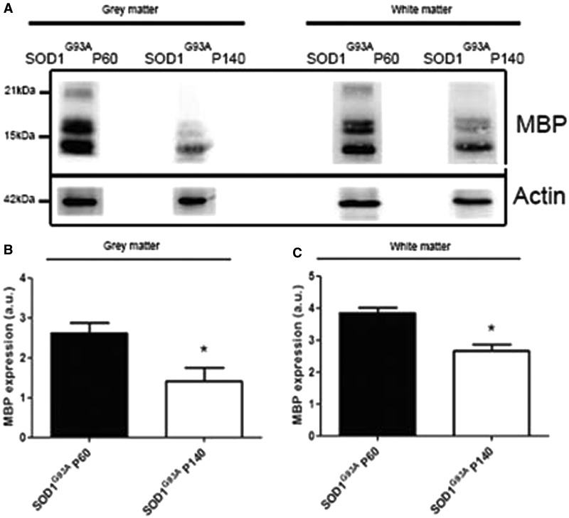
Figure 8.
Western blot of laser capture microscopy-collected ventral grey and white matter spinal cord fractions of mice at the asymptomatic (post-natal Day 60, P60) and symptomatic (post-natal Day 140, P140) disease stage. (A–C) At the symptomatic disease stage, the expression level of MBP was significantly lower compared with the asymptomatic disease stage in the ventral grey matter as well as the white matter (two-tailed unpaired t-test, n = 3, *P < 0.05). Data represent mean ± SEM.

