Abstract
A 76-year-old woman with hypertension was admitted to the hospital with complaints of chest pain and dyspnea. An echocardiogram and pulmonary computed tomography angiography showed right atrial myxoma complicated with pulmonary thromboembolism. An operation to resect the right atrial myxoma and pulmonary embolism was recommended; however, the patient refused and was discharged with anticoagulation therapy. Two years later, she developed dyspnea. Radiological studies and echocardiography showed similar results with the previous findings. The patient underwent mediastinotomy with resection of the right atrial myxoma and pulmonary embolectomy. As there are few reports on right atrial myxoma complicated with pulmonary embolism, we report a successful case of surgical removal of right atrial myxoma and pulmonary embolism.
Keywords: Myxoma, Pulmonary embolism, Right atrial tumor, Heart neoplasm
CASE REPORT
A 76-year-old woman with hypertension was admitted to the hospital with complaints of chest pain and dyspnea. Arterial blood gas analysis revealed the following findings of hypoxemia and hypocapnia: PO2, 36.7 mmHg; and PCO2, 30.4 mmHg. The chest radiographs exhibited findings suggestive of pulmonary edema. The patient's cardiac enzyme values were: creatine kinase-MB, 105 ng/mL; and troponin I, 0.38 ng/mL. The B-type nitriuretic peptide test result was 64. A diagnosis of recent myocardial infarction was made based on the following electrocardiographic findings: T-wave inversions and Q waves in leads II and III and a VF, and T-wave inversions in leads V1 to V4. Transthoracic echocardiography revealed akinesia in the apex free wall of the right ventricle, moderate pulmonary hypertension (right ventricular systolic pressure [RVSP], 55 mmHg), and a 2.7×2.8 cm mobile mass in the free wall of the right atrium. Pulmonary computed tomography (CT) angiography revealed pulmonary emboli occluding the pulmonary arteries in the upper and lower lobes on both sides, and a suspicious intra-atrial cardiac tumor with pulmonary thromboembolism was revealed according to the radiologic findings (Fig. 1). We initiated systemic administration of heparin. To detect other possible causes of the pulmonary embolism, abdominal CT and tumor marker tests were performed, the results of which were all normal. The patient was diagnosed with pulmonary embolism due to the mass in the right atrium based on the result that the patient's D-dimer was <0.35. However, the patient refused surgical treatment. The patient was discharged from the hospital and received coumadin drug therapy. After 26 months, she was intubated due to severe dyspnea and was then referred to Ajou University Hospital. Transthoracic echocardiography revealed: 1) hypokinesia in the right ventricle, 2) a dilated right ventricle, 3) mild pulmonary hypertension, 4) a 3.5×2.8 cm mass in the free wall of the right atrium, which had grown compared to previous findings, and 5) tricuspid regurgitation.
Fig. 1.
On first admission, transthoracic echocardiography (TTE) and pulmonary computed tomography (CT) angiography were performed. (A) TTE shows a 2.5×2.6 cm in diameter mass (white arrow) in the right atrium. The mass was attached to the atrial free wall by a short pedicle. (B, C) Pulmonary CT angiography shows thromboembolism wedged in the RPA&LPA. Ao, ascending aorta; RPA, right main pulmonary artery; LPA, left main pulmonary artery.
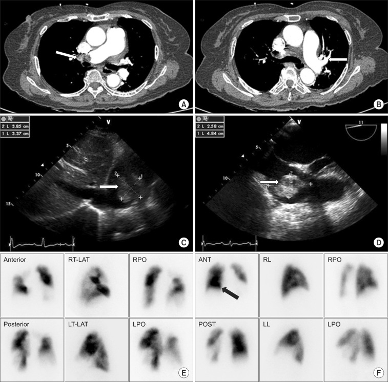
Pulmonary CT angiography revealed emboli occluding the right pulmonary artery and segmental arteries in the upper, middle, and lower lobes with a right atrial mass. The radiological characteristics were similar to that of a previous study (an intra-atrial cardiac tumor with pulmonary thromboembolism) (Fig. 2A, B). Pulmonary perfusion imaging showed decreased perfusion in the majority of the upper and lower right lobes, in part of the anterior segment of the left upper lobe, and in part of the superior and posterior basal segments of the left lower lobe (Fig. 2E, F). Transesophageal echocardiography revealed a 4.8×2.6 cm hypermobile mass with multilobular heterogeneous echogenicity in the upper lateral aspect of the free wall of the right atrium, which was not affecting the blood flow in the superior vena cava, inferior vena cava, or tricuspid valves (Fig. 2C, D). Coronary angiography showed findings of moderate coronary artery disease, which was treated with drug therapy alone. We decided to perform removal of the right pulmonary arterial emboli and the mass in the right atrium. The emboli in the left pulmonary artery decreased in size compared to the previous CT findings, and it was not considered a critical lesion because it was confined to the inferior lingular segmental artery. A standard sternotomy incision was made. The patient's body temperature was lowered to 23℃ (moderate hypothermia) through cardiopulmonary bypass, and then the myxoma with a 2 to 3 mm pedicle on the free wall of the right atrium was excised along with adjacent normal tissue. At 23℃, we opened the right pulmonary artery, which revealed that the myxoma segment was not adhered to the intima of the artery (Fig. 3A). An embolus within the right pulmonary artery was removed en bloc in a bloodless surgical field (Fig. 3B). After we confirmed that there were no further masses in the arteries supplying each segment, the right pulmonary artery and atrium were closed. Histopathological examination revealed that both the mass resected from the right atrium (4.3×3.5×3 cm) (Fig. 3C, white arrow) and the embolus removed from the right pulmonary artery (7×2×1.5 cm) (Fig. 3C, black arrow) were myxomas. On transthoracic echocardiography, we found that there were no residual masses. In addition, the size of the right atrium had decreased and the pulmonary artery pressure had slightly decreased to 35 mmHg. However, tricuspid regurgitation remained stationary (grade 2/4). Pulmonary CT angiography showed no further findings of pulmonary embolism. In the pulmonary perfusion imaging studies, the right lung had improved, but the left lung showed no changes. The patient was uneventfully discharged from the hospital on postoperative day 17. The patient is currently on out-patient follow-up without any significant symptoms. According to the echocardiography performed in December 2010, there was no evidence of pulmonary embolism, and the RVSP was 35 mmHg.
Fig. 2.
On second admission, pulmonary computed tomography (CT) angiography, transthoracic echocardiogram (TTE), transesophageal echocardiography (TEE), a lung perfusion preoperative scan, and postoperative scan were performed. (A) Pulmonary CT angiography shows thromboembolism wedged in the right main pulmonary artery and that (B) it disappeared in the left main pulmonary artery. (C) TTE shows a 3.5×2.8 cm mass (white arrow). (D) TEE shows a 4.8×2 cm mass (white arrow). (E) Bilateral large perfusion defects are shown in a lung perfusion preoperative scan, and (F) scan shows improvement of right lung perfusion postoperative scan (black arrow). RT-LAT, right-lateral; LT-LAT, left-lateral; RPO, right posterior oblique; LPO, left posterior oblique.
Fig. 3.
Intraoperative photograph and a pathologic photograph. (A) Picture shows the mass (white arrow) attached to the atrial free wall by a short pedicle (black arrow). (B) Picture shows fragmentation of right atrial myxoma in the right main pulmonary artery. (C) Picture shows right atrial myxoma (white arrow, 4.5×3.5×3 cm), which was ovoid, whitish gray, and myxoid. Fragmentation from the right main pulmonary artery (black arrow, 7×2×1.5 cm) is an elongated grayish soft gelatinous mass with a marked papillary surface. SVC, superior vena cava; RPA, right main pulmonary artery.
DISCUSSION
Pulmonary emboli are mainly caused by deep vein thromboses and embolus in the right heart or at the tip of a catheter. The first case of myxoma in the right atrium was reported in 1908. Chitwood [1] were the first to perform a successful resection of a myxoma through cardiopulmonary bypass. The recurrence rate of pulmonary embolism after resection has been reported to be 0.4% to 5%. Pulmonary emboli frequently recur when 1) surgical resection is incomplete, 2) emboli detached during surgery adhere to the intima of the heart, 3) emboli are of multicentric origin, 4) there is a family history of myxoma, or 5) a new myxoma develops from myxoma precursor cells. Because myxoma tissue is extremely friable, it is frequently detached and it adheres to a new site [2-5].
Approximately 75% of all the myxoma cases occur in the left atrium, 23% in the right atrium, and 2% in the ventricle. Myxoma can manifest obstruction symptoms due to blood flow blockade, symptoms due to embolism, arrhythmia, and other generalized symptoms. Syncope or sudden death can develop when pulmonary or systemic circulation is disturbed or when blood flow to the atrioventricular valve is blocked. Thrombosis can be induced by myxoma segments, thrombi that has formed within the tumor, or infected lesions within the tumor. Embolism occurs in 30% to 45% of the patients with myxoma of the left atrium. Emboli originating from myxoma in left atrium are mainly found in the brain, kidney, and branches of the aorta and lower extremities. Embolism occurs in approximately 10% of the patients with myxoma in the right atrium, and even pulmonary embolism can occur as in our case. In our case, the pulmonary embolism that had been diagnosed in May 2008 was different from the one detected by pulmonary CT angiography in March 2006. The former is thought to have been induced by thrombi formed by the myxoma in the right atrium, but not by myxoma segments, because the CT density (region of interest [ROI] property) was significantly higher in the myxoma detected in May 2008 (ROI, -20 for the myxoma in the right atrium as well as for the myxoma in the right pulmonary artery) than in the myxoma detected in March 2006 (ROI, 80). In March 2006, heparin administration at admission and maintenance anticoagulant therapy with coumadin resolved thrombi in the pulmonary artery on both sides were performed; however, in May 2008, the myxoma mass in the right atrium was separated and occluded the right pulmonary artery, causing severe dyspnea that required endotracheal intubation.
Removal of myxoma in the right side is important for preventing pulmonary embolism, maintaining contractibility, and restoring dilation functions of bilateral atria [6]. Special precautions should be taken during tumor resection in order to prevent thrombus formation and pulmonary embolism. Jones et al. [7] proposed that complete resection of myxoma should be performed under adequate exposure and minimal manipulation. Furthermore, pulmonary embolectomy should be considered in cases of myxoma associated with pulmonary embolism in order to improve patient symptoms. We reported a case of a 76-year-old woman with myxoma in the free wall of the right atrium. The patient's initial bilateral pulmonary embolism was developed due to thrombi formed by the myxoma; however, the embolism that occurred again two years later was due to myxoma segments. The patient was successfully treated with surgery and was uneventfully discharged from the hospital.
Footnotes
No potential conflict of interest relevant to this article was reported.
References
- 1.Chitwood WR., Jr Clarence Crafoord and the first successful resection of a cardiac myxoma. Ann Thorac Surg. 1992;54:997–998. doi: 10.1016/0003-4975(92)90676-u. [DOI] [PubMed] [Google Scholar]
- 2.Hermans K, Jaarsma W, Plokker HW, Cramer MJ, Morshuis WJ. Four cardiac myxomas diagnosed three times in one patient. Eur J Echocardiogr. 2003;4:336–338. doi: 10.1016/s1525-2167(03)00018-0. [DOI] [PubMed] [Google Scholar]
- 3.Shinfeld A, Katsumata T, Westaby S. Recurrent cardiac myxoma: seeding or multifocal disease? Ann Thorac Surg. 1998;66:285–288. doi: 10.1016/s0003-4975(98)00481-0. [DOI] [PubMed] [Google Scholar]
- 4.Kim SH, Kim JW, Jang IS, Choi JY, Hwang JY, Seo BK. Biatrial myxoma: a case report. Korean J Thorac Cardiovasc Surg. 1998;31:1094–1096. [Google Scholar]
- 5.Song H, Baek WK, Ahn H, Chae H, Kim CW. Surgical excision of intracardiac myxoma: a 15-year experience. Korean J Thorac Cardiovasc Surg. 1992;25:176–182. [Google Scholar]
- 6.Shimizu M, Okuri H, Yokoyama K, Kawada H, Takizawa T, Kikawada R. A case of right atrial myxoma: effect of large myxoma in the right atrium studied by M-mode and Doppler echocardiography. Jpn Circ J. 1995;59:579–586. doi: 10.1253/jcj.59.579. [DOI] [PubMed] [Google Scholar]
- 7.Jones DR, Warden HE, Murray GF, Hill RC, Graeber GM, Cruzzavala JL, et al. Biatrial approach to cardiac myxomas: a 30-year clinical experience. Ann Thorac Surg. 1995;59:851–855. doi: 10.1016/0003-4975(95)00064-r. [DOI] [PubMed] [Google Scholar]


