Abstract
This aim of this study was to evaluate the progression of grip, tip pinch, key (lateral) pinch, and tripod pinch strengths in patients suffering from carpal tunnel syndrome with thenar atrophy following surgery. Between October 2008 and May 2010, 46 patients (49 hands) with carpal tunnel syndrome associated with thenar atrophy underwent surgery. Thenar atrophy was assessed by clinical inspection. Evaluations for grip strength and for tip, key, and tripod pinch strengths were made using a hydraulic hand dynamometer grip and a hydraulic pinch gauge, respectively. These measurements were taken before surgery and at 3 and 6 months after the procedure. When we compared the averages of all forces measured in the affected hand before the surgery with all forces measured at 3 months postoperative, we found no significant differences. However, after 6 months, we found significant differences for all four strength tests as compared with those measurements taken preoperatively and at the 3 month time point. Our results suggest that patients with thenar atrophy show increased grip strength and pinch strength by the sixth month after surgical treatment.
Keywords: Carpal tunnel syndrome, Thenar atrophy, Hand strength, Grip, Pinch
Introduction
Carpal tunnel syndrome (CTS) is defined as numbness, nocturnal paresthesia, and hypoesthesia in the skin innervated by the median nerve among others symptoms and clinical signs caused by compression of the median nerve within the carpal tunnel. Various authors have investigated the role of specific clinical signs in the diagnosis of CTS. Studies have shown that thenar atrophy has a specificity of 90–99 % for this diagnosis; however, the sensitivity of thenar atrophy has been determined to be only 12.6 % [8]. The objective of this study was to evaluate the progression of grip strength, tip pinch strength, key pinch strength, and tripod pinch strength in patients affected by CTS associated with thenar atrophy, who underwent surgical decompression of carpal tunnel.
Materials and Methods
Patients and Inclusion Criteria
Between October 2008 and May 2010, 46 patients (49 hands) had surgical release of the carpal tunnel. The time between symptom onset and surgery ranged from 12 to 240 months, with an average of 65 months. The cohort comprised three (6.5 %) males and 43 (93.5 %) females, with an average age of 55 years, with a range of 37 to 85 years. The right hand was dominant in 44 patients (95.6 %) and left one was dominant in two (4.3 %).
For inclusion in the study, patients must have presented with a diagnosis of idiopathic CTS and the presence of thenar atrophy. For clinical diagnosis of CTS, patients were considered to have CTS if their history and physical examination revealed three or more in a list of six clinical criteria (CTS-6) [9]. For thenar atrophy, the apparent flattening of the thenar eminence was assessed at the first medical appointment, by a hand therapist at a preoperative evaluation, and by the surgeon in the operating room. The thenar atrophy was considered positive when present at each of these three investigations.
Patients who had previously undergone surgical treatment were excluded from the study. The study received full approval from the Research Ethics Committee of Universidade Federal de São Paulo and Hospital São Paulo. All participants gave written informed consent.
Preoperative Assessments, Surgical Intervention, and Follow-up
Patients were evaluated before surgery, as well as at 3 and 6 months after the procedure. Nerve conduction studies and electromyogram were performed preoperatively in all cases. Of the 49 hands with thenar atrophy, 12 hands showed normal distal motor latency (DML) and abnormal sensory velocity conduction (SVC), 11 hands showed abnormal DML and normal SVC, and 26 hands showed abnormal DML and SVC.
The measurement of grip strength was performed using a hydraulic grip dynamometer (Baseline, Irvington, NY, USA), which was adjusted to the second position. Grip, tip pinch, key pinch, and tripod pinch strength were first assessed preoperatively and then at 3 and 6 months postoperatively. The measurements of tip pinch, key pinch, and tripod pinch strengths were performed using a hydraulic gauge pinch dynamometer from Baseline (Irvington, NY, USA). To perform the measurements, the subjects sat with his or her arm raised in a position parallel to the body with the elbow flexed at 90°, and the forearm and wrist in a neutral position. Three measurements with the maximum possible force were made per test, and the average values calculated in kilograms-force.
Statistical Analysis
Data from each force measurement were averaged, and standard deviations were calculated for both hands. The Student’s t test was used to analyze numerical variables. The significance level was set at 5 %. The null hypothesis (H0) for the first analysis held that there would be no difference in strength in the operated hand between the pre- and postoperative periods. The alternate hypothesis (H1) was that there would be an increase in strength in the operated hand between the pre- and postoperative periods.
Results
In our study of 49 wrists with CTS, 31 surgeries (63 %) were performed on the right wrist, which is the dominant hand in 29 of these cases, and 18 (37 %) surgeries were performed on the left hand, which was the dominant hand for only one case. Three patients underwent operation on both sides. Endoscopic release was performed in 35 cases, open procedure was performed in 12 cases, and release by retinaculotome was performed in 2 cases.
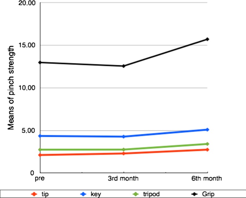
Table 1 shows the means, standard deviations, and minimum and maximum values of each of the strength test measured in the operated hands at each time point. Figure 1 shows the evolution of pinch and grip strengths before and after the surgical treatment.
Table 1.
Hand strength measurements preoperatively, and at 3 and 6 months after surgery
| Preoperative | 3 months | 6 months | ||
|---|---|---|---|---|
| Grip | Means | 12.98 | 12.56 | 15.71 |
| Standard deviation | 5.86 | 4.30 | 5.66 | |
| Minimum | 2.60 | 4.60 | 4.00 | |
| Maximum | 28.00 | 25.30 | 30.00 | |
| Tip | Means | 2.11 | 2.29 | 2.74 |
| Standard deviation | 0.78 | 0.82 | 1.03 | |
| Minimum | 1.00 | 1.00 | 1.00 | |
| Maximum | 4.00 | 4.30 | 5.50 | |
| Key | Means | 4.35 | 4.27 | 5.09 |
| Standard deviation | 1.72 | 1.54 | 1.59 | |
| Minimum | 1.00 | 1.00 | 1.30 | |
| Maximum | 8.00 | 8.00 | 8.00 | |
| Tripod | Means | 2.74 | 2.75 | 3.41 |
| Standard deviation | 1.11 | 0.97 | 1.29 | |
| Minimum | 1.00 | 1.00 | 1.00 | |
| Maximum | 5.30 | 5.60 | 7.50 | |
Fig. 1.
Evolution of the means of pinch and grip strength
When comparing the forces measured in the preoperative period and 3 months after surgery, we found an increase of 9 % in tip pinch strength, no change in the tripod pinch strength, but decreases of 3 and 2 % for the grip and key pinch strengths, respectively. There were no statistically significant differences between the mean strengths.
Between the preoperative period and 6 months after surgery, we identified a 30 % increase in the tip pinch strength, a 24 % increase in the tripod pinch strength, a 21 % increase in grip strength, and a 17 % increase in key pinch strength, for which all increases were statistically significant. We then compared the increases in strength between the 3 and 6 month time points, postoperatively. As expected, we identified a 25 % increase in grip strength, a 24 % increase in the tripod pinch strength, a 20 % increase in tip pinch strength, and a 19 % increase in key pinch strength. These results were also statistically significant.
Discussion
Currently, there is no gold standard for establishing a diagnosis of CTS. Previous diagnoses have been based on both clinical findings and the results of electrodiagnostic testing [10]. For the diagnosis of CTS in our study, patients needed to have presented with three or more from a list of six clinical criteria (CTS-6) [9].
In this study, we performed endoscopic release in 35 cases, open procedure in 12 cases, and release by retinaculotome in 2 cases. Brown et al. [1] studied the differences between the results of endoscopic and open surgery in the treatment of patients with CTS and patients with and without thenar atrophy but found no significant differences in the results between the two types of surgeries. Fernandes et al. [4], Paula et al. [15], and Meirelles et al. [13] evaluated the surgical treatment for CTS with Paine’s retinaculotome and showed excellent results with this surgical technique.
Although visual thenar evaluation has limited value as a diagnostic tool, it was still used in several studies. Thenar muscle atrophy and impaired sensibility in the distribution of the median nerve are signs of severe and prolonged median nerve compression, according to Eversmann [2]. In 1970, Phalen [16] noted that thenar atrophy is an absolute indication for surgery. Some authors agree that any degree of hypotrophy is an indication for CTS surgery [6, 17], and good results can be obtained after surgical release. Another useful method to assess hand function is the measurement of grip, tip pinch strength, key pinch strength, and tripod pinch strength. A Jamar hydraulic dynamometer was used in another study [5], but we chose baseline dynamometers in this study [4, 5]. Force evaluation has also been used by many authors [11, 12], but only two studies reported both grip and pinch strength with the means and standard deviations or 95 % confidence intervals for pre- and postoperative assessments [5].
Eversmann [3] reported that in patients undergoing carpal tunnel release, hand strength begins to return between weeks 2 and 3, postoperatively. At 6 weeks postoperatively, patients had 50 % of their preoperative strength, and between 8 and 10 weeks, patients had 75 % of preoperative strength. After 6 months, the study revealed that the maximum force had returned; however, 15 to 20 % of patients never regained their original strength because of changes in the configuration of the carpal bones and/or loss of the pulley effect of the retinaculum. By comparison, Gellman et al. [7], who used open release surgery on CTS, evaluated patients preoperatively and at postoperatively at 3 weeks, 6 weeks, 3 months, and 6 months and found that grip strength of 28, 73, 99, and 116 %, respectively; by the same comparisons, tip pinch strength was 74, 96, 108, and 126 %, respectively.
Nagaoka et al. [14] concluded that endoscopic release surgery in cases of CTS accompanied by thenar atrophy can achieve good results with respect to atrophy recovery. The average key pinch strength preoperatively was 3.6 ± 1.3 kg and increased to 5.1 ± 1.5 kg after surgery; tip pinch strength was 2.6 ± 1.0 kg preoperatively and 3.9 ± 1.4 kg after surgery. Follow-up time was 12 to 88 months, with an average age at time of surgery of 59.6 years.
In our study, we observed a 21 % improvement in grip strength, a 30 % increase in tip pinch strength, a 24 % increase in tripod pinch strength, and a 17 % increase in key pinch strength at 6 months postoperatively in relation to the preoperative period. These results showed that by 6 months, the patients showed a significant improvement in hand strength.
Acknowledgments
Conflict of interest
The authors declare that they have no conflicts of interest, commercial associations, or intent of financial gain regarding this research.
References
- 1.Brown RA, Gelberman RH, Seiler JG, 3rd, et al. Carpal tunnel release. A prospective, randomized assessment of open and endoscopic methods. Carpal tunnel release: a prospective, randomized assessment of open and endoscopic methods. J Bone Joint Surg Am. 1993;75(9):1265–1275. doi: 10.2106/00004623-199309000-00002. [DOI] [PubMed] [Google Scholar]
- 2.Eversmann WW., Jr Compression and entrapment neuropathies of the upper extremity. J Hand Surg. 1983;8:759–766. doi: 10.1016/s0363-5023(83)80266-4. [DOI] [PubMed] [Google Scholar]
- 3.Eversmann WW., Jr . Entraptament and compression neuropathies. In: Green DP, editor. Operative hand surgery. New York: Churchill Livingstone; 1988. pp. 1336–1366. [Google Scholar]
- 4.Fernandes CH, Meirelles LM, Carneiro RS, et al. Tratamento cirúrgico da síndrome do canal do carpo por incisão palmar e utilização do instrumento de Paine. Rev Bras Ortop. 1999;34:260–270. [Google Scholar]
- 5.Geere J, Chester R, Kale S, et al. Power grip, pinch grip, manual muscle testing or thenar atrophy—which should be assessed as a motor outcome after carpal tunnel decompression? A systematic review. BMC Musculoskelet Disord. 2007;8:114–123. doi: 10.1186/1471-2474-8-114. [DOI] [PMC free article] [PubMed] [Google Scholar]
- 6.Gelberman RH, Pfeffer GB, Galbraith RT, et al. Results of treatment of severe carpal-tunnel syndrome without internal neurolysis of the median nerve. J Bone Joint Surg Am. 1987;69(6):896–903. [PubMed] [Google Scholar]
- 7.Gellman H, Kan D, Gee V, et al. Analysis of pinch and grip strength after carpal tunnel release. J Hand Surg Am. 1989;14(5):863–864. doi: 10.1016/S0363-5023(89)80091-7. [DOI] [PubMed] [Google Scholar]
- 8.Gomes I, Becker J, Ehlers JA, et al. Prediction of the neurophysiological diagnosis of carpal tunnel syndrome from the demographic and clinical data. Clin Neurophysiol. 2006;117(5):964–971. doi: 10.1016/j.clinph.2005.12.020. [DOI] [PubMed] [Google Scholar]
- 9.Graham B, Regehr G, Naglie G, et al. Development and validation of diagnostic criteria for carpal tunnel syndrome. J Hand Surg Am. 2006;31(6):919–924. [PubMed] [Google Scholar]
- 10.Keith MW, Masear V, Chung KC, et al. American Academy of Orthopaedic Surgeons clinical practice guideline on diagnosis of carpal tunnel syndrome. J Bone Joint Surg Am. 2009;91(10):2478–2479. doi: 10.2106/JBJS.I.00643. [DOI] [PMC free article] [PubMed] [Google Scholar]
- 11.Leit ME, Weiser RW, Tomaino MM. Patient reported outcome after carpal tunnel release for advanced disease: a prospective and longitudinal assessment in patients older than age 70. J Hand Surg Am. 2004;29(3):379–383. doi: 10.1016/j.jhsa.2004.02.003. [DOI] [PubMed] [Google Scholar]
- 12.Mathiowetz V, Kashman N, Volland G, et al. Grip and pinch strength: normative data for adults. Arch Phys Med Rehabil. 1985;66(2):69–74. [PubMed] [Google Scholar]
- 13.Meirelles LM, Santos JBG, Santos LL, et al. Evaluation of the Boston questionnaire applied at late post-operative period of carpal tunnel syndrome operated with the Paine retinaculotome through palmar port. Acta Ortop Bras. 2006;14:126–132. doi: 10.1590/S1413-78522006000300002. [DOI] [Google Scholar]
- 14.Nagaoka M, Nagao S, Matsuzaki H. Endoscopic release for carpal tunnel syndrome accompanied by thenar muscle atrophy. Arthroscopy. 2004;20(8):848–850. doi: 10.1016/j.arthro.2004.06.033. [DOI] [PubMed] [Google Scholar]
- 15.Paula SEC, Meirelles LM, Santos JBG, et al. Long-term clinical evaluation—by Phalen, Tinel sign and night paresthesia—of patients submitted to carpal tunnel release surgery with Paine retinaculotome. Acta Ortop Bras. 2006;14:213–216. [Google Scholar]
- 16.Phalen GS. Reflections on 21 years’ experience with the carpal-tunnel syndrome. JAMA. 1970;212:1365–1367. doi: 10.1001/jama.1970.03170210069012. [DOI] [PubMed] [Google Scholar]
- 17.Santos LL, Meirelles LM, Santos JBG, et al. Carpal tunnel syndrome: reassessment of long-term outcomes with the use of Paine retinaculotome during surgery through a palmar incision. Acta Ortop Bras. 2005;13:225–228. doi: 10.1590/S1413-78522005000500002. [DOI] [Google Scholar]


