Abstract
Primary bone healing fails to occur in 5–15 % of scaphoid bones that undergo fracture fixation. Untreated, occult fractures result in nonunion up to 12 % of the time. Conventional bone grafting is the accepted management in the treatment algorithm of scaphoid nonunion if the proximal pole is vascularized. Osteonecrosis of the proximal scaphoid pole intuitively suggests a need for transfer of the vascularized bone to the nonunion site. Scaphoid nonunion treatment aims to prevent biological and mechanical subsidence of the involved bone, destabilization of the carpus, and early degenerative changes associated with scaphoid nonunion advanced collapse. Pedicled distal radius and free vascularized bone grafts (VBGs) offer hand surgeons an alternative treatment option in the management of carpal bone nonunion. VBGs are also indicated in the treatment of avascular necrosis of the scaphoid (Preiser’s disease), lunate (Kienböck’s disease), and capitate. Relative contraindications to pedicled dorsal radius vascularized bone grafting include humpback deformity, carpal instability, or collapse. The free medial femoral condyle bone graft has offered a novel treatment option for the humpback deformity to restore geometry of the carpus, otherwise not provided by pedicled grafts. In general, VBGs are contraindicated in the setting of a carpal bone without an intact cartilaginous shell, in advanced carpal collapse with degenerative changes, and in attempts to salvage small or collapsed bone fragments. Wrist salvage procedures are generally accepted as the more definitive treatment option under such circumstances. This manuscript offers a current review of the techniques and outcomes of VBGs to the carpal bones.
Keywords: Vascular bone grafts, VBG’s, Kienböck’s, Scaphoid nonunions, Preiser’s
Nonvascularized vs. Vascularized Bone Grafts
Several advantages of the use of vascularized bone grafts (VBGs) over nonvascularized bone grafts (non-VBGs) have been clarified through basic science and clinical research [66]. Transfer of vascularized bone preserves osteocytes. Superior biological and mechanical properties result secondary to minimization of postimplantation remodeling. Vascularized grafts have also demonstrated increased perfusion over time, accelerated graft consolidation, and rapid repopulation by cells [62, 90]. Furthermore, the provision of osteogenic and angiogenic factors into sites of avascular necrosis (AVN) has been postulated as an additional benefit [65, 83].
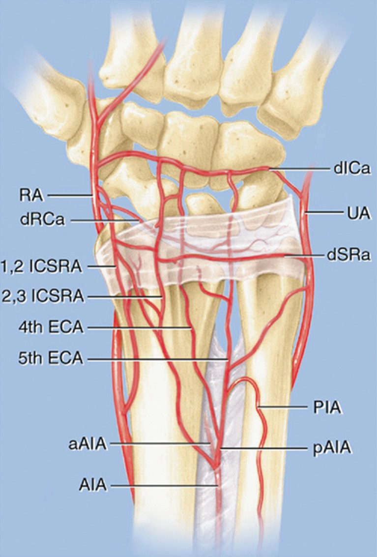
The first reported use of a VBG in the wrist was by Roy-Camille, who transferred the scaphoid tubercle on an abductor pollicis brevis muscle pedicle [69]. This innovation was followed by clarification of the detailed distal radius wrist vascular anatomy and its assortment of potential vascular pedicles to the bone (Fig. 1). Canine studies subsequently generated a reproducible model for the study of vascularized bone grafting and comparison studies between VBGs and non-VBGs in the treatment of scaphoid nonunion with and without proximal pole AVN [89, 90]. Sunagawa and colleagues [83] demonstrated 73 and 0 % scaphoid union rates with the use of VBGs and non-VBGs, respectively, in the treatment of scaphoid nonunion. Blood flow rate in the proximal pole, at 6 weeks postoperatively, was significantly higher with the use of VBG. This difference in rate of flow equilibrated at 8 weeks, but suggested quicker revascularization of avascular bone segments through the use of VBGs.
Fig. 1.
Dorsal distal radius arterial anatomy. The four main pedicles, upon which VBGs from this location are based, include the 1,2-ICSRA, 2,3-ICSRA, 4th ECA, and 5th ECA. RA radial artery, UA ulnar artery, AIA anterior interosseous artery, aAIA anterior branch of anterior interosseous artery, pAIA posterior branch of anterior interosseous artery, PIA posterior interosseous artery, dRCa dorsal radiocarpal arch, dICa dorsal intercarpal arch, dSRa dorsal supraretinacular arch, 1,2-ICSRA 1,2-intercompartmental supraretinacular artery, 2,3-ICSRA 2,3-intercompartmental supraretinacular artery, 4th ECA fourth extensor compartment artery, 5th ECA fifth extensor compartment artery. Adapted for use by permission of the Mayo Foundation for Medical Education and Research, Rochester, MN, USA. All rights reserved
Scaphoid Nonunion
The scaphoid is the most commonly fractured of all carpal bones, representing 60 % of fractures to the carpus [11, 45]. Nonunion occurs 5–15 % of the time, despite treatment [11]. Factors that commonly increase the risk for scaphoid nonunion include delay in diagnosis or treatment, proximal pole fracture, carpal instability, or displacement of fracture fragments more than 1 mm [11, 68, 81]. Scaphoid nonunions are traditionally and effectively treated with conventional grafting techniques from the distal radius or iliac crest. Barring AVN of the proximal pole, a recent meta-analysis demonstrated successful union in the treatment of scaphoid nonunion with non-VBGs, citing union rates of 77 and 94 % with K-wire or screw fixation, respectively [59]. Other studies have cited union rates of 40–67 and 70–90 % for treatment of scaphoid nonunion with non-VBG and K-wire fixation [40, 81]. Other authors suggest that the union rate after treatment of scaphoid nonunion with non-VBG and screw fixation rarely falls below 90 % [40], but no comparative studies have been performed comparing the two methods of graft fixation and their impact on union outcome [59].
Osteonecrosis of the scaphoid occurs in 3 % of patients after scaphoid fracture nonunion [40, 66]. Fractures involving the proximal third of the scaphoid are particularly predisposed, attributable to the scaphoid’s retrograde intraosseous blood supply. Because plain films are unreliable in establishing the diagnosis of AVN, magnetic resonance imaging (MRI) should be considered preoperatively as an adjunct, understanding that it too is not without fault [59]. MRI is 68 % accurate in confirming the diagnosis; with the use of gadolinium, accuracy increases to 83 % [40]. Established MRI criteria for AVN include areas of low signal intensity on T1-weighted images and high signal intensity on T2-weighted images [19]. Green originally suggested that the presence or absence of intraoperative punctate bone bleeding be the definitive gold standard in determining bone vascularity [28].
A recent meta-analysis cited union rates of 47 % when scaphoid nonunion was treated with non-VBG and screw fixation in the presence of proximal pole necrosis [59].VBGs increased this rate of union to 88 % under the same conditions. These findings, however, were predominantly extracted from the compiled data of retrospective case series.
Treatment of Scaphoid Nonunion with VBGs
Many authors have implemented VBGs, commonly from the dorsal or volar distal radius, in their treatment of avascular carpal bone segments [24, 53, 87, 96]. Judet intuitively validated VBG use over non-VBG use for the treatment of nonunion when he stated that “dead bone added to dead bone does not produce live bone” [59]. Indications for the use of VBGs in the treatment of scaphoid nonunion include a failed previous conventional graft or AVN of the proximal pole [66]. Some grafts are more effective than others in reestablishing normal scaphoid geometry (i.e., intrascaphoid angle, scapholunate angle, revised carpal height ratio).
In Zaidemberg’s initial report of the use of the 1,2-intercompartmental supraretinacular artery (1,2-ICSRA) for scaphoid nonunion, 11 out of 11 patients achieved radiographic union at an average of 6 weeks. All patients experienced complete resolution of pain, while pain with motion was entirely eliminated in 6 out of 11. Grip strength improved in all patients [40, 101]. Steinmann et al. [81] demonstrated similar results, with 14 out of 14 patients achieving union with the use of the 1,2-ICSRA as a dorsal onlay or interpositional palmar-based wedge graft. Average healing time was 11 weeks. Four out of four patients with proximal pole AVN went on to heal. Waitayawinyu et al. [94] also reviewed their experience, noting union in 28 out of 30 patients with proximal pole AVN, who had not undergone prior conventional grafting. Average time to union was 5 months. Significant improvements in grip strength, satisfaction scores (Likert scale 1–5), scaphoid height-to-length ratio, and Disabilities of the Arm, Shoulder, and Hand scores were noted. These outcomes are supported by cadaveric models that indicate that postoperative motion is not inhibited by the 1,2-ICSRA bone graft [30].
There are few exceptions to the reported 90 % union rate when using VBGs in the treatment of scaphoid nonunion with proximal pole AVN. Boyer et al. treated patients with 1,2-ICSRA VBGs and reported a union rate of 60 % in patients with proximal pole AVN [7]. Their four failures were in fractures that received a prior, unsuccessful conventional grafting procedure. Thus, prior surgery was considered an adverse factor that may lead to poorer outcomes following vascularized bone grafting. Suboptimal results were also reported by Straw and colleagues, who used the 1,2-ICSRA for the treatment of scaphoid nonunion and had a 27 % union rate [82]. Sixteen of their 22 patients had proximal pole AVN. Notably, they removed a single K-wire (used for fixation) at 8 weeks regardless of union. They concluded that the 1,2-ICSRA may not offer any advantage over conventional bone grafting in the treatment of scaphoid proximal pole AVN, but acknowledged that operator error in procedure performance or inadequate scaphoid construct postoperative immobilization may have contributed. Their high nonunion rate may have also been attributable to the high prevalence of proximal pole AVN in their series.
Discrepancies in reported outcomes suggest the need for randomized, comparative studies between VBGs and conventional grafts for the treatment of scaphoid nonunion. Standards for postoperative immobilization, type of fixation, timeframe until final assessment for union, and the means by which union is confirmed (i.e., plain films, computed tomography, or MRI) have not been clarified. Chang and colleagues elaborated upon factors contributing to scaphoid nonunion in 14 patients for whom use of the 1,2-ICSRA failed to achieve union [11]. These factors included female gender, proximal pole AVN, smoking status, K-wire fixation, and presence of humpback deformity. In their series, 34 out of 48 scaphoid fractures healed at an average of 15.6 weeks (71 %). Six of the 14 failures were in patients thought to be poor candidates for the 1,2-ICSRA due to the presence of humpback deformity. The conclusions of this large series were (1) the 1,2-ICRSA was still a viable graft with excellent union rates given that careful patient and fracture selection occurred and (2) nonunions with AVN and humpback deformity needed a large volar vascularized graft and the 1,2-ICRSA was not suitable for this type of nonunion.
Proximal third fracture nonunions of the scaphoid have been treated by a variety of VBG techniques. Donor sites include the distal radius [11, 15, 47, 56, 77, 79, 81, 101], ulna [29], metacarpal [6, 55], along with free vascularized grafts [19, 39]. The earliest reported use of a VBG from the volar distal radius was from Braun et al. [8]. Zaidemberg et al. [101] introduced the 1,2-ICSRA at approximately the same time as Sheetz et al. [77] anatomically described, in detail, the arterial supply of the distal radius. The 1,2-ICSRA, 2,3-intercompartmental supraretinacular artery (2,3-ICSRA), fourth extensor compartment artery (4th ECA), and fifth extensor compartment artery (5th ECA) were present 94, 100, 100, and 100 % of the time, respectively, in their study, each with nutrient vessels into the radius [77].
Zaidemberg and colleagues initially described the “ascending irrigating branch of the radial artery” as a periosteal vessel originating distally from the radial artery in the anatomic snuffbox and lying on the surface of extensor retinaculum between the first and second dorsal compartments [101]. This vessel was later identified as a supraretinacular vessel and not a periosteal vessel (the 1,2-ICSRA) by Sheetz et al. [77]. Sheetz et al. and Waitayawinyu et al. have provided objective anatomic landmarks of the 1,2-ICSRA to assist the operating surgeon in locating/elevating the pedicle and selecting a graft donor site from the dorsal radius (Fig. 2) [77, 95]. Their findings are summarized in Table 1.
Fig. 2.
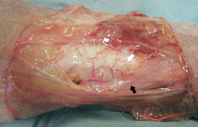
First (black arrowhead) and second (white arrowhead) extensor compartments are indicated on the dorsoradial aspect of this cadaver dissection. Note the 1,2-ICSRA overlying the extensor retinaculum between these two compartments. Adapted from Waitayawinyu et al. [95]. Permission for use courtesy of the American Society for Surgery of the Hand, http://www.assh.org
Table 1.
Anatomic landmarks of the 1,2-ICSRA VBG
| Artery present 94 % of the time |
| Located superficial to the extensor retinaculum, where the retinaculum is adherent to the bony tubercle separating the tendons of compartments 1 and 2 |
| Average distance from the tip of the radial styloid to its distal origin from the radial artery = 1.9 mm (the “ascending irrigating branch of the radial artery”) |
| Average distance from the tip of the radial styloid to its proximal origin from the radial artery = 48 mm |
| Average internal diameter = 0.3 mm |
| Average pedicle length = 22.5 mm |
| Average number of periosteal perforators along length of artery = 5.5 |
| Percent of periosteal perforators that penetrate cancellous bone = 6 % |
| Donor site distance, proximal to the articular surface of the distal radius, that incorporates the greatest number of periosteal perforators = 8–18 mm, with 15 mm being the mean distance from the radiocarpal joint that the periosteal nutrient arteries penetrate bone |
Elevation of the 1,2-ICSRA VBG
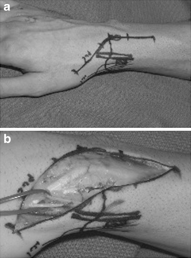
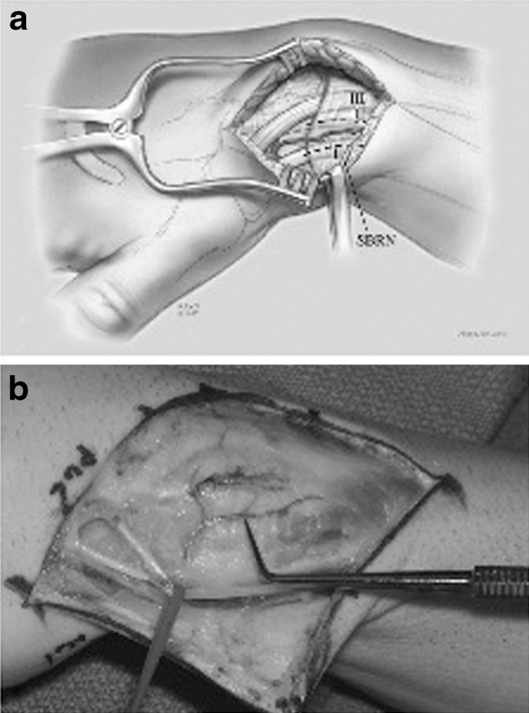
Tourniquet is insufflated after simple elevation of the extremity for exsanguination to facilitate easier identification of the small vascular pedicle. Previously, a dorsoradial curved incision was made overlying the snuff box, along the interval between the first and second compartments [11]. Current technique advises an incision overlying the extensor pollicis longus tendon, as this facilitates scaphoid exposure and fixation as well as graft donor site exposure (Fig. 3) [48]. Care is exercised to preserve the superficial radial nerve. The 1,2-ICSRA can be seen overlying the extensor retinaculum, between the tendons of the first and second dorsal compartments (Fig. 4). Care is taken to preserve a retinacular cuff around the vessel, as it is dissected distally towards its origin from the radial artery. Skeletonizing the vessel is not needed and only risks vascular injury. Further retraction of the first and second compartment tendons facilitates exposure of the dorsal wrist capsule. Before graft design and elevation, a transverse linear incision is made, through the capsule, atop the scaphoid from a point just ulnar to the 1,2-ICSRA to the fibers of the dorsal intercarpal ligament. If the nonunion is nondisplaced, it is often wiser initially to place a cannulated compression screw and then create the recipient site cavity. When displaced, the nonunion site can be debrided, followed by trough creation for the graft, followed by cannulated compression screw insertion after fragment reduction and graft placement. The 1,2-ICSRA is no longer recommended for use as a wedge graft, and simple press fit techniques for graft placement will often result in loosening. The trough is made with a dovetail to allow the graft to fit appropriately. The graft donor is then harvested on the dorsal distal radius, utilizing parameters described by Waitayawinyu et al. [95] and Sheetz et al. [77]. The graft is elevated using osteotomes, with care not to transect the pedicle on the distal cut, and the tourniquet is released to assess the graft for punctate bleeding. The graft is then inset into the dorsal scaphoid trough. While Waitayawinyu et al. [94] note that a radial styloidectomy can be performed to decrease tension on the vascular pedicle, prevent radiocarpal impingement, and improve exposure, in the Mayo Clinic experience, this has not been necessary in over 70 cases (A.Y. Shin, personal communication).
Fig. 3.
Elevation of the 1,2-ICSRA. a An incision is made following the course of the EPL tendon, with care to preserve the superficial radial nerve. b Soft tissues are elevated off the extensor retinaculum. Adapted from Larson et al. [48]. By permission of the Mayo Foundation for Medical Education and Research, Rochester, MN, USA. All rights reserved
Fig. 4.
Elevation of the 1,2-ICSRA. a The 1,2-ICSRA can be seen overlying the extensor retinaculum, between the tendons of the first and second extensor compartments dorsal compartment. b The 1,2-ICSRA is delineated at the tip of the instrument. Adapted from Larson et al. [48]. By permission of the Mayo Foundation for Medical Education and Research, Rochester, MN, USA. All rights reserved
Volar Radius-Based VBGs and Free VBGs
Restoration of carpal geometry and treatment of the humpback deformity are emphasized as essential components in the management of scaphoid nonunion with carpal collapse [40]. Collapse is defined as the presence of either humpback deformity or dorsal intercalated segmental instability (DISI). Objective parameters of collapse include a revised carpal height ratio ≤1.52 (normal, ±1.57), lateral intrascaphoid angle ≥45° (normal, ≤35°), or a radiolunate angle ≥15° (normal, ≤10°) [38]. The limitations of grafts from the dorsal radius to correct the humpback deformity and promote healing of scaphoid nonunion have led to the search for more appropriate donor sites.
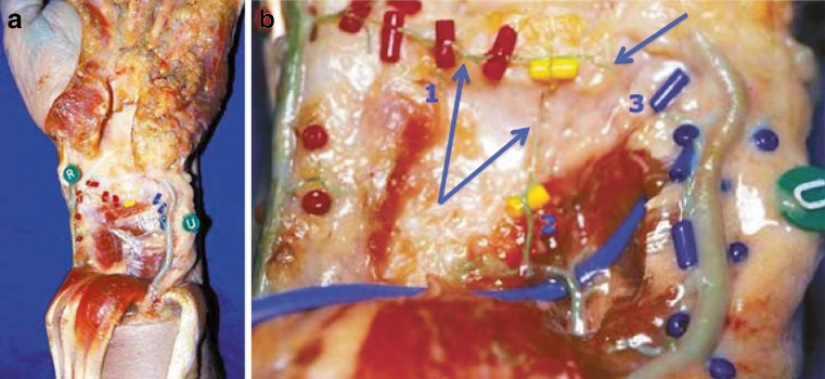
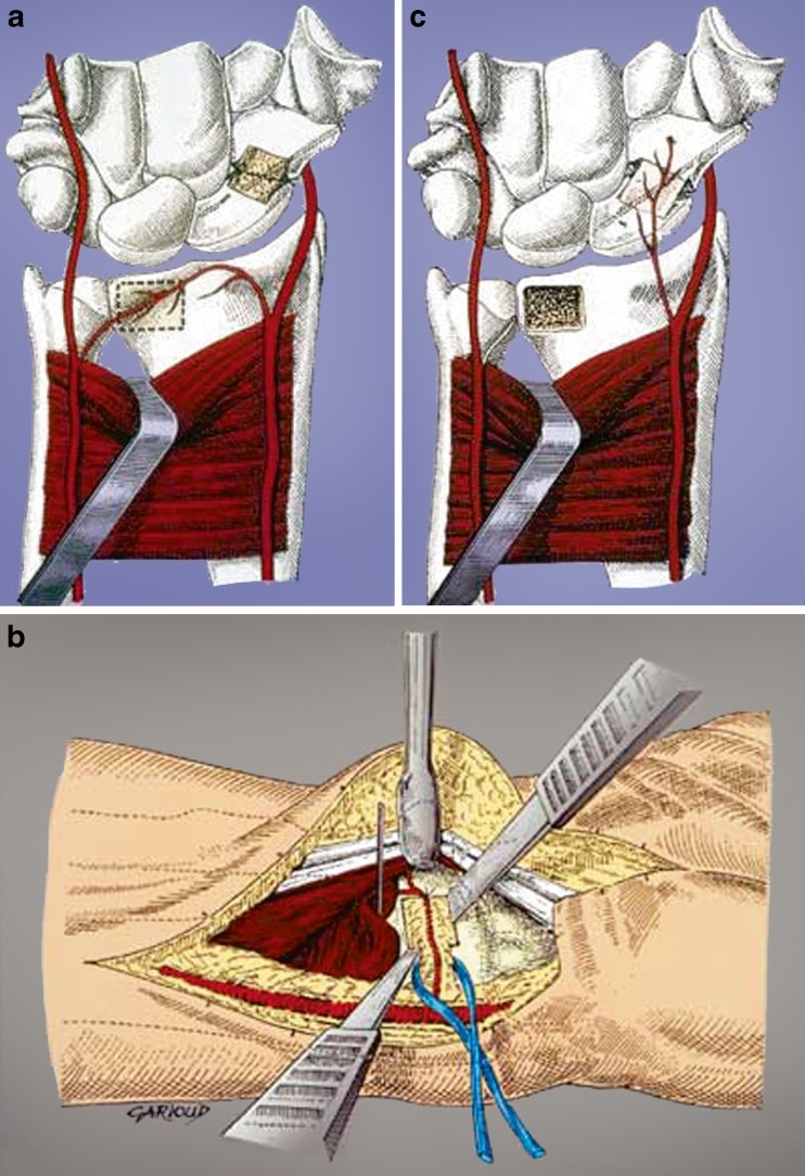
Grafts from the volar distal radius were first described by Richard Braun [8] and further delineated by Mathoulin and Haerle [56]. Volar wrist VBGs are based on the radial carpal artery (Fig. 5). This artery serves as the radial contribution to the palmar carpal arch and is located just distal to the pronator quadratus [66]. Mathoulin and Wahegaonkar [57] have described the dissection of the volar-based VBG pedicle and graft harvest in detail (Fig. 6).
Fig. 5.
a Cadaver dissection demonstrating the radial (R) and ulnar (U) contributions to the transverse volar carpal artery. b Closer view demonstrating the radial artery contribution to the volar carpal artery upon which the graft is based (1), a distal branch of the AIA (2), and the ulnar contribution to the transverse volar carpal artery (3). Vessels are identified by blue arrows. Adapted with permission from Mathoulin C. Treatment of scaphoid nonunion with a vascularized bone graft harvested from the volar aspect of the radius. Maîtrise Orthopédique no. 105, June 2001
Fig. 6.
Harvest and inset of the volar distal radius VBG based on the radial contribution to the volar carpal artery. Donor site (a), graft elevation with small osteotomes (b), and inset (c) are noted. A small pyramidal shaped graft is harvested, and a small rim of fascia is taken around the pedicle during its subperiosteal dissection from the distal radius. Adapted with permission from Mathoulin C. Treatment of scaphoid nonunion with a vascularized bone graft harvested from the volar aspect of the radius. Maîtrise Orthopédique no. 105, June 2001
One hundred percent union rates have been reported with the use of volar radius-based VBGs for scaphoid nonunion [9, 15, 43, 56]. Dailiana et al. demonstrated complete abolition of pain in all nine of their patients with a waist nonunion and a significant improvement in the scapholunate angle and carpal height at an average 24-month follow-up [15]. Their study represents the second largest case series presenting treatment results of long-standing scaphoid nonunion without AVN, managed with VBGs from the volar distal radius. They also advocate that the palmar approach provides the best visualization of the waist of the scaphoid, preserves the dorsally dominant blood supply of scaphoid, and leads to minimum loss of wrist flexion–extension, with prognostic factors supported by other studies [76]. These reported results of 100 % union, utilizing volar-based grafts for the treatment of scaphoid nonunion, need to be considered in the context they are presented—scaphoid nonunion treatment without AVN. This may explain the discrepancy seen in union rates when these results are compared to those of other vascularized dorsal grafts used to treat scaphoid nonunion in the presence of AVN [11, 94]. Furthermore, it bears mentioning that volar-based grafts are often placed as an inlay rather than as a structural wedge graft.
Options for the treatment of scaphoid nonunion with free VBGs also exist, notably from the iliac crest and supracondylar region of the medial femur [19, 23, 39, 49]. Indications for the use of the free medial femoral condyle (MFC) graft in the treatment of scaphoid nonunion include correction of humpback deformity or carpal collapse with AVN of the proximal pole. In the absence of proximal pole AVN, nonvascularized wedge grafts are still considered appropriate [40].
Sakai et al. originally described a free vascularized thin corticoperiosteal graft from the medial femoral condylar region [72]. The graft has two source pedicles, either the descending genicular artery (specifically the articular branch) or the superomedial genicular artery. The descending genicular vessel is longer and larger than the latter source and also includes a saphenous branch to the skin, allowing the surgeon to incorporate a skin paddle with the transfer. Six patients with fracture nonunion of the upper extremity were treated successfully in their series. The authors emphasize the importance of preserving the cambium layer of the periosteum (the layer intimately in contact with the bone cortex via Sharpey’s fibers), during graft elevation, to maintain its osteogenic capacity.
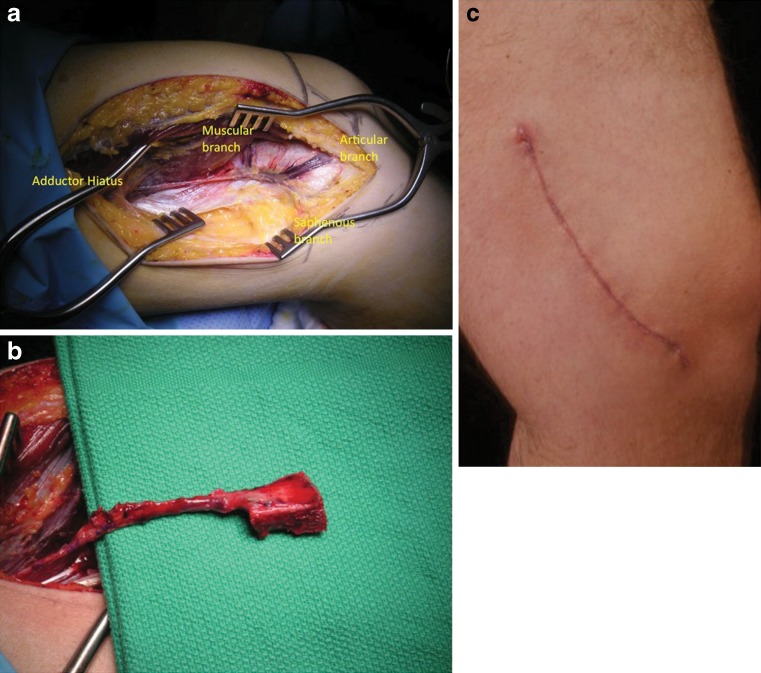
Doi et al. [19] initially described the use of the MFC vascularized periosteal bone graft for treatment of scaphoid nonunion as an onlay graft. The descending genicular vessel emerges from the medial aspect of the femoral vessel just proximal to Hunter’s canal in the medial aspect of the adductor magnus. The vessel is typically revealed after anterior traction of the vastus medialis muscle (Fig. 7a). The vessel is then mobilized, and the superomedial genicular vessels ligated if the former is considered of adequate size. The graft is designed over the supracondylar region of the medial femur and is then carefully elevated in order to not injure the medial collateral ligament or joint capsule of the knee [19]. Bleeding from the bone graft margins is verified prior to pedicle division (Fig. 7b). The donor site is closed primarily, leaving a linear scar (Fig. 7c)
Fig. 7.
Anatomy of the MFC vascularized periosteal bone graft. With anterior retraction of the vastus medialis muscle, the three branches of the descending genicular artery are identified (a). The graft just prior to pedicle division (b). Typical appearing donor site scar (c). Photos courtesy of Dr. Michael W. Neumeister, Division of Plastic Surgery, Southern Illinois University, Springfield, IL, USA
Doi and colleagues [19] and Jones and colleagues [39] reported complete healing in 10 out of 10 patients and 12 out of 12 patients, respectively, at an average of 12 and 13 weeks, respectively, when using the MFC VBG for the treatment of scaphoid nonunion. Range of motion and grip strength has been shown to be comparable to the contralateral wrist, with patients returning to work at an average of 4 months postoperatively [19]. Carpal collapse was significantly improved in the review by Jones et al., with mean lateral intrascaphoid angle improving from 66° to 28°, scaphoid height-to-length ratio improving from 0.78 to 0.65, and the scapholunate angle improving from 63° to 49°. Their preferred anastomotic sites, in the proximal wrist, were to the radial artery in end-to-side fashion and end-to-end to the venae comitantes.
Suggested attributes of the graft have included its ability to allow for a volar inset into the scaphoid, assisting in the correction of the humpback deformity and DISI. The graft has a consistent and robust vascular pedicle with excellent cancellous bone [40]. Yamamoto and colleagues defined the blood supply to the MFC via dissection of 19 fresh cadaveric lower limbs [98]. The descending genicular artery (present in 89 % of specimens) and superior medial genicular artery take its origins from the superficial femoral artery with an average distance of 13.7 and 5.2 cm from the joint line, respectively, and have an internal diameter averaging 1.5 and 0.78 mm, respectively. On average, 30 corticoperiosteal perforators fed the MFC and penetrated the cortical bone, on average, to a depth of 1.3 cm. The majority of perforators were located in the posterior distal quadrant of the MFC, with an average of 6.4 perforators present in this location [98].
Jones et al. also conducted a retrospective comparative study assessing the treatment outcomes of scaphoid nonunion with proximal pole AVN and carpal collapse [38]. Either a 1,2-ICSRA VBG or a MFC VBG was used. Four out of 10 patients healed using the pedicled graft at 19 weeks and 12 out of 12 patients healed using the MFC VBG at 13 weeks. This suggests, again, the importance of restoration of blood supply in the setting of AVN and correction of altered scaphoid geometry to obtain union in this difficult subset of scaphoid nonunions.
Summary of Results
Nonunion occurs in 10–15 % of scaphoid fractures, with risk factors including displacement, carpal instability, and fracture of the proximal pole. Conventional bone grafting with screw fixation has the ability to provide bony union, in the absence of proximal pole AVN. If the initial non-VBG fails or if proximal pole necrosis is present, consideration should be given to transfer of a VBG, either pedicled or free, to the nonunion site after debridement. In terms of correcting the humpback deformity/carpal collapse, volar radius-based grafts and free periosteal bone grafts from the supracondylar region of the medial femur have offered good results.
Preiser’s Disease
Preiser’s disease is an uncommon entity described as osteonecrosis of the scaphoid. Preiser initially described five cases of “rarefying osteitis” in 1910 after scaphoid fracture [91]. He stated that the pathology resulted from an alteration in bone nutrition, secondary to trauma, and made correlations with the lunatomalacia of Kienböck [91]. Not all patients with Preiser’s disease have antecedent trauma, however, and it has been suggested by some that Preiser’s disease, as it was initially described, should be a name reserved for those cases of posttraumatic AVN of the scaphoid, not for scaphoid AVN in the absence of antecedent trauma [21]. De Smet [16] offered the following parameters for diagnosis of Preiser’s: (1) no prior trauma or operative intervention to the involved wrist, (2) plain radiographic changes in at least 80 % of the scaphoid bone, and (3) MRI changes involving the entire scaphoid, excluding the tubercle. Predisposing factors include corticosteroid use, autoimmune diseases affecting the microvasculature (i.e., systemic sclerosis and lupus), and trauma [68]. Herbert and Lanzetta [33] staged Preiser’s disease based on radiographic appearance (a system comparable to that of Kienböck’s disease staging) (Table 2).
Table 2.
Herbert and Lanzetta classification of Preiser’s disease
| Stage I—absence of scaphoid changes on plain film |
| Stage II—scaphoid sclerosis on plain film |
| Stage III—scaphoid fragmentation |
| Stage IV—scaphoid collapse with or without arthritic changes |
Kalainov et al. [42] suggested the possibility of two patterns of avascularity in the scaphoid, based on MRI signal. Type I refers to AVN and/or ischemia involving the entire scaphoid, whereas type II refers to more limited disease, affecting on average 42 % of the scaphoid. Type II patients tended to have more favorable outcomes, with scaphoid bones less likely to fragment.
Because of the rarity of the condition, defined treatment algorithms do not exist. Treatment options have included observation, arthroscopic debridement [58], closing radial wedge osteotomy [32], scaphoid excision with four-bone fusion, proximal row carpectomy, total wrist arthrodesis, transfer of a vascular pedicle, and VBGs from the dorsal and volar distal radius [42, 61]. If a VBG is selected as treatment, it is essential to confirm the presence of an intact cartilaginous shell and the absence of arthritic changes at the radiocarpal and midcarpal joints (Herbert stage I or II) [66].
Results of Treatment with VBGs
There is no universally accepted VBG for the treatment of Preiser’s. For a description of graft elevation and inset technique, the reader is directed to previous discussion of VBG elevation and inset for scaphoid nonunion, as the techniques are comparable. Postoperative radioscaphoid joint unloading with external fixation or scaphocapitate pinning can be considered, but has not been proven as beneficial [42, 61]. Intraoperative conversion to a salvage procedure should be discussed with the patient prior to signed consent. Kalainov and colleagues reported on 9 out of 19 patients with Preiser’s that received a VBG [42]. Of these patients, fragmentation and scaphoid collapse could not be prevented in any with type I disease (four out of nine). Disease progression was halted in three out of five patients with type II disease, at an average follow-up of 17 months. The other two patients with type II disease demonstrated progression to stage III, with one patient requiring a proximal row carpectomy (PRC) after the VBG failed to provide pain relief.
Moran et al. reviewed their results of 1,2-ICSRA and 2,3-ICSRA VBG treatment for type I Preiser’s in eight patients [61]. At 36 months average follow-up, pain resolution was incomplete. Wrist flexion–extension and radial–ulnar deviation decreased, but not to a statistically significant level. Postoperative grip strength increased from 62 to 66 % of the contralateral extremity. This increase was not noted to be statistically significant. Despite encouraging evidence of revascularization in all MRIs obtained postoperatively, the proximal pole consistently remained incompletely revascularized.
Summary of Results
Results of VBGs for the treatment for Preiser’s have led some authors to consider Preiser’s disease as a contraindication to VBG [66]. Until prospective, comparative studies of treatment are conducted, VBGs can be considered as a potential intervention for early Herbert stage (I/II) disease and may potentially offer patients pain relief for multiple years prior to performance of salvage procedures.
Kienböck’s Disease
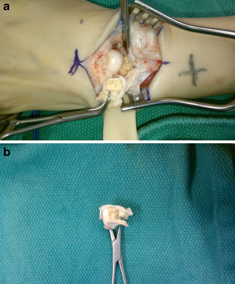
Kienböck’s disease, also known as idiopathic lunate osteonecrosis, has an etiology and pathogenesis that are incompletely understood. Figure 8 offers an intraoperative gross depiction of the appearance of the avascular lunate during a salvage procedure for Lichtman stage IIIb disease. Peste originally described the pathology in 1843, but it was Robert Kienböck, an early Viennese pioneer in radiology, who published the classic description in 1910 [74]. Variations in the osseous construct of the lunate and its vascular anatomy are considered to place the lunate blood supply at risk to trauma [18, 26, 88]. Ulnar minus variance places increased load transmission across the lunate [75].
Fig. 8.
Gross depiction of AVN of the lunate in Kienböck’s disease. Specimen obtained during salvage procedure for stage IIIb disease (a, b). Distal extremity is to the left and proximal to the right in a. Specimen removed in b. Photos courtesy of Dr. Reuben A. Bueno, Division of Plastic Surgery, Southern Illinois University, Springfield, IL, USA
While a predictable, progressively worsening pattern of lunate osteonecrosis occurs, including fragmentation, collapse, and subsequent radiocarpal/midcarpal arthritis, clinical symptoms do not necessarily correlate [17]. The natural history of the disease and surgery’s ability to alter its progression are not well-defined [17, 18]. Treatment algorithms have been suggested, but a recent systematic review demonstrated that no treatment is more advantageous than another in early and late stage Kienböck’s treatment [37]. Further, because the majority of this data was consolidated from retrospective studies, the authors were unable to suggest that outcomes of any treatment were better than placebo or the natural course of the disease.
Stahl originally developed a staging system for Kienböck’s based on findings from anteroposterior radiographs [80]. Lichtman revised this system [50]. The Stahl–Lichtman classification system describes four stages of progressive degeneration of the carpus secondary to AVN of the lunate [34, 68] (Table 3).
Table 3.
Stahl–Lichtman classification of Kienböck’s disease
| Stage I—lunate normal on plain film, MRI positive |
| Stage II—lunate sclerosis on plain film, no collapse |
| Stage III—lunate collapse with (a) normal proximal row alignment/carpal stability or (b) abnormal proximal row alignment/carpal instability |
| Stage IV—degenerative changes in the radiocarpal and/or midcarpal joint, fixed scaphoid rotation |
Some authors question the reliability of Lichtman’s classification system [3]. Through the use of diagnostic arthroscopy, Bain and Begg developed an articular-based classification system for Kienböck’s disease. In support of its use, they suggested that procedure selection should be based on the principle of leaving the carpus articulating with functional articular surfaces identified through arthroscopy. They suggest that treatment should be individually designed for the patient, based on a direct assessment of the pathoanatomy of the articular cartilage.
Goldfarb et al. [27] defined carpal instability as a radioscaphoid angle >60° to improve distinction between stages III(a) and III(b). Saunders and Lichtman [74] introduced an additional subclassification of stage III, known as III(c), which includes a coronal fracture of the lunate. Poor clinical outcomes after III(c) led to their suggestion that a coronal lunate fracture, regardless of carpal stability, is best managed with a salvage procedure. Stage IV’s similar radiographic appearance to scapholunate advanced collapse and scaphoid nonunion advanced collapse led to its potential designation as Kienböck’s disease advanced collapse, recognizing its unique etiology [74]. Prior to a discussion of treatment, the reader is directed to Dias and Lunn’s manuscript reviewing several unanswered inquiries about Kienböck’s disease [74]. Lluch and Garcia-Elias [52] describe a vicious circle of focal osteolysis, fractures, and necrosis at the root of Kienböck’s disease, the initial trigger of which is unknown.
Stage, ulnar variance, and status of the cartilaginous shell serve to currently guide treatment. Treatment options conceptually seek to alter mechanical load on the lunate, improve its devascularized biological status, or salvage wrist motion in advanced disease.
Mechanical: immobilization, joint leveling/unloading (radial shortening/wedge osteotomy) [10, 64, 65, 73, 97], capitate shortening osteotomy (CSO) with [93] or without [1, 25, 92] VBG, arthroscopic core decompression of the lunate [4], and metaphyseal core decompression of the radius or ulna [36]
Biological: revascularization-free iliac bone grafting [22], pedicled dorsal distal radius [34, 60], direct implantation of an arteriovenous pedicle [35, 78, 85], pronator quadratus pedicle flap [57], pisiform transfer [14], and index metacarpal [5]
Salvage: wrist denervation, lunate excision with [71] or without replacement [44], PRC [63], limited [86, 99] or total wrist fusion [51], and total wrist arthroplasty
Almost all reports on treatment are retrospective, suggesting the need for prospective, comparative studies. Joint leveling/unloading procedures, in patients with ulnar-negative variance, are performed under the premise of changing the abnormal radioulnar geometry to decrease excessive pressure through the lunate [54, 64]. By increasing radiolunate contact area, radial shortening decreases axial load through the lunate, predominantly along its radial border, the typical location of osteonecrosis of the lunate in Kienböck’s [64]. Satisfactory results from radial shortening procedures have been reported for treatment of stage II–IIIa disease [64, 84, 93]. A shortening of only 3 mm is recommended [84]. Yasuda et al. [99] reported quick pain relief and excellent function after scaphotrapeziotrapezoid (STT) arthrodesis for stage III(b) patients. Comparable results between intercarpal arthrodesis and joint leveling procedures in the treatment of the ulnar-negative wrist with Kienböck’s have been reported [12].
VBGs conceptually aim to improve the reduced blood supply of the lunate in Kienböck’s disease. Beck first described use of a vascularized os pisiform to a cored-out lunate for the treatment of Kienböck’s in 1971 [14]. More commonly, the VBG based on the fourth and fifth extensor compartmental arteries (4 + 5 ECA) is used [66]. The 4 + 5 ECA VBG relies on retrograde flow from the 5th ECA, which subsequently sends blood orthograde along the 4th ECA after ligation of its communication with the posterior branch of the anterior interosseous artery (AIA). Because it provides a large, long pedicle, the 4 + 5 ECA VBG is a preferred treatment for early stage Kienböck’s [68].
VBGs can be utilized for stage II disease and potentially for stage III(a) disease [66]. Kakar and colleagues offer a historical and current summary of the revascularization techniques offered for Kienböck’s disease [41]. Accepted principles dictate that the integrity of the lunate’s cartilaginous shell and the absence of degenerative changes are imperative to confirm prior to VBG transfer. In such circumstances (i.e., stage IV disease), VBG would be contraindicated. Previous surgery, including wrist arthroscopy via the 4–5 portal, that has potentially violated the underlying 4 + 5 ECA vascular pedicle, should be considered a relative contraindication for use of this graft.
Elevation of the 4 + 5 ECA VBG
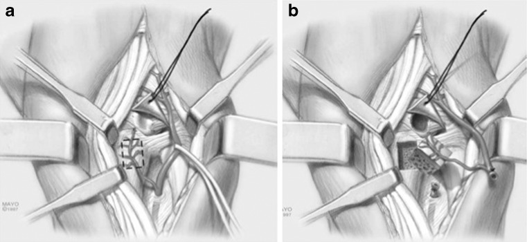
Simple extremity elevation, without the use of an Esmarch band, facilitates identification of the pedicle during dissection. Of the four dorsal distal radius pedicles available for a VBG (i.e., 1,2-ICSRA, 2,3-ICSRA, 4ECA, 5ECA), the 5th ECA is the largest [20]. A dorsal longitudinal skin incision is made overlying the radius and lunate. The extensor retinaculum is incised overlying the 5th compartment and elevated as a radially based flap to the second compartment. The 5th ECA is identified at the radial base of the fifth compartment, either partially within or against the septum between the fourth and fifth compartments. It is traced proximally to its origin from the posterior branch of the AIA (Fig. 9a). The 4th ECA takes origin from the posterior branch of the AIA, near its junction with the 5th ECA. The 4th ECA is followed distally along the floor of the fourth compartment, radial to the posterior interosseous nerve.
Fig. 9.
Elevation of the 4 + 5 ECA VBG. The 5th ECA is identified and traced proximally to its origin from the AIA. The 4th ECA is traced distally from this location to the dorsal, distal radius. Bone graft harvest donor site is typically 1 cm from the radiocarpal joint, underlying the 4th ECA pedicle (a). After radiocarpal ligament sparing capsulotomy, the lunate core is debrided, AIA is ligated, and the VBG is elevated (b). Adapted from Moran et al. [60]. By permission of the Mayo Foundation for Medical Education and Research, Rochester, MN, USA. All rights reserved
Next, a dorsal radiocarpal ligament sparing capsulotomy is performed and the lunate is visualized. Assessment of the lunate’s cartilaginous shell dictates the appropriateness of proceeding with the VBG or conversion to a salvage procedure. Assessment for midcarpal arthritis and radiocarpal arthritis is required, as their presence would be contraindications to a VBG. If VBG is considered appropriate, necrotic bone is cored out of the lunate via a dorsal window. The bone graft is marked out along the 4th ECA, approximately 1 cm from the radiocarpal joint. Dimensions of the graft coincide with the design needed to fill the lunate after debridement of its necrotic bone. The AIA is ligated (proximal to the 5th and 4th ECA origins), and the graft is elevated and press fit into position after confirmation of its vascularity with release of the tourniquet (Fig. 9b). Cancellous bone graft can be inserted into the lunate to fill an excessive void prior to graft insertion.
The wrist is unloaded for 8–12 weeks with casting, external fixator, or scaphocapitate pinning to avoid early loading of the graft which might inhibit revascularization of the lunate [34, 60]. Bone revascularization may lead to ongoing collapse of the lunate due to increased osteoclast activity during the early stages of bone healing [2].
Results of Treatment with VBGs
Daecke et al. reported the results of long-term follow-up on patients with either early [14] (23 patients) or late [13] (21 patients) stage Kienböck’s disease treated with either a pedicled pisiform vascularized graft (early stage disease) or complete replacement of the lunate by the pisiform [70] (late stage disease). At 12 years follow-up, 14 out of 20 patients with early stage disease (who had preoperative films for comparison) did not experience disease progression, a finding comparable to that of other studies [46]. Pedicled pisiform VBGs result in high patient satisfaction for both early and late stage Kienböck’s, with late stage disease more likely to progress towards osteoarthritis despite treatment [13, 14]. Significant increases in range of motion are noted for early stage disease treated with pedicle pisiform VBGs, but not for late stage disease treated with complete lunate replacement by the pedicled pisiform graft (Saffar procedure).
Eleven patients in the study of Daecke et al. [14], with ulnar-negative variance, also received a radial shortening procedure in conjunction with a vascularized os pisiform graft. The results from this group, however, demonstrated no significant difference among subjective, objective, or combined assessment parameters. Quenzer and colleagues [67] commented on improved radiographic appearance of the lunate in 55 % of patients that received a VBG in conjunction with a joint leveling procedure, in comparison to only 20 % who received a joint leveling procedure alone.
Results of treatment of Kienböck’s disease with combination procedures have been reported [57, 93]. Waitayawinyu et al. reviewed their results of decompressive CSO with a VBG from the base of the second or third metacarpal, based on the dorsal metacarpal arcade [93]. They treated 14 patients with Lichtman stage II or IIIa, ulnar-neutral or ulnar-positive wrists, with an average follow-up of 41 months. Significant improvements in grip strength and satisfaction were noted; satisfactory total arc of motion was maintained (89 % of contralateral side). Radial shortening potentially contributes to the development of ulnar abutment syndrome in the ulnar-neutral or ulnar-positive wrist and, therefore, was not selected as the procedure for carpus unloading in their patient population [93]. Viola et al. and Gay et al. have demonstrated the decompressive benefit of CSO [25, 92]. The consequences of CSO upon the STT joint, secondary to load transfer, are still unknown [25]. Moran et al. utilized the 4 + 5 ECA VBG in their treatment of 26 patients with stage II, IIIa, or IIIb Kienböck’s disease [60]. Twenty-four patients (92 %) acknowledged significant improvement in pain 3 months after surgery. Stage progression was halted in 20 patients, and grip strength improved significantly from 50 to 89 % of the contralateral side. Twelve out of 17 patients (71 %) who received an MRI at a mean 20-month follow-up visit demonstrated improved lunate signal and return of trabecular structure, suggesting evidence of revascularization. All patients with evidence of revascularization returned to their prior positions in the workplace.
Comparison of different VBG procedures for treatment of Kienböck’s is limited. Volar and dorsal distal radius VBGs for the treatment of Kienböck’s disease do not appear to differ in their subjective and objective outcome measures.
Uncommon Carpal Bone AVN Treated With VBGs
Successful treatment of AVN of the capitate and trapezium with use of the 4 + 5 ECA and 1,2-ICSRA, respectively, has been reported [31, 100].
Summary of Findings
Kienböck’s disease, or AVN of the lunate, has an unknown etiology. Its staging correlates with progressive collapse of the lunate, carpal instability, and ultimately, pancarpal arthritis. Patients do not necessarily experience the degree of symptoms that would be expected from the radiographic exam. Comparable results, between joint leveling and VBG interventions, have been demonstrated [66]. Early stage disease (II, IIIa) remains amenable to VBG procedures (i.e., 4 + 5 ECA, vascularized pisiform). Some authors advocate VBGs for IIIb disease. Temporary unloading of the graft is often recommended after the procedure (i.e., scaphocapitate pinning, external fixator). Advanced stage disease (IV) necessitates a salvage procedure. Future studies are needed to clarify the more optimal VBG for treatment of Kienböck’s disease and whether a VBG in combination with joint leveling procedures provides better results.
Acknowledgments
Conflict of Interest
The authors declare that they have no conflict of interest.
References
- 1.Almquist E. Capitate shortening in the treatment of Kienbock’s disease. Hand Clin. 1993;9:505–12. [PubMed] [Google Scholar]
- 2.Aspenberg P, Wang J, Jonsson K, et al. Experimental osteonecrosis of the lunate. Revascularization may cause collapse. J Hand Surg Br. 1994;19B:565–9. doi: 10.1016/0266-7681(94)90116-3. [DOI] [PubMed] [Google Scholar]
- 3.Bain G, Durrant A. An articular-based approach to Kienbock avascular necrosis of the lunate. Tech Hand Up Extrem Surg. 2011;15(1):41–7. doi: 10.1097/BTH.0b013e31820e82e8. [DOI] [PubMed] [Google Scholar]
- 4.Bain G, Smith M, Watts A. Arthoscopic core decompression of the lunate in early stage Kienbock disease of the lunate. Tech Hand Up Extrem Surg. 2011;15(1):66–9. doi: 10.1097/BTH.0b013e3181e1d2b4. [DOI] [PubMed] [Google Scholar]
- 5.Bengoechea-Beeby M, Cepeda-Una J, Avascal-Zuloaga A. Vascularized bone graft from the index metacarpal for Kienbock’s disease: a case report. J Hand Surg Am. 2001;26A:437–43. doi: 10.1053/jhsu.2001.24137. [DOI] [PubMed] [Google Scholar]
- 6.Bertelli J, Tacca C, Rost J. Thumb metacarpal vascularized bone graft in long-standing scaphoid nonunion—a useful graft via dorsal or palmar approach: a cohort study of 24 patients. J Hand Surg Am. 2004;29A:1089–97. doi: 10.1016/j.jhsa.2004.06.007. [DOI] [PubMed] [Google Scholar]
- 7.Boyer M, von Schroeder H, Axelrod T. Scaphoid nonunion with avascular necrosis of the proximal pole: treatment with a vascularized bone graft from the dorsum of the distal radius. J Hand Surg Br. 1998;23B:686–90. doi: 10.1016/s0266-7681(98)80029-6. [DOI] [PubMed] [Google Scholar]
- 8.Braun RM. Proximal pedicle bone grafting in the forearm and proximal carpal row. Orthop Trans. 1983;7:35. [Google Scholar]
- 9.Braun R. Viable pedicle bone grafting in the wrist. In: Urbaniak JR, editor. Microsurgery for major limb reconstruction. Mosby: St. Louis; 1987. pp. 220–9. [Google Scholar]
- 10.Calfee R, Van Steyn M, Gyuricza C, et al. Joint leveling for advanced Kienbock’s disease. J Hand Surg Am. 2010;35A:1947–54. doi: 10.1016/j.jhsa.2010.08.017. [DOI] [PMC free article] [PubMed] [Google Scholar]
- 11.Chang M, Bishop A, Moran S, et al. The outcomes and complications of 1,2-intercompartmental supraretinacular artery pedicled vascularized bone grafting of scaphoid nonunions. J Hand Surg Am. 2006;31:387–96. doi: 10.1016/j.jhsa.2005.10.019. [DOI] [PubMed] [Google Scholar]
- 12.Coe M, Trumble T. Biomechanical comparison of methods used to treat Kienbock’s disease. Hand Clin. 1993;9:417–29. [PubMed] [Google Scholar]
- 13.Daecke W, Lorenz S, Wieloch P, et al. Lunate resection and vascularized os pisiform transfer in Kienbock disease: an average of 10 years of follow-up study after Saffar’s procedure. J Hand Surg Am. 2005;30A:677–84. doi: 10.1016/j.jhsa.2005.02.015. [DOI] [PubMed] [Google Scholar]
- 14.Daecke W, Lorenz S, Wieloch P, et al. Vascularized os pisiform for reinforcement of the lunate in Kienböck’s disease: an average of 12 years of follow-up study. J Hand Surg Am. 2005;30A:915–22. doi: 10.1016/j.jhsa.2005.03.019. [DOI] [PubMed] [Google Scholar]
- 15.Dailiana Z, Malizos K, Zachos V, et al. Vascularized bone grafts from the palmar radius for the treatment of waist nonunions of the scaphoid. J Hand Surg Am. 2006;31:397–404. doi: 10.1016/j.jhsa.2005.09.021. [DOI] [PubMed] [Google Scholar]
- 16.De Smet L. Avascular nontraumatic necrosis of the scaphoid. Preiser’s disease? Chir Main. 2000;19:82–5. doi: 10.1016/S1297-3203(00)73464-0. [DOI] [PubMed] [Google Scholar]
- 17.Delaere O, Dury M, Molderez A, et al. Conservative versus operative treatment for Kienbock’s disease: a retrospective study. J Hand Surg Br. 1998;23B:33–6. doi: 10.1016/s0266-7681(98)80214-3. [DOI] [PubMed] [Google Scholar]
- 18.Dias J, Lunn P. Ten questions on kienbock’s disease of the lunate. J Hand Surg Eur. 2010;35B:538–43. doi: 10.1177/1753193410373703. [DOI] [PubMed] [Google Scholar]
- 19.Doi K, Oda T, Soo-Heong T, et al. Free vascularized bone graft for nonunion of the scaphoid. J Hand Surg Am. 2000;25A:507–19. doi: 10.1053/jhsu.2000.5993. [DOI] [PubMed] [Google Scholar]
- 20.Elhassan B, Shin A. Vascularized bone grafting for treatment of Kienbock’s disease. J Hand Surg Am. 2009;34A:146–54. doi: 10.1016/j.jhsa.2008.10.014. [DOI] [PubMed] [Google Scholar]
- 21.Ferlic D, Morin P. Idiopathic avascular necrosis of the scaphoid: Preiser’s disease? J Hand Surg Am. 1989;14A:13–6. doi: 10.1016/0363-5023(89)90053-1. [DOI] [PubMed] [Google Scholar]
- 22.Gabl M, Lutz M, Reinhart C, et al. Stage 3 Kienbock’s: reconstruction of the fractured lunate using a free vascularized iliac bone graft and external fixation. J Hand Surg Br. 2002;27B:369–73. doi: 10.1054/jhsb.2002.0766. [DOI] [PubMed] [Google Scholar]
- 23.Gabl M, Reinhart C, Lutz M, et al. Vascularized bone graft from the iliac crest for the treatment of nonunion of the proximal part of the scaphoid with an avascular segment. J Bone Joint Surg Am. 1999;81:1414–28. doi: 10.2106/00004623-199910000-00006. [DOI] [PubMed] [Google Scholar]
- 24.Garcia-Elias M, Vall A, Salo J, et al. Carpal alignment after different surgical approaches to the scaphoid: a comparative study. J Hand Surg Am. 1988;13A:604–12. doi: 10.1016/S0363-5023(88)80106-0. [DOI] [PubMed] [Google Scholar]
- 25.Gay A, Parratte S, Glard Y, et al. Isolated capitate shortening osteotomy for the early stage of Kienbock disease with neutral ulnar variance. Plast Reconstr Surg. 2009;124:560–6. doi: 10.1097/PRS.0b013e3181addc50. [DOI] [PubMed] [Google Scholar]
- 26.Gelberman R, Bauman T, Menon J, et al. The vascularity of the lunate bone and Kienbock’s disease. J Hand Surg Am. 1980;5A:272–8. doi: 10.1016/s0363-5023(80)80013-x. [DOI] [PubMed] [Google Scholar]
- 27.Goldfarb C, Hsu J, Gelberman R, et al. The Lichtman classification for Kienbock’s disease: an assessment of reliability. J Hand Surg Am. 2003;28A:74–80. doi: 10.1053/jhsu.2003.50035. [DOI] [PubMed] [Google Scholar]
- 28.Green D. The effect of avascular necrosis on Russe bone grafting for scaphoid nonunion. J Hand Surg Am. 1985;10A:597–605. doi: 10.1016/s0363-5023(85)80191-x. [DOI] [PubMed] [Google Scholar]
- 29.Guimberteau J, Panconi B. Recalcitrant non-union of the scaphoid treated with a vascularized bone graft based on the ulnar artery. J Bone Joint Surg Am. 1990;72A:88–97. [PubMed] [Google Scholar]
- 30.Hankins C, Budoff J. Analysis of wrist motion following vascularized bone graft to the proximal scaphoid. J Hand Surg Am. 2011;36A:583–6. doi: 10.1016/j.jhsa.2010.12.035. [DOI] [PubMed] [Google Scholar]
- 31.Hattori Y, Doi K, Sakamoto S, et al. Vascularized pedicled bone graft for avascular necrosis of the capitate: case report. J Hand Surg Am. 2009;34A:1303–7. doi: 10.1016/j.jhsa.2009.04.012. [DOI] [PubMed] [Google Scholar]
- 32.Hayashi O, Sawaizumi T, Nambu A, et al. Closing radial wedge osteotomy for Preiser’s disease: a case report. J Hand Surg Am. 2006;31A:1154–6. doi: 10.1016/j.jhsa.2006.04.010. [DOI] [PubMed] [Google Scholar]
- 33.Herbert T, Lanzetta M. Idiopathic osteonecrosis of the scaphoid. J Hand Surg Br. 1994;19B:174–82. doi: 10.1016/0266-7681(94)90159-7. [DOI] [PubMed] [Google Scholar]
- 34.Hermans S, Degreef I, De Smet L. Vascularised bone graft for Kienbock’s disease: preliminary results. Scand J Plast Reconstr Surg Hand Surg. 2007;41:77–81. doi: 10.1080/02844310601127441. [DOI] [PubMed] [Google Scholar]
- 35.Hori Y, Tamai S. Blood vessel transplantation to bone. J Hand Surg Am. 1979;4A:23–33. doi: 10.1016/s0363-5023(79)80101-x. [DOI] [PubMed] [Google Scholar]
- 36.Illarramendi A, De Carli P. Radius decompression for treatment of Kienbock disease. Tech Hand Up Extrem Surg. 2003;7(3):110–3. doi: 10.1097/00130911-200309000-00007. [DOI] [PubMed] [Google Scholar]
- 37.Innes L, Strauch R. Systematic review of the treatment of Kienbock’s disease in its early and late stages. J Hand Surg Am. 2010;35A:713–7. doi: 10.1016/j.jhsa.2010.02.002. [DOI] [PubMed] [Google Scholar]
- 38.Jones D, Burger H, Bishop A, et al. Treatment of scaphoid waist nonunions with an avascular proximal pole and carpal collapse: a comparison of two vascularized bone grafts. J Bone Joint Surg Am. 2008;90:2616–25. doi: 10.2106/JBJS.G.01503. [DOI] [PubMed] [Google Scholar]
- 39.Jones D, Moran S, Bishop A, et al. Free-vascularized medial femoral condyle bone transfer in the treatment of scaphoid nonunions. Plast Reconstr Surg. 2010;125:1176–84. doi: 10.1097/PRS.0b013e3181d1808c. [DOI] [PubMed] [Google Scholar]
- 40.Kakar S, Bishop A, Shin A. Role of vascularized bone grafts in the treatment of scaphoid nonunions associated with proximal pole avascular necrosis and carpal collapse. J Hand Surg Am. 2011;36A:722–5. doi: 10.1016/j.jhsa.2010.10.015. [DOI] [PubMed] [Google Scholar]
- 41.Kakar S, Giuffre J, Shin A. Revascularization procedures for Kienbock disease. Tech Hand Up Extrem Surg. 2011;15(1):55–65. doi: 10.1097/BTH.0b013e318206f428. [DOI] [PubMed] [Google Scholar]
- 42.Kalainov D, Cohen M, Hendrix R, et al. Preiser’s disease: identification of two patters. J Hand Surg Am. 2003;28A:767–78. doi: 10.1016/S0363-5023(03)00260-0. [DOI] [PubMed] [Google Scholar]
- 43.Kawai H, Yamamoto K. Pronator quadratus pedicled bone graft for old scaphoid fractures. J Bone Joint Surg Br. 1988;70B:829–31. doi: 10.1302/0301-620X.70B5.3192587. [DOI] [PubMed] [Google Scholar]
- 44.Kawai H, Yamamoto K, Yamamoto T, et al. Excision of the lunate in Kienböck's disease. Results after long-term follow-up. J Bone Joint Surg Br. 1988;70B(2):287–92. doi: 10.1302/0301-620X.70B2.3346307. [DOI] [PubMed] [Google Scholar]
- 45.Kawamura K, Chung K. Treatment of scaphoid fractures and nonunions. J Hand Surg Am. 2008;33:988–97. doi: 10.1016/j.jhsa.2008.04.026. [DOI] [PMC free article] [PubMed] [Google Scholar]
- 46.Kuhlmann J, Kron C, Boabighi A, et al. Vascularised pisiform bone graft. Indications, technique and long-term results. Acta Orthop Belg. 2003;69:311–6. [PubMed] [Google Scholar]
- 47.Kuhlmann J, Mimoun M, Boabighi A, et al. Vascularized bone graft pedicled on the volar carpal artery for non-union of the scaphoid. J Hand Surg Br. 1987;16B:203–10. doi: 10.1016/0266-7681_87_90014-3. [DOI] [PubMed] [Google Scholar]
- 48.Larson A, Bishop A, Shin A. Dorsal distal radius vascularized pedicled bone grafts for scaphoid nonunions. Tech Hand Up Extrem Surg. 2006;10:212–23. doi: 10.1097/01.bth.0000231579.32406.17. [DOI] [PubMed] [Google Scholar]
- 49.Larson A, Bishop A, Shin A. Free medial femoral condyle bone grafting for scaphoid nonunions with humpback deformity and proximal pole avascular necrosis. Tech Hand Up Extrem Surg. 2007;11:246–58. doi: 10.1097/bth.0b013e3180cab17c. [DOI] [PubMed] [Google Scholar]
- 50.Lichtman D, Mack G, MacDonald R, et al. Kienbock’s disease: the role of silicone replacment arthroplasty. J Bone Joint Surg Am. 1977;59A:899–908. [PubMed] [Google Scholar]
- 51.Lin H, Stern P. “Salvage” procedures in the treatment of Kienbock’s disease: proximal row carpectomy and total wrist arthrodesis. Hand Clin. 1993;9:521–6. [PubMed] [Google Scholar]
- 52.Lluch A, Garcia-Elias M. Etiology of Kienbock disease. Tech Hand Up Extrem Surg. 2011;15(1):33–7. doi: 10.1097/BTH.0b013e3182107329. [DOI] [PubMed] [Google Scholar]
- 53.Malizos K, Dailiana Z, Kirou M, et al. Longstanding nonunions of scaphoid fractures with bone loss: successful reconstruction with vascularized bone grafts. J Hand Surg Br. 2001;26B:330–4. doi: 10.1054/jhsb.2001.0570. [DOI] [PubMed] [Google Scholar]
- 54.Masear V, Zook E, Pichora D, et al. Strain-gauge evaluation of lunate unloading procedures. J Hand Surg Am. 1992;17A:437–43. doi: 10.1016/0363-5023(92)90344-O. [DOI] [PubMed] [Google Scholar]
- 55.Mathoulin C, Brunelli F. Further experience with the index metacarpal vascularized bone graft. J Hand Surg Br. 1998;23B:311–7. doi: 10.1016/s0266-7681(98)80048-x. [DOI] [PubMed] [Google Scholar]
- 56.Mathoulin C, Haerle M. Vascularized bone graft from the palmar carpal artery for treatment of scaphoid nonunion. J Hand Surg Br. 1998;23B:318–23. doi: 10.1016/s0266-7681(98)80049-1. [DOI] [PubMed] [Google Scholar]
- 57.Mathoulin C, Wahegaonkar A. Revascularization of the lunate by a volar vascularized bone graft and an osteotomy of the radius in treatment of the Kienbock’s disease. Microsurgery. 2009;29:373–8. doi: 10.1002/micr.20657. [DOI] [PubMed] [Google Scholar]
- 58.Menth-Chiari W, Poehling G. Preiser’s disease: arthoscopic treatment of avascular necrosis of the scaphoid. Arthroscopy. 2000;16(2):208–13. doi: 10.1016/S0749-8063(00)90038-0. [DOI] [PubMed] [Google Scholar]
- 59.Merrell G, Wolfe S, Slade J. Treatment of scaphoid nonunions: quantitative meta-analysis of the literature. J Hand Surg Am. 2002;27A:685–91. doi: 10.1053/jhsu.2002.34372. [DOI] [PubMed] [Google Scholar]
- 60.Moran S, Cooney W, Berger R, et al. The use of the 4 + 5 extensor compartmental vascularized bone graft for the treatment of Kienböck’s disease. J Hand Surg Am. 2005;30A:50–8. doi: 10.1016/j.jhsa.2004.10.002. [DOI] [PubMed] [Google Scholar]
- 61.Moran S, Cooney W, Shin A. The use of vascularized grafts from the distal radius for the treatment of Preiser’s disease. J Hand Surg Am. 2006;31A:705–10. doi: 10.1016/j.jhsa.2006.02.002. [DOI] [PubMed] [Google Scholar]
- 62.Muramatsu K, Bishop A. Cell repopulation in vascularized bone grafts. J Orthop Res. 2002;20:772–8. [DOI] [PubMed]
- 63.Nakamura R, Horii E, Watanabe K, et al. Proximal row carpectomy versus limited wrist arthrodesis for advanced Kienböck’s disease. J Hand Surg Br. 1998;23B:741–5. doi: 10.1016/s0266-7681(98)80087-9. [DOI] [PubMed] [Google Scholar]
- 64.Nakamura R, Nakao E, Nishizuka T, et al. Radial osteotomy for Kienbock’s disease. Tech Hand Up Extrem Surg. 2011;15(1):48–54. doi: 10.1097/BTH.0b013e31820baa36. [DOI] [PubMed] [Google Scholar]
- 65.Nakamura R, Tsuge S, Watanabe K, et al. Radial wedge osteotomy for Kienbock disease. J Bone Joint Surg Am. 1991;73A(9):1391–6. [PubMed] [Google Scholar]
- 66.Payatakes A, Sotereanos DG. Pedicled vascularized bone grafts for scaphoid and lunate reconstruction. J Am Acad of Orthop Surg. 2009;17:744–55. doi: 10.5435/00124635-200912000-00003. [DOI] [PubMed] [Google Scholar]
- 67.Quenzer D, Dobyns J, Linscheid R, et al. Radial recession osteotomy for Kienbock’s disease. J Hand Surg Am. 1997;22A:386–95. doi: 10.1016/S0363-5023(97)80003-2. [DOI] [PubMed] [Google Scholar]
- 68.Rizzo M, Moran S. Vascularized bone grafts and their applications in the treatment of carpal pathology. Semin Plast Surg. 2008;22(3):213–27. doi: 10.1055/s-2008-1081404. [DOI] [PMC free article] [PubMed] [Google Scholar]
- 69.Roy-Camille R. Fractures et pseudoarthroses du scapphoide moyen: utilisation d’un greffo pedicule. Actualites de Chirurgie Orthopedique. 1965;4:197–214. [Google Scholar]
- 70.Saffar P. Replacement of the semilunar bone by the pisiform. Description of a new technique for the treatment of Kienboeck's disease. Ann Chir Main. 1982;1:276–9. doi: 10.1016/S0753-9053(82)80027-6. [DOI] [PubMed] [Google Scholar]
- 71.Sakai A, Toba N, Oshige T, et al. Kienböck disease treated by excisional arthroplasty with a palmaris longus tendon ball: a comparative study of cases with or without bone core. Hand Surg. 2004;9(2):145–9. doi: 10.1142/S0218810404002315. [DOI] [PubMed] [Google Scholar]
- 72.Sakai K, Kazuteru D, Kawai S. Free vascularized thin corticoperiosteal graft. Plast Reconstr Surg. 1991;87(2):290–8. doi: 10.1097/00006534-199102000-00011. [DOI] [PubMed] [Google Scholar]
- 73.Salmon J, Stanley J, Trail I. Kienbock’s disease: conservative management versus radial shortening. J Bone Joint Surg Br. 2000;82B:820–3. doi: 10.1302/0301-620X.82B6.10570. [DOI] [PubMed] [Google Scholar]
- 74.Saunders B, Lichtman D. A classification-based treatment algorithm for Kienbock’s disease: current and future considerations. Tech Hand Up Extrem Surg. 2011;15:38–40. doi: 10.1097/BTH.0b013e31820e82d2. [DOI] [PubMed] [Google Scholar]
- 75.Schuind F, Eslami S, Ledoux P. Kienbock’s disease. J Bone Joint Surg Br. 2008;90B:133–9. doi: 10.1302/0301-620X.90B2.20112. [DOI] [PubMed] [Google Scholar]
- 76.Schuind F, Haentjens P, Van Innis F, et al. Prognostic factors in the treatment of carpal scaphoid nonunion. J Hand Surg Am. 1999;24A:761–76. doi: 10.1053/jhsu.1999.0430. [DOI] [PubMed] [Google Scholar]
- 77.Sheetz K, Bishop A, Berger R. The arterial blood supply of the distal radius and ulna and its potential use in vascularized pedicled bone grafts. J Hand Surg Am. 1995;20:902–14. doi: 10.1016/S0363-5023(05)80136-4. [DOI] [PubMed] [Google Scholar]
- 78.Simmons S, Tobias B, Lichtman D. Lunate revascularization with artery implantation and bone grafting. J Hand Surg Am. 2009;34A:155–60. doi: 10.1016/j.jhsa.2008.10.015. [DOI] [PubMed] [Google Scholar]
- 79.Sotereanos D, Darlis N, Dailiana Z, et al. A capsular-based vascularized distal radius graft for proximal pole scaphoid pseudarthrosis. J Hand Surg Am. 2006;31A:580–7. doi: 10.1016/j.jhsa.2006.01.005. [DOI] [PubMed] [Google Scholar]
- 80.Stahl F. On lunatomalacia (Kienbock’s disease): clinical and roentgenological study, especially on its pathogenesis and late results of immobilization treatment. Acta Chir Scand. 1947;126:1–133. [Google Scholar]
- 81.Steinmann S, Bishop A, Berger R. Use of the 1,2 intercompartmental supraretinacular artery as a vascularized pedicle bone graft for difficult scaphoid nonunion. J Hand Surg Am. 2002;27A:391–401. doi: 10.1053/jhsu.2002.32077. [DOI] [PubMed] [Google Scholar]
- 82.Straw R, Davis T, Dias J. Scaphoid nonunion: treatment with a pedicled vascularized bone graft based on the 1,2-intercomparmental supraretinacular branch of the radial artery. J Hand Surgery Br. 2002;27B:413–6. doi: 10.1054/jhsb.2002.0808. [DOI] [PubMed] [Google Scholar]
- 83.Sunagawa T, Bishop AT, Muramatsu K. Role of conventional and vascularized bone grafts in scaphoid nonunion with avascular necrosis: a canine experimental study. J Hand Surg Am. 2000;25:849–59. doi: 10.1053/jhsu.2000.8639. [DOI] [PubMed] [Google Scholar]
- 84.Takahara M, Watanabe T, Tsuchida H, et al. Long-term follow-up of radial shortening osteotomy for Kinebock disease. Surgical technique. J Bone Joint Surg Am. 2009;91(Suppl 2):184–90. [DOI] [PubMed]
- 85.Tamai S. Bone revascularization by vessel implantation for the treatment of Kienbock’s disease. Tech Hand Up Extrem Surg. 1999;3(3):154–61. doi: 10.1097/00130911-199909000-00002. [DOI] [PubMed] [Google Scholar]
- 86.Tambe A, Ali F, Trail I, et al. Is radiolunate fusion a viable option in advanced Kienbock disease? Acta Orthop Belg. 2007;73:598–603. [PubMed] [Google Scholar]
- 87.Tsai T, Chao E, Tu Y, et al. Management of scaphoid nonunion with avascular necrosis using 1,2 intercompartmental supraretinacular artery bone grafts. Chang Gung Med J. 2002;25:321–8. [PubMed] [Google Scholar]
- 88.Tsuge S, Nakamura R. Anatomical risk factors for Kienbock’s disease. J Hand Surg Br. 1993;18B:70–5. doi: 10.1016/0266-7681(93)90201-p. [DOI] [PubMed] [Google Scholar]
- 89.Tu YK, Bishop AT, Kato T, et al. Experimental carpal reverse-flow pedicle vascularized bone grafts. Part I: the anatomical basis of vascularized pedicle bone grafts based on the canine distal radius and ulna. J Hand Surg Am. 2000;25:34–45. doi: 10.1053/jhsu.2000.jhsu025a0034. [DOI] [PubMed] [Google Scholar]
- 90.Tu YK, Bishop AT, Kato T, et al. Experimental carpal reverse-flow pedicle vascularized bone grafts. Part II: bone blood flow measurement by radioactive labeled microspheres in a canine model. J Hand Surg Am. 2000;25:46–54. doi: 10.1053/jhsu.2000.jhsu025a0046. [DOI] [PubMed] [Google Scholar]
- 91.Vidal M, Linscheid R, Amadio P, et al. Preiser’s disease. Ann Chir Main Memb Super. 1991;10(3):227–36. doi: 10.1016/S0753-9053(05)80287-X. [DOI] [PubMed] [Google Scholar]
- 92.Viola R, Kiser P, Bach A, et al. Biomechanical analysis of capitate shortening with capitate hamate fusion in the treatment of Kienbock’s disease. J Hand Surg Am. 1998;23A:395–401. doi: 10.1016/S0363-5023(05)80456-3. [DOI] [PubMed] [Google Scholar]
- 93.Waitayawinyu T, Chin S, Luria S, et al. Capitate shortening osteotomy with vascularized bone grafting for the treatment of Kienbock’s disease in the ulnar positive wrist. J Hand Surg Am. 2008;33A:1267–73. doi: 10.1016/j.jhsa.2008.04.006. [DOI] [PubMed] [Google Scholar]
- 94.Waitayawinyu T, McCallister W, Katolik L, et al. Outcome after vascularized bone grafting of scaphoid nonunions with avascular necrosis. J Hand Surg Am. 2009;34A:387–94. doi: 10.1016/j.jhsa.2008.11.023. [DOI] [PubMed] [Google Scholar]
- 95.Waitayawinyu T, Robertson C, Chin S, et al. The detailed anatomy of the 1,2 intercompartmental supraretinacular artery for vascularized bone grafting of scaphoid nonunions. J Hand Surg Am. 2008;33A:168–74. doi: 10.1016/j.jhsa.2007.08.021. [DOI] [PubMed] [Google Scholar]
- 96.Waters P, Stewart S. Surgical treatment of nonunion and avascular necrosis of the proximal part of the scaphoid in adolescents. J Bone Joint Surg Am. 2002;84A:915–20. doi: 10.2106/00004623-200206000-00004. [DOI] [PubMed] [Google Scholar]
- 97.Weiss A, Weiland A, Moore J, et al. Radial shortening for Kienbock disease. J Bone Joint Surg Am. 1991;73A(3):384–91. [PubMed] [Google Scholar]
- 98.Yamamoto H, Jones DB, Moran SL, et al. The arterial anatomy of the medial femoral condyle and its clinical implications. J Hand Surg. 2010;35E:569–74. doi: 10.1177/1753193410364484. [DOI] [PubMed] [Google Scholar]
- 99.Yasuda M, Masada K, Takeuchi E, et al. Scaphotrapeziotrapezoid arthrodesis for the treatment of man stage 3B Kienbock disease. Scand J Plast Reconstr Surg Hand Surg. 2005;39:242–6. doi: 10.1080/02844310510006204. [DOI] [PubMed] [Google Scholar]
- 100.Zafra M, Carpintero P, Cansino D. Osteonecrosis of the trapezium treated with a vascularized distal radius bone graft. J Hand Surg Am. 2004;29A:1098–101. doi: 10.1016/j.jhsa.2004.06.009. [DOI] [PubMed] [Google Scholar]
- 101.Zaidemberg C, Siebert J, Angrigiani C. A new vascularized bone graft for scaphoid nonunion. J Hand Surg Am. 1991;16A:474–8. doi: 10.1016/0363-5023(91)90017-6. [DOI] [PubMed] [Google Scholar]


