Abstract
Obesity is associated with an increased risk of nonalcoholic fatty liver disease (NAFLD). Steatosis, the hallmark feature of NAFLD, occurs when the rate of hepatic fatty acid uptake from plasma and de novo fatty acid synthesis is greater than the rate of fatty acid oxidation and export (as triglyceride within VLDL). Therefore, an excessive amount of intrahepatic triglyceride represents an imbalance between complex interactions of metabolic events. The presence of steatosis is associated with a constellation of adverse alterations in glucose, fatty acid and lipoprotein metabolism. It is likely that abnormalities in fatty acid metabolism, in conjunction with adipose tissue, hepatic, and systemic inflammation, are key factors involved in the development of insulin resistance, dyslipidemia and other cardiometabolic risk factors associated with NAFLD. However, it is not clear whether NAFLD causes metabolic dysfunction or whether metabolic dysfunction is responsible for IHTG accumulation, or possibly both. Understanding the precise factors involved in the pathogenesis and pathophysiology of NAFLD will provide important insights into the mechanisms responsible for the cardiometabolic complications of obesity.
Obesity is associated with a spectrum of liver abnormalities, known as nonalcoholic fatty liver disease (NAFLD), characterized by an increase in intrahepatic triglyceride (IHTG) content (i.e. steatosis) with or without inflammation and fibrosis (i.e. steatohepatitis). NAFLD has become an important public health problem because of its high prevalence, potential progression to severe liver disease, and association with serious cardiometabolic abnormalities, including type 2 diabetes mellitus (T2DM), the metabolic syndrome and coronary heart disease (CHD).1 In addition, the presence of NAFLD is associated with a high risk of developing T2DM, dyslipidemia (high plasma TG and/or low plasma HDL-cholesterol concentrations), and hypertension.2 The purpose of this review is to provide a comprehensive assessment of the complex clinical and physiological interactions among NAFLD, adiposity, and metabolic dysfunction.
DIAGNOSIS AND PREVALENCE
The hallmark feature of NAFLD is steatosis. Excessive intrahepatic triglyceride (IHTG), or steatosis, has been chemically defined as IHTG content >5% of liver volume or liver weight,3 or histologically defined when 5% or more of hepatocytes contain visible intracellular triglycerides (TG).4 Recently, data obtained from two studies, which evaluated IHTG content by using magnetic resonance spectroscopy (MRS) in large numbers of subjects, provide additional insights into defining “normal” IHTG content.5,6 The results from one study, conducted in a cohort of Hispanic and non-Hispanic Caucasians and African American subjects, who were considered to be at low-risk for NAFLD (i.e. BMI<25 kg/m2, no diabetes, and normal fasting serum glucose and alanine aminotransferase concentrations), suggest the threshold for a normal amount of IHTG should be 5.6% of liver volume, because this value represented the 95th percentile for this “normal” population.6 Data from the second study, found the 95th percentile for IHTG content was 3% in lean, young adult, and Caucasian men and women who had normal oral glucose tolerance.5 However, none of the values proposed for diagnosing steatosis are based on the relationship between IHTG and a rigorous assessment of either metabolic or clinical outcome. In fact, the relationship between insulin sensitivity and IHTG content in obese subjects is monotonic, without evidence of an obvious threshold that can be used to define normality.7
The prevalence rate of NAFLD increases with increasing body mass index (BMI).8 An analysis of liver histology obtained from liver donors,9 automobile crash victims,10 autopsy findings,11 and clinical liver biopsies 12 suggests that the prevalence rates of steatosis and steatohepatitis are approximately 15% and 3%, respectively, in non-obese persons, 65% and 20%, respectively, in persons with class I and II obesity (BMI 30.0–39.9 kg/m2), and 85% and 40%, respectively, in extremely obese patients (BMI ≥40 kg/m2). The relationship between BMI and NAFLD is influenced by racial/ethnic background and genetic variation in specific genes.5,13,14
LIVER PHYSIOLOGY AND PATHOPHYSIOLOGY
The liver is a metabolic workhorse that performs a diverse array of biochemical functions necessary for whole-body metabolic homeostasis. The metabolic activities of the liver require a rich blood supply for delivery and export of substrates, hormones, and nutrients. The hepatic vascular network consists of a dual contribution from the hepatic artery, which delivers ~30%, and the portal vein, which delivers ~70%, of the blood reaching the liver.15 During basal conditions, 1.5 L of blood are transported to the liver every min, which deliver a large load of compounds that require metabolic processing. Excessive accumulation of IHTG is associated with alterations in glucose, fatty acid (FA) and lipoprotein metabolism and inflammation, which have adverse consequences on health. However, it is not clear whether NAFLD causes these abnormalities or whether these metabolic abnormalities cause IHTG accumulation. In addition, the relationship between NAFLD and metabolic complications is often confounded by concomitant increases in visceral adipose tissue and intramyocellular TG, which are also risk factors for metabolic dysfunction.7,16,17 Therefore, persons with increased IHTG often have increased ectopic fat accumulation in other organs and increased visceral fat mass.17
Hepatic Lipid Metabolism
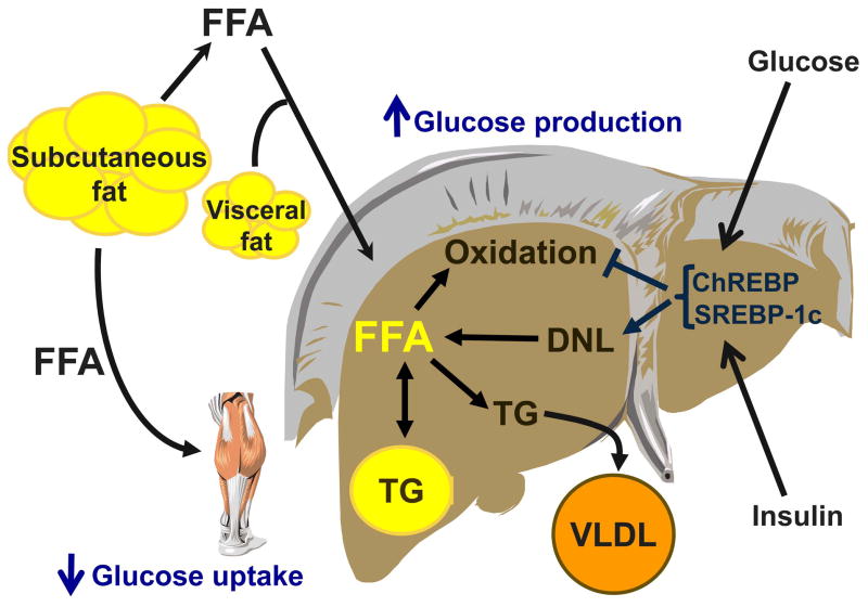
Steatosis develops when the rate of FA input (uptake and synthesis with subsequent esterification to TG) is greater than the rate of FA output (oxidation and secretion). Therefore, the amount of TG present in hepatocytes represents a complex interaction among: 1) hepatic fatty acid uptake, derived from plasma free fatty acid (FFA) released from hydrolysis of adipose tissue TG and FFA released from hydrolysis of circulating TG, 2) de novo fatty acid synthesis (de novo lipogenesis [DNL]), 3) fatty acid oxidation (FAO), and 4) fatty acid export within VLDL-TG (Figure 1).
Figure 1.
Physiological interrelationships among fatty acid metabolism, insulin resistance, dyslipidemia, and intrahepatic triglyceride content in nonalcoholic fatty liver disease (NAFLD). The rate of release of FFA from adipose tissue and delivery to the liver and skeletal muscle is increased in obese persons with NAFLD, which results in an increase in hepatic and muscle FFA uptake. In addition, intrahepatic de novo lipogenesis (DNL) of fatty acids is greater in subjects with NAFLD than in those with normal intrahepatic triglyceride (IHTG), which further contributes to the accumulation of intracellular fatty acids. The production and secretion of TG in VLDL is increased in subjects with NAFLD, which provides a mechanism for removing IHTG; however, the rate of secretion does not adequately compensate for the rate of TG production. Increased plasma glucose and insulin associated with NAFLD stimulate DNL and inhibit fatty acid oxidation, by affecting sterol regulatory element binding protein (SREBP-1c) and carbohydrate responsive element binding protein (ChREBP). These metabolic processes lead to an increase in intracellular fatty acids that are not oxidized or exported within VLDL-TG, and are esterified to TG and stored within lipid droplets. Certain lipid intermediates of fatty acid metabolism can impair insulin signaling and cause tissue insulin resistance. Therefore, this scheme illustrates how alterations in fatty acid metabolism can lead to an accumulation of intrahepatic (and intramuscular) TG, stimulate VLDL-TG secretion with subsequent hypertriglyceridemia, and cause insulin resistance in the liver and skeletal muscle.
Fatty acid uptake
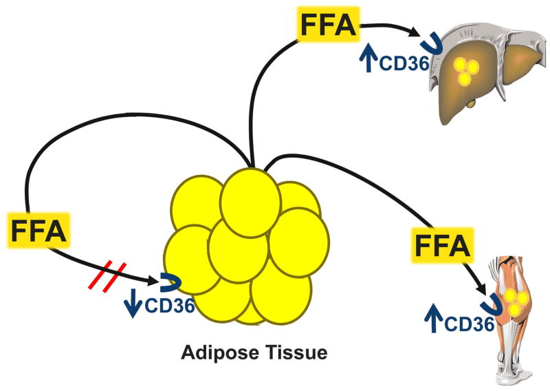
The rate of hepatic FFA uptake depends on the delivery of FFA to the liver and the liver’s capacity for FFA transport. During postabsorptive conditions, the major source of FFA delivered to the liver is derived from FFA released from subcutaneous adipose tissue, which enter the systemic circulation and are then transported to the liver by the hepatic artery and portal vein, after passage through splanchnic tissues. Although lipolysis of visceral adipose tissue TG releases additional FFA directly into the portal system, the relative contribution of portal vein FFA derived from visceral fat is small compared with FFA derived from subcutaneous fat; only about 5% and 20% of portal vein FFA originate from visceral fat in lean and obese subjects, respectively.18 We have found that the rate of FA release into the systemic circulation increases directly with increasing fat mass in both men and women, so that the rate of FFA release in relationship to fat-free mass is greater in obese than lean subjects.19 In addition, gene expression of hepatic lipase and hepatic lipoprotein lipase (LPL) are higher in obese subjects with NAFLD than subjects without NAFLD, suggesting that FFA released from lipolysis of circulating TG also contribute to hepatocellular FFA accumulation and steatosis.20,21 It is possible that these increases in hepatic lipase and hepatic LPL, along with higher postprandial lipemia and FFA concentrations reported in subjects with NAFLD,22 are responsible for the increased postprandial incorporation of dietary fatty acids into IHTG observed in obese subjects with T2DM.23 Membrane proteins that direct trafficking of FFA from plasma into tissues are also likely involved in increased hepatic FFA uptake. Gene expression and/or protein content of FAT/CD36, which is an important regulator of tissue FFA uptake from plasma, are increased in liver and skeletal muscle but decreased in adipose tissue in obese subjects with NAFLD compared with obese subjects who have normal IHTG content,24,25 suggesting that membrane fatty acid transport proteins redirect the uptake of plasma FFA from adipose tissue toward other tissues. Therefore, the summation of these data suggests that alterations in adipose tissue lipolytic activity, regional hepatic lipolysis of circulating TG, and tissue FFA transport proteins are involved in the pathogenesis of steatosis and ectopic fat accumulation (Figure 2).
Figure 2.
Alterations in cellular fatty acid transport facilitate ectopic fat accumulation in the liver and skeletal muscle. The fatty acid translocase, CD36, regulates tissue FFA uptake from plasma. CD36 expression and protein content is decreased in adipose tissue, but increased in the liver and skeletal muscle of insulin-resistant animals and human subjects who have increased intrahepatic and intramyocellular triglyceride content. These findings suggest that alterations in tissue fatty acid transport could be involved in the pathogenesis of ectopic triglyceride accumulation by redirecting plasma fatty acid uptake from adipose tissue toward other tissues.
De novo lipogenesis
The liver synthesizes fatty acids de novo through a complex cytosolic polymerization in which acetyl-CoA is converted to malonyl-CoA by acetyl-CoA carboxylase (ACC), and undergoes several cycles metabolic reactions to form one palmitate molecule. The rate of DNL is regulated by the fatty acid synthase (FAS) complex, ACC 1 and 2, diacylglycerol acyltransferase (DGAT) 1 and 2, stearoyl-CoA desaturase 1 (SCD1), and several nuclear transcription factors (SREBPs, ChREBP, liver X receptor α [LXRα], farnesoid X receptor [FXR], and peroxisome proliferator-activated receptors [PPARs]).26 Hepatic DNL is regulated independently by insulin and glucose, through the activation of SREBP-1c 27 and ChREBP,28 which transcriptionally activate nearly all genes involved in DNL. Data from studies conducted in mouse models demonstrate that hepatic overexpression of SREBP-1c or hyperinsulinemia stimulate lipogenesis and cause hepatic steatosis,29,30 whereas the levels of all the enzymes involved in DNL are reduced in ChREBP knockout mice.31 In humans, NAFLD is associated with increased hepatic expression of several genes involved in DNL.32,33
The contribution of DNL to total IHTG production in normal subjects is small and accounts for less than 5% of fatty acids incorporated into secreted VLDL-TG (~1–2 g/d) 34. However, the contribution of DNL to total IHTG production in subjects with NAFLD is much higher and accounts for 15%–23% of the fatty acids within IHTG and secreted in VLDL-TG.34,35 Moreover, data from a study that used sophisticated magnetic resonance spectroscopy techniques to evaluate postprandial glucose metabolism in vivo suggest that the increase in DNL precedes the development of NAFLD.36 Compared with insulin-sensitive subjects, consumption of a high-carbohydrate meal was associated with a much lower rate of muscle glycogen synthesis and a diversion of most of the ingested glucose toward hepatic DNL and IHTG synthesis in insulin-resistant subjects who had normal IHTG content. These data suggest that insulin resistance in skeletal muscle could promote IHTG accumulation by diverting ingested carbohydrate away from storage as muscle glycogen and toward de novo fatty acid synthesis.
Although hepatic DNL is a quantitatively minor pathway for TG synthesis, the rate of DNL might have important metabolic regulatory functions. For example, intrahepatic fatty acids that have been synthesized de novo activate PPARα to maintain glucose and lipid homeostasis.37 In addition, malonyl-CoA, the first intermediate of DNL, inhibits carnitine palmitoyltransferase 1(CPT1) activity, thereby preventing the entry of FFA into the mitochondrion and inhibiting FAO.38 The notion of potential allosteric inhibition of FAO by DNL is supported by data that found hepatic CPT-1 expression is decreased in subjects with NAFLD.33
Fatty acid oxidation
The complex metabolic processes performed by the liver require a considerable amount of energy; the metabolic rate of liver tissue (~0.28 kcal/g of tissue per day) is similar to that of the brain, and is nearly 20 times greater than the metabolic rate of resting skeletal muscle and 50 times greater than the metabolic rate of adipose tissue.39 Therefore, although the liver weighs only ~1.5 kg in adults, representing a small portion of total body weight (~2.5% in lean persons), it consumes ~450 kcal/d and accounts for ~20% of total resting energy expenditure.39 The mix of fuels used by the liver in vivo is difficult to quantify accurately because of the complicated exchange of metabolites between multiple biochemical pathways and technical limitations. It is estimated that fatty acid and amino acid oxidation provide ~90% of the fuel for basal hepatic energy requirements, and that the use of FFA as a fuel decreases during the fed state.40
The oxidation of intrahepatocellular fatty acid occurs primarily within mitochondria, and to a much lesser extent by peroxisomes and microsomes. Transport of FA inside the mitochondrial matrix is regulated by a carnitine-dependent enzyme shuttle, sequentially CPT1, carnitine translocase, and CPT2. Mitochondrial β-oxidation progressively shortens the fatty acyl-CoA by two carbon units at each cycle (released as acetyl-CoA), through a series of dehydrogenation, hydration, and cleavage reactions that involve a membrane-bound and soluble enzymes, which are transcriptionally regulated by PPAR-α.41 Acetyl-CoA derived from FAO can either enter the tricarboxylic acid cycle for complete oxidation and energy production for the liver, or can be condensed to form ketone bodies (acetoacetate and beta-hydroxybutyrate) which are exported to provide fuel for other tissues.38
Data from studies conducted in rodent models demonstrate that inhibition or activation of intrahepatic FAO can influence IHTG content. Genetic or experimentally-induced deficiencies in mitochondrial oxidative enzymes lead to hepatic steatosis,42,43 whereas increasing the expression or activity of hepatic enzymes involved in FAO, reduces IHTG accumulation.44–47 However, it is not known whether FAO is defective in human subjects with NAFLD, because there are currently no reliable methods for measuring hepatic FAO in vivo. Indirect measures of hepatic mitochondrial FAO, assessed by plasma ketone body concentrations, suggest that hepatic FAO is either increased or normal in subjects with NAFLD.48–51 In addition, although CPT1 expression is decreased, gene expression of other hepatic fatty acid oxidative enzymes are generally greater in subject with NAFLD than in those with normal IHTG content 24,33 In contrast, subjects with NAFLD have evidence of hepatic mitochondrial structural and functional abnormalities, including loss of mitochondrial cristae and paracrystalline inclusions,49,52 a decrease in mitochondrial respiratory chain activity,53 impaired ability to resynthesize ATP after a fructose challenge,54 and increased hepatic uncoupling protein 2,33 which affect energy production but not FAO. These abnormalities could represent an adaptive uncoupling of FAO and ATP production, which allows the liver to oxidize excessive FA substrates without generating unneeded ATP.
VLDL kinetics
Very-low-density lipoproteins are complex lipoprotein particles that are produced by the liver and secreted into the systemic circulation. The formation of VLDL provides an important mechanism for converting water-insoluble TG into a water-soluble form that can be exported from the liver and delivered to peripheral tissues. Hepatic VLDL assembly involves the fusion of a newly synthesized apolipoprotein B-100 (apoB-100) molecule with a TG droplet through the action of microsomal triglyceride transfer protein (MTP); each VLDL particle contains a single molecule of apoB-100. The fatty acids that are esterified into TG and secreted as VLDL are derived from several sources. In subjects with normal IHTG content, ~70% of FA incorporated into VLDL-TG originate from systemic plasma and the remaining are derived from several non-systemic FA sources, including hepatic DNL, lipolysis of IHTG, and lipolysis of visceral adipose tissue.55
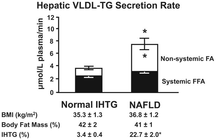
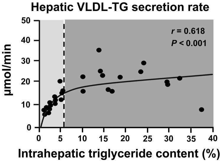
Intrahepatocellular fatty acids that are not oxidized are esterified to TG, which can either be incorporated into VLDL and secreted into the circulation or stored within the liver. Therefore, the secretion of VLDL provides a mechanism for reducing IHTG content. In fact, an impairment in hepatic VLDL secretion, caused by genetic defects, such as familial hypobetalipoproteinemia, 56 or pharmacological agents that inhibit MTP,57 are associated with an increase in IHTG content. However, data from most 58,59 but not all 34 studies have found that VLDL-TG secretion rate is greater in subjects with NAFLD than in those with normal IHTG content. We found that the rate of VLDL-TG secretion was twice as great in non-diabetic obese subjects with NAFLD than in those with normal IHTG content, who were matched on BMI and percent body fat (Figure 3). The increase in VLDL-TG secretion was almost entirely accounted for by a marked increase in the contribution of non-systemic FA, presumably derived from lipolysis of intrahepatic and visceral fat and DNL, to VLDL-TG secretion.59 In addition, the relationship between VLDL-TG secretion and IHTG content differed between the two groups; VLDL-TG secretion increased linearly with increasing IHTG content in subjects with normal IHTG, but appeared to reach a plateau in subjects with NAFLD, independent of IHTG content (Figure 4). Therefore, the increase in VLDL-TG secretion rate in subjects with NAFLD is not able to adequately compensate for the increased rate of IHTG production, so steatosis is maintained.
Figure 3.
Total VLDL-TG secretion rate (sum of grey and white bars) in subjects with normal and increased (nonalcoholic fatty liver disease [NAFLD]) intrahepatic triglyceride (IHTG) content, who were matched on BMI and percent body fat. White bars represent fatty acids in VLDL-TG that originated from systemic plasma free fatty acids, presumably derived primarily from lipolysis of subcutaneous fat, whereas black bars represent fatty acids in VLDL-TG that originated from non-systemic fatty acids, presumably derived primarily from lipolysis of intrahepatic and visceral fat and de novo lipogenesis. *Value significantly different from corresponding value in the Normal IHTG group, P < 0.05. (Adapted from: Fabbrini E, Mohammed BS, Magkos F, Korenblat KM, Patterson BW, Klein S. Alterations in adipose tissue and hepatic lipid kinetics in obese men and women with nonalcoholic fatty liver disease. Gastroenterology 2008;134:424–431).
Figure 4.
Relationship between VLDL-TG secretion rate and intrahepatic triglyceride content (IHTG) in subjects with normal IHTG (triglyceride content <5.6% of liver volume) and nonalcoholic fatty liver disease (NAFLD) (triglyceride content >10% of liver volume). (Adapted from: Fabbrini E, Mohammed BS, Magkos F, Korenblat KM, Patterson BW, Klein S. Alterations in adipose tissue and hepatic lipid kinetics in obese men and women with nonalcoholic fatty liver disease. Gastroenterology 2008;134:424–431).
The mechanism responsible for the inadequate increase in hepatic TG export is not known, but might be related to physical limitations in the liver’s ability to secrete large VLDL particles. In contrast to VLDL-TG kinetics, the secretion rate of VLDL-apoB-100 was not different between subjects with high and low IHTG content, so the molar ratio of VLDL-TG to VLDL-apoB-100 secretion rates, an index of the TG content of nascent VLDL, was more than two-fold greater in those with NAFLD.59 Data from a study conducted in transgenic mice that overexpress SREBP-1a and develop massive steatosis found that very large VLDL particles cannot be secreted from the liver because they exceed the diameter of the sinusoidal endothelial pores, resulting in an accumulation of IHTG.60 Therefore, the composite of these data suggest that the failure to upregulate VLDL-apoB secretion rate in obese subjects with NAFLD leads to the production of large VLDL particles, which cannot penetrate sinusoidal endothelial pores for export out of the liver.
Insulin sensitivity
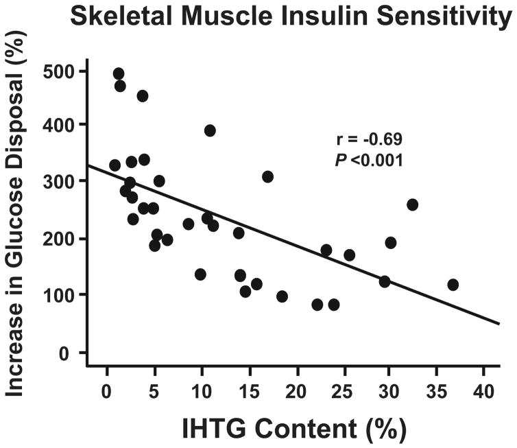
Insulin has important metabolic effects in multiple organ systems. Although the term “insulin resistance” is usually used to describe impaired insulin-mediated glucose uptake in skeletal muscle, insulin resistance associated with obesity and NAFLD also involves the liver (impaired insulin-mediated suppression of glucose production) and adipose tissue (impaired insulin-mediated suppression of lipolysis). The presence of steatosis is an important marker of multi-organ insulin resistance, independent of BMI, percent body fat, and visceral fat mass, 7,16,25,48,61 Moreover, insulin resistance in liver, adipose tissue and skeletal muscle is directly related to percent liver fat (Figure 5).7,48,49,61 However, it is not known whether NAFLD causes or is a consequence of insulin resistance, or possibly both.
Figure 5.
Relationship between intrahepatic triglyceride (IHTG) content and skeletal muscle insulin sensitivity, defined as the percent increase in the rate of glucose disposal in response to insulin infusion during a hyperinsulinemic-euglycemic clamp procedure. (Adapted from: Korenblat KM, Fabbrini E, Mohammed BS, Klein S. Liver, Muscle, and Adipose Tissue Insulin Action Is Directly Related to Intrahepatic Triglyceride Content in Obese Subjects. Gastroenterology 2008).
Fatty acid metabolism
Whole-body lipolytic rates, expressed as the rate of FFA release per unit of fat-free mass, is usually greater in obese than lean persons and is directly related with body fat mass.19 The presence of NAFLD in obese persons is associated with adipose tissue insulin resistance and even greater rates of adipose tissue lipolysis than in obese persons without NAFLD. 7,16,48,61 Excessive rates of release of FFA from adipose tissue into the circulation increases the delivery of FFA to the liver and skeletal muscle, which can simultaneously lead to an increase in IHTG and cause insulin resistance in liver and skeletal muscle.62 Skeletal muscle insulin resistance and hyperinsulinemia can further increase the accumulation of IHTG by stimulating hepatic DNL and TG synthesis. 36 An increase in IHTG content itself could be involved in the pathogenesis of hepatic insulin resistance by releasing FA into the cytoplasm, which can have adverse effects on insulin signaling.62
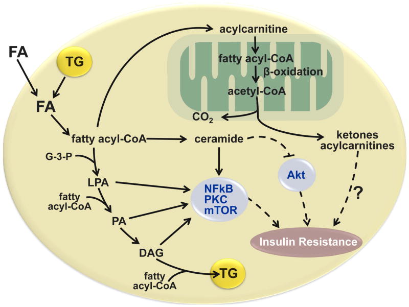
The cellular mechanism(s) responsible for fatty-acid induced insulin resistance in muscle and liver is not completely clear. A large volume of data from studies conducted in animal models and human subjects suggest that excessive intracellular lipid intermediates generated by fatty acid metabolism, particularly diacylglycerol (DAG), long chain fatty acyl-CoA, ceramide, lysophosphatidic acid, and phosphatidic acid, can interfere with insulin action by activating protein kinase C and mTOR, and inhibiting Akt, which have direct adverse effects on insulin signaling, and by activating the nuclear factor kinase B (NFκB) system which can cause insulin resistance through activation of inflammatory pathways (Figure 6).63,64 However, these conclusions are based primarily on studies that have simply demonstrated an association between these lipid intermediates and impaired insulin action, and not a cause-and-effect relationship. Moreover, the results from some studies have found that an increase in these lipid intermediates is not associated with insulin resistance.65–67 The ability to identify the cellular mediators responsible for FA-induced insulin resistance is further complicated by the possibility that the mechanism might not be the same among all tissues. Transgenic mice that overexpress muscle diacylglycerol acyltransferase (DGAT2), which catalyzes the final step of TG synthesis by adding fatty acyl-CoA to DAG, have high intramyocellular levels of DAG, long chain fatty acyl-CoA, and ceramide and have abnormal hepatic insulin sensitivity, impaired insulin signaling and insulin-mediated glucose uptake.68 In contrast, transgenic mice that overexpress hepatic DGAT2 have high intrahepatocellular levels of DAG, long chain fatty acyl-CoA, and ceramide, but do not have abnormal hepatic insulin sensitivity.69
Figure 6.
Potential cellular mechanisms responsible for the relationship between fatty acid metabolism and insulin resistance in the liver and skeletal muscle. Obese persons with nonalcoholic fatty liver disease have increased rates of adipose tissue lipolysis and fatty acid (FA) release into plasma and increased intrahepatic and intramyocellular triglyceride (TG) content. Intracellular FA delivered from plasma or derived from lipolysis of intracellular triglyceride (TG) can be transported to the mitochondria for oxidation, esterified to TG or partially metabolized to several lipid intermediates, long chain fatty acyl-CoA, ceramide, lysophosphatidic acid (LPA), and phosphatidic acid (PA), and diacylglycerol (DAG). These lipid intermediates can interfere with insulin signaling by activating protein kinase C (PKC), mammalian target of rapamycin (mTOR), and nuclear factor kinase B (NFκB), and inhibiting Akt (also known as protein kinase B). The oxidation of intracellular FAs involve the conversion of long-chain fatty acyl-CoAs to acylcarnitines, which enter the mitochondria, and are progressively shortened by β-oxidation, which produces acetyl-CoA that can enter the tricarboxylic acid cycle for complete oxidation. The incomplete oxidation of fatty acyl-CoA generates ketone bodies and acylcarnitines, which might also have adverse effects on insulin action. (Adapted from: Schenk S, Saberi M, Olefsky JM. Insulin sensitivity: modulation by nutrients and inflammation. J Clin Invest 2008;118:2992–3002, and Nagle CA, Klett EL, Coleman RA. Hepatic triacylglycerol accumulation and insulin resistance. J Lipid Res 2009;50 Suppl:S74–79).
Adipose tissue inflammation
Adipose tissue contains several different cell types, including adipocytes and macrophages that produce cytokines (e.g. IL-6 and TNF-α), and chemokines (e.g. CCL2 [also known as monocyte chemoattractant protein 1]), which can cause inflammation and insulin resistance.70 Adipose tissue macrophage content and production of cytokines and chemokines are greater in obese than lean subjects.71 Moreover, macrophage infiltration and inflammatory markers are greater in adipose tissue of subjects with NAFLD than BMI-matched subjects with normal IHTG content.72 Therefore, the secretion of adipose tissue inflammatory proteins in obese persons with NAFLD is likely involved in the pathogenesis of insulin resistance, but the relative contribution of adipose tissue inflammation in comparison with other potential factors that can cause insulin resistance is not known.
Intrahepatic inflammation
Diet-induced and genetically-induced obesity in rodent models cause steatosis, insulin resistance, and increased hepatic NF-κB activity.73,74 In addition, selective activation of hepatocellular NF-κB causes hepatic inflammation without steatosis, and results in both hepatic and skeletal muscle insulin resistance.74 These animals have increased hepatocyte expression of IL-6 and plasma IL-6 concentrations, suppression of IL-6 activity by administering neutralizing IL-6 antibodies resulted in a decrease in both hepatic and peripheral insulin resistance. These data suggest that steatosis can cause both hepatic and systemic insulin resistance by activating NF-κB, which upregulates the production of proinflammatory cytokines that affect both local and systemic insulin action.
Adipocyte-derived hormones
Adipocytes produce a series of peptide hormones, which are associated with insulin action (e.g. resistin, retinol-binding protein 4, adiponectin, leptin). Among these proteins, adiponectin, which is the most abundant secretory protein produced by adipose tissue, is the most closely related with insulin action. Plasma adiponectin concentrations are inversely associated with hepatic steatosis,22,75 insulin resistance,76 T2DM,77 and the metabolic syndrome. Delivery of recombinant adiponectin into mice with liver steatosis markedly reduced hepatomegaly and IHTG content.44
Endoplasmic reticulum stress
The endoplasmic reticulum (ER) is a critical intracellular organelle that coordinates synthesis, folding and trafficking of proteins. Transmembrane and secreted proteins are folded into the ER and then directed to cellular destinations. Unfolded or misfolded proteins are detected, removed from the ER and degraded by proteasome system.78 Under stress conditions, such as hypoxia, alterations in energy and substrates, toxins, viral infections, unfolded proteins accumulate in the ER and initiate an adaptive response known as the unfolded protein response (UPR) in an effort to restore organelle function.79 The UPR is initiated by three ER transmembrane sensors, PKR-like endoplasmic-reticulum kinase (PERK), inositol-requiring enzyme 1 (IRE-1), and activating transcription factor 6 (ATF6). These transmembrane sensors activate an adaptive response that results in cessation of protein synthesis, increase of protein-folding chaperones, and increase in ER-associated degradation genes. The UPR is also able to induce activation of the c-Jun NH2-terminal kinase (JNK) pathway and thereby inhibit insulin signaling through the subsequent phosphorylation and/or degradation of IRS1.80 Recent data from experimental models indicate that ER stress is critical to the initiation and integration of pathways of inflammation and insulin action in obesity, T2DM, and NAFLD. ER stress response can be induced in the liver by saturated FA in rats,81 and this activation can lead to activation of JNK and insulin resistance.80 Activation of ER stress in the liver has also been shown in human subjects with NAFLD, as documented by activation of PERK and an increase in the ER chaperone GRP78 mRNA.82 Data from a study conducted in extremely obese patients, found that ER stress is associated with NAFLD and improves with weight loss and resolution of steatosis. Bariatric surgery-induced weight loss increased insulin sensitivity in multiple organs and decreased IHTG content and both liver and adipose tissue activation of all three ER stress pathways.83
Hepatic steatosis in the absence of insulin resistance
The complexity of the relationship between NAFLD and insulin resistance is underscored by the observation that steatosis is not always associated with insulin resistance. A dissociation between steatosis and insulin resistance has been reported in selected genetically-altered or pharmacologically-manipulated animal models and human subjects. Overexpression of hepatic DGAT,69 blockade of hepatic VLDL secretion,66 and pharmacological blockade of β-oxidation84 in mice causes hepatic steatosis, but not hepatic or skeletal muscle insulin resistance, whereas inhibiting hepatocyte TG synthesis in obese mice decreases hepatic steatosis but does not improve insulin sensitivity.85 In addition, hepatic steatosis caused by genetic deficiency of apoB synthesis and decreased VLDL hepatic secretion in patients with familial hypobetalipoproteinemia is not accompanied by hepatic or peripheral insulin resistance.(S. Klein unpublished observations). These data support the notion that hepatic accumulation of TG does not necessarily cause insulin resistance. In fact, it is possible that the esterification of excessive FA to inert TG molecules protects the hepatocyte by preventing the accumulation of potentially toxic intracellular fatty acids.86 Inhibiting hepatocyte TG synthesis by treatment with DGAT2 antisense oligonucleotide in obese mice decreased hepatic steatosis, but increased hepatic free FA, markers of lipid peroxidation/oxidant stress, lobular necroinflammation, and fibrosis.87 The mechanism(s) responsible for the marked differences in the relationship between hepatic steatosis and insulin resistance across different studies is not known, but suggest that other factors associated with steatosis, such as inflammation, circulating adipokines, ER stress, or as yet unidentified lipid metabolites, affect insulin sensitivity but are not necessarily directly related with IHTG content. It is also possible that there is a temporal dissociation between steatosis and insulin resistance, so that IHTG accumulation is secondary to a primary defect in skeletal muscle insulin action, by diverting ingested carbohydrates away from muscle glycogen storage to DNL.36
ENERGY BALANCE AND NAFLD
Calorie restriction and weight loss is an effective therapy for obese patients with NAFLD. A marked decrease in IHTG content and improvement in hepatic insulin sensitivity occurs very rapidly, within 48 h, of calorie restriction (~1100 kcal/d diet).88 A comprehensive review of 14 studies that evaluated the effect of lifestyle weight loss therapy on NAFLD/NASH,89 and data from recent prospective diet intervention studies,88,90 found that a 5–10% weight loss improved liver biochemistries, liver histology (steatosis and inflammation) and IHTG content, in conjunction with an increase in hepatic and skeletal muscle insulin sensitivity, and decrease in hepatic VLDL-TG secretion rate.91 Bariatric surgery is the most effective available weight loss therapy. There has been concern that the large and rapid weight loss, induced by bariatric surgery, can actually worsen NAFLD by increasing hepatic inflammation and fibrosis.92 However, data from more recent surgical series suggest that weight loss induced by bariatric surgery decreases steatosis, inflammation and fibrosis.93,94 In addition, bariatric surgery induced weight loss has considerable beneficial metabolic effects in the liver manifested by a decrease in: 1) hepatic glucose production, 2) hepatic VLDL-triglyceride secretion rate, and 3) hepatic gene expression of factors that regulate hepatic inflammation and fibrogenesis.95 These data suggest that bariatric surgery-induced weight loss is an effective therapy for NAFLD in patients with morbid obesity, by normalizing the metabolic abnormalities involved in the pathogenesis and pathophysiology of NAFLD, and by preventing the progression of hepatic inflammation and fibrosis.
The effect of overfeeding on IHTG content and metabolic function has not been carefully studied in human subjects. Data from a study conducted in rodents suggest that overfeeding first has metabolic effect on the liver followed by an effect on muscle; insulin resistance in liver was observed after 3 d and in muscle after 7 days of overfeeding.96 Four wks of overfeeding in lean men and women, designed to cause a 5%-15% increase in body weight, resulted in a significant increase in IHTG content (from 1.1% to 2.8%), a decline in insulin sensitivity, and an increase in serum transaminase concentrations.97
CONCLUSIONS
Although obesity is associated with multiple metabolic risk factors for cardiovascular disease, including insulin resistance, diabetes, and dyslipidemia, about 30% of obese adults are “metabolically normal”, usually defined by some measure of insulin sensitivity or having ≤1 cardiometabolic abnormality.98–100 Excessive intrahepatic triglyceride (IHTG) content in obese persons is a robust marker of metabolic abnormalities (insulin resistance in liver, muscle and adipose tissue, alterations in FFA metabolism, and increased VLDL-TG secretion rate), independent of BMI, percent body fat, and visceral fat mass. Conversely, obese persons who have normal IHTG content appear to be resistant to developing obesity-related metabolic complications. However, it is not known whether NAFLD is a cause or a consequence of metabolic dysfunction. A better understanding of the mechanisms responsible for the pathogenesis and pathophysiology of NAFLD will potentially identify both novel biomarkers for metabolic risk and unique targets for therapeutic intervention.
Acknowledgments
This publication was made possible by National Institutes of Health grants UL1 RR024992 (Clinical and Translational Science Award), DK 56341 (Clinical Nutrition Research Unit), and DK 37948.
References
- 1.Marchesini G, Bugianesi E, Forlani G, Cerrelli F, Lenzi M, Manini R, et al. Nonalcoholic fatty liver, steatohepatitis, and the metabolic syndrome. Hepatology. 2003;37:917–923. doi: 10.1053/jhep.2003.50161. [DOI] [PubMed] [Google Scholar]
- 2.Adams LA, Lymp JF, St Sauver J, Sanderson SO, Lindor KD, Feldstein A, et al. The natural history of nonalcoholic fatty liver disease: a population-based cohort study. Gastroenterology. 2005;129:113–121. doi: 10.1053/j.gastro.2005.04.014. [DOI] [PubMed] [Google Scholar]
- 3.Hoyumpa AM, Jr, Greene HL, Dunn GD, Schenker S. Fatty liver: biochemical and clinical considerations. Am J Dig Dis. 1975;20:1142–1170. doi: 10.1007/BF01070758. [DOI] [PubMed] [Google Scholar]
- 4.Kleiner DE, Brunt EM, Van Natta M, Behling C, Contos MJ, Cummings OW, et al. Design and validation of a histological scoring system for nonalcoholic fatty liver disease. Hepatology. 2005;41:1313–1321. doi: 10.1002/hep.20701. [DOI] [PubMed] [Google Scholar]
- 5.Petersen KF, Dufour S, Feng J, Befroy D, Dziura J, Dalla Man C, et al. Increased prevalence of insulin resistance and nonalcoholic fatty liver disease in Asian-Indian men. Proc Natl Acad Sci U S A. 2006;103:18273–18277. doi: 10.1073/pnas.0608537103. [DOI] [PMC free article] [PubMed] [Google Scholar]
- 6.Szczepaniak LS, Nurenberg P, Leonard D, Browning JD, Reingold JS, Grundy S, et al. Magnetic resonance spectroscopy to measure hepatic triglyceride content: prevalence of hepatic steatosis in the general population. Am J Physiol Endocrinol Metab. 2005;288:E462–468. doi: 10.1152/ajpendo.00064.2004. [DOI] [PubMed] [Google Scholar]
- 7.Korenblat KM, Fabbrini E, Mohammed BS, Klein S. Liver, Muscle, and Adipose Tissue Insulin Action Is Directly Related to Intrahepatic Triglyceride Content in Obese Subjects. Gastroenterology. 2008 doi: 10.1053/j.gastro.2008.01.07. [DOI] [PMC free article] [PubMed] [Google Scholar]
- 8.Ruhl CE, Everhart JE. Determinants of the association of overweight with elevated serum alanine aminotransferase activity in the United States. Gastroenterology. 2003;124:71–79. doi: 10.1053/gast.2003.50004. [DOI] [PubMed] [Google Scholar]
- 9.Marcos A, Fisher RA, Ham JM, Olzinski AT, Shiffman ML, Sanyal AJ, et al. Selection and outcome of living donors for adult to adult right lobe transplantation. Transplantation. 2000;69:2410–2415. doi: 10.1097/00007890-200006150-00034. [DOI] [PubMed] [Google Scholar]
- 10.Hilden M, Christoffersen P, Juhl E, Dalgaard JB. Liver histology in a ‘normal’ population--examinations of 503 consecutive fatal traffic casualties. Scand J Gastroenterol. 1977;12:593–597. doi: 10.3109/00365527709181339. [DOI] [PubMed] [Google Scholar]
- 11.Lee RG. Nonalcoholic steatohepatitis: a study of 49 patients. Hum Pathol. 1989;20:594–598. doi: 10.1016/0046-8177(89)90249-9. [DOI] [PubMed] [Google Scholar]
- 12.Gholam PM, Kotler DP, Flancbaum LJ. Liver pathology in morbidly obese patients undergoing Roux-en-Y gastric bypass surgery. Obes Surg. 2002;12:49–51. doi: 10.1381/096089202321144577. [DOI] [PubMed] [Google Scholar]
- 13.Romeo S, Kozlitina J, Xing C, Pertsemlidis A, Cox D, Pennacchio LA, et al. Genetic variation in PNPLA3 confers susceptibility to nonalcoholic fatty liver disease. Nat Genet. 2008;40:1461–1465. doi: 10.1038/ng.257. [DOI] [PMC free article] [PubMed] [Google Scholar]
- 14.Browning JD, Szczepaniak LS, Dobbins R, Nuremberg P, Horton JD, Cohen JC, et al. Prevalence of hepatic steatosis in an urban population in the United States: impact of ethnicity. Hepatology. 2004;40:1387–1395. doi: 10.1002/hep.20466. [DOI] [PubMed] [Google Scholar]
- 15.Sase S, Monden M, Oka H, Dono K, Fukuta T, Shibata I. Hepatic blood flow measurements with arterial and portal blood flow mapping in the human liver by means of xenon CT. J Comput Assist Tomogr. 2002;26:243–249. doi: 10.1097/00004728-200203000-00014. [DOI] [PubMed] [Google Scholar]
- 16.Vega GL, Chandalia M, Szczepaniak LS, Grundy SM. Metabolic correlates of nonalcoholic fatty liver in women and men. Hepatology. 2007;46:716–722. doi: 10.1002/hep.21727. [DOI] [PubMed] [Google Scholar]
- 17.Hwang JH, Stein DT, Barzilai N, Cui MH, Tonelli J, Kishore P, et al. Increased intrahepatic triglyceride is associated with peripheral insulin resistance: in vivo MR imaging and spectroscopy studies. Am J Physiol Endocrinol Metab. 2007;293:E1663–1669. doi: 10.1152/ajpendo.00590.2006. [DOI] [PubMed] [Google Scholar]
- 18.Nielsen S, Guo Z, Johnson CM, Hensrud DD, Jensen MD. Splanchnic lipolysis in human obesity. J Clin Invest. 2004;113:1582–1588. doi: 10.1172/JCI21047. [DOI] [PMC free article] [PubMed] [Google Scholar]
- 19.Mittendorfer B, Magkos F, Fabbrini E, Mohammed BS, Klein S. Relationship between body fat mass and free fatty acid kinetics in men and women. Obesity. 2009 doi: 10.1038/oby.2009.224. (in press) [DOI] [PMC free article] [PubMed] [Google Scholar]
- 20.Pardina E, Baena-Fustegueras JA, Catalan R, Galard R, Lecube A, Fort JM, et al. Increased Expression and Activity of Hepatic Lipase in the Liver of Morbidly Obese Adult Patients in Relation to Lipid Content. Obes Surg. 2008 doi: 10.1007/s11695-008-9739-9. [DOI] [PubMed] [Google Scholar]
- 21.Westerbacka J, Kolak M, Kiviluoto T, Arkkila P, Siren J, Hamsten A, et al. Genes involved in fatty acid partitioning and binding, lipolysis, monocyte/macrophage recruitment, and inflammation are overexpressed in the human fatty liver of insulin-resistant subjects. Diabetes. 2007;56:2759–2765. doi: 10.2337/db07-0156. [DOI] [PubMed] [Google Scholar]
- 22.Musso G, Gambino R, Durazzo M, Biroli G, Carello M, Faga E, et al. Adipokines in NASH: postprandial lipid metabolism as a link between adiponectin and liver disease. Hepatology. 2005;42:1175–1183. doi: 10.1002/hep.20896. [DOI] [PubMed] [Google Scholar]
- 23.Ravikumar B, Carey PE, Snaar JE, Deelchand DK, Cook DB, Neely RD, et al. Real-time assessment of postprandial fat storage in liver and skeletal muscle in health and type 2 diabetes. Am J Physiol Endocrinol Metab. 2005;288:E789–797. doi: 10.1152/ajpendo.00557.2004. [DOI] [PubMed] [Google Scholar]
- 24.Greco D, Kotronen A, Westerbacka J, Puig O, Arkkila P, Kiviluoto T, et al. Gene expression in human NAFLD. Am J Physiol Gastrointest Liver Physiol. 2008;294:G1281–1287. doi: 10.1152/ajpgi.00074.2008. [DOI] [PubMed] [Google Scholar]
- 25.Fabbrini EMF, Mohammed BS, Pietka T, Abumrad NA, Patterson BW, Okunade A, Klein S. Intrahepatic fat, not visceral fat, is linked with metabolic complications of obesity. Proc Natl Acad Sci U S A. 2009 doi: 10.1073/pnas.0904944106. Published online before print August 24, 2009. [DOI] [PMC free article] [PubMed] [Google Scholar]
- 26.Musso G, Gambino R, Cassader M. Recent insights into hepatic lipid metabolism in non- alcoholic fatty liver disease (NAFLD) Prog Lipid Res. 2009;48:1–26. doi: 10.1016/j.plipres.2008.08.001. [DOI] [PubMed] [Google Scholar]
- 27.Shimomura I, Bashmakov Y, Ikemoto S, Horton JD, Brown MS, Goldstein JL. Insulin selectively increases SREBP-1c mRNA in the livers of rats with streptozotocin-induced diabetes. Proc Natl Acad Sci U S A. 1999;96:13656–13661. doi: 10.1073/pnas.96.24.13656. [DOI] [PMC free article] [PubMed] [Google Scholar]
- 28.Yamashita H, Takenoshita M, Sakurai M, Bruick RK, Henzel WJ, Shillinglaw W, et al. A glucose-responsive transcription factor that regulates carbohydrate metabolism in the liver. Proc Natl Acad Sci U S A. 2001;98:9116–9121. doi: 10.1073/pnas.161284298. [DOI] [PMC free article] [PubMed] [Google Scholar]
- 29.Shimano H, Horton JD, Shimomura I, Hammer RE, Brown MS, Goldstein JL. Isoform 1c of sterol regulatory element binding protein is less active than isoform 1a in livers of transgenic mice and in cultured cells. J Clin Invest. 1997;99:846–854. doi: 10.1172/JCI119248. [DOI] [PMC free article] [PubMed] [Google Scholar]
- 30.Shimomura I, Bashmakov Y, Horton JD. Increased levels of nuclear SREBP-1c associated with fatty livers in two mouse models of diabetes mellitus. J Biol Chem. 1999;274:30028–30032. doi: 10.1074/jbc.274.42.30028. [DOI] [PubMed] [Google Scholar]
- 31.Iizuka K, Bruick RK, Liang G, Horton JD, Uyeda K. Deficiency of carbohydrate response element-binding protein (ChREBP) reduces lipogenesis as well as glycolysis. Proc Natl Acad Sci U S A. 2004;101:7281–7286. doi: 10.1073/pnas.0401516101. [DOI] [PMC free article] [PubMed] [Google Scholar]
- 32.Mitsuyoshi H, Yasui K, Harano Y, Endo M, Tsuji K, Minami M, et al. Analysis of hepatic genes involved in the metabolism of fatty acids and iron in nonalcoholic fatty liver disease. Hepatol Res. 2009;39:366–373. doi: 10.1111/j.1872-034X.2008.00464.x. [DOI] [PubMed] [Google Scholar]
- 33.Kohjima M, Enjoji M, Higuchi N, Kato M, Kotoh K, Yoshimoto T, et al. Re-evaluation of fatty acid metabolism-related gene expression in nonalcoholic fatty liver disease. Int J Mol Med. 2007;20:351–358. [PubMed] [Google Scholar]
- 34.Diraison F, Moulin P, Beylot M. Contribution of hepatic de novo lipogenesis and reesterification of plasma non esterified fatty acids to plasma triglyceride synthesis during non-alcoholic fatty liver disease. Diabetes Metab. 2003;29:478–485. doi: 10.1016/s1262-3636(07)70061-7. [DOI] [PubMed] [Google Scholar]
- 35.Donnelly KL, Smith CI, Schwarzenberg SJ, Jessurun J, Boldt MD, Parks EJ. Sources of fatty acids stored in liver and secreted via lipoproteins in patients with nonalcoholic fatty liver disease. J Clin Invest. 2005;115:1343–1351. doi: 10.1172/JCI23621. [DOI] [PMC free article] [PubMed] [Google Scholar]
- 36.Petersen KF, Dufour S, Savage DB, Bilz S, Solomon G, Yonemitsu S, et al. The role of skeletal muscle insulin resistance in the pathogenesis of the metabolic syndrome. Proc Natl Acad Sci U S A. 2007;104:12587–12594. doi: 10.1073/pnas.0705408104. [DOI] [PMC free article] [PubMed] [Google Scholar]
- 37.Chakravarthy MV, Pan Z, Zhu Y, Tordjman K, Schneider JG, Coleman T, et al. “New” hepatic fat activates PPARalpha to maintain glucose, lipid, and cholesterol homeostasis. Cell Metab. 2005;1:309–322. doi: 10.1016/j.cmet.2005.04.002. [DOI] [PubMed] [Google Scholar]
- 38.McGarry JD, Foster DW. Regulation of hepatic fatty acid oxidation and ketone body production. Annu Rev Biochem. 1980;49:395–420. doi: 10.1146/annurev.bi.49.070180.002143. [DOI] [PubMed] [Google Scholar]
- 39.Klein S, Jeejeebhoy K. The malnourished patient: nutritional assessment and management. In: Feldman M, Freidman L, Sleisenger M, editors. Sleisenger & Fordtran’s Gastrointestinal and Liver Disease. 7. Philadelphia: W.B. Saunders Co; 2002. pp. 265–285. [Google Scholar]
- 40.Muller MJ. Hepatic fuel selection. Proc Nutr Soc. 1995;54:139–150. doi: 10.1079/pns19950043. [DOI] [PubMed] [Google Scholar]
- 41.Desvergne B, Wahli W. Peroxisome proliferator-activated receptors: nuclear control of metabolism. Endocr Rev. 1999;20:649–688. doi: 10.1210/edrv.20.5.0380. [DOI] [PubMed] [Google Scholar]
- 42.Zhang D, Liu ZX, Choi CS, Tian L, Kibbey R, Dong J, et al. Mitochondrial dysfunction due to long-chain Acyl-CoA dehydrogenase deficiency causes hepatic steatosis and hepatic insulin resistance. Proc Natl Acad Sci U S A. 2007;104:17075–17080. doi: 10.1073/pnas.0707060104. [DOI] [PMC free article] [PubMed] [Google Scholar]
- 43.Ibdah JA, Perlegas P, Zhao Y, Angdisen J, Borgerink H, Shadoan MK, et al. Mice heterozygous for a defect in mitochondrial trifunctional protein develop hepatic steatosis and insulin resistance. Gastroenterology. 2005;128:1381–1390. doi: 10.1053/j.gastro.2005.02.001. [DOI] [PubMed] [Google Scholar]
- 44.Xu A, Wang Y, Keshaw H, Xu LY, Lam KS, Cooper GJ. The fat-derived hormone adiponectin alleviates alcoholic and nonalcoholic fatty liver diseases in mice. J Clin Invest. 2003;112:91–100. doi: 10.1172/JCI17797. [DOI] [PMC free article] [PubMed] [Google Scholar]
- 45.Seo YS, Kim JH, Jo NY, Choi KM, Baik SH, Park JJ, et al. PPAR agonists treatment is effective in a nonalcoholic fatty liver disease animal model by modulating fatty-acid metabolic enzymes. J Gastroenterol Hepatol. 2008;23:102–109. doi: 10.1111/j.1440-1746.2006.04819.x. [DOI] [PubMed] [Google Scholar]
- 46.Stefanovic-Racic M, Perdomo G, Mantell BS, Sipula IJ, Brown NF, O’Doherty RM. A moderate increase in carnitine palmitoyltransferase 1a activity is sufficient to substantially reduce hepatic triglyceride levels. Am J Physiol Endocrinol Metab. 2008;294:E969–977. doi: 10.1152/ajpendo.00497.2007. [DOI] [PubMed] [Google Scholar]
- 47.Savage DB, Choi CS, Samuel VT, Liu ZX, Zhang D, Wang A, et al. Reversal of diet-induced hepatic steatosis and hepatic insulin resistance by antisense oligonucleotide inhibitors of acetyl-CoA carboxylases 1 and 2. J Clin Invest. 2006;116:817–824. doi: 10.1172/JCI27300. [DOI] [PMC free article] [PubMed] [Google Scholar]
- 48.Bugianesi E, Gastaldelli A, Vanni E, Gambino R, Cassader M, Baldi S, et al. Insulin resistance in non-diabetic patients with non-alcoholic fatty liver disease: sites and mechanisms. Diabetologia. 2005;48:634–642. doi: 10.1007/s00125-005-1682-x. [DOI] [PubMed] [Google Scholar]
- 49.Sanyal AJ, Campbell-Sargent C, Mirshahi F, Rizzo WB, Contos MJ, Sterling RK, et al. Nonalcoholic steatohepatitis: association of insulin resistance and mitochondrial abnormalities. Gastroenterology. 2001;120:1183–1192. doi: 10.1053/gast.2001.23256. [DOI] [PubMed] [Google Scholar]
- 50.Chalasani N, Gorski JC, Asghar MS, Asghar A, Foresman B, Hall SD, et al. Hepatic cytochrome P450 2E1 activity in nondiabetic patients with nonalcoholic steatohepatitis. Hepatology. 2003;37:544–550. doi: 10.1053/jhep.2003.50095. [DOI] [PubMed] [Google Scholar]
- 51.Kotronen A, Seppala-Lindroos A, Vehkavaara S, Bergholm R, Frayn KN, Fielding BA, et al. Liver fat and lipid oxidation in humans. Liver Int. 2009 doi: 10.1111/j.1478-3231.2009.02076.x. [DOI] [PubMed] [Google Scholar]
- 52.Caldwell SH, Swerdlow RH, Khan EM, Iezzoni JC, Hespenheide EE, Parks JK, et al. Mitochondrial abnormalities in non-alcoholic steatohepatitis. J Hepatol. 1999;31:430–434. doi: 10.1016/s0168-8278(99)80033-6. [DOI] [PubMed] [Google Scholar]
- 53.Perez-Carreras M, Del Hoyo P, Martin MA, Rubio JC, Martin A, Castellano G, et al. Defective hepatic mitochondrial respiratory chain in patients with nonalcoholic steatohepatitis. Hepatology. 2003;38:999–1007. doi: 10.1053/jhep.2003.50398. [DOI] [PubMed] [Google Scholar]
- 54.Cortez-Pinto H, Chatham J, Chacko VP, Arnold C, Rashid A, Diehl AM. Alterations in liver ATP homeostasis in human nonalcoholic steatohepatitis: a pilot study. JAMA. 1999;282:1659–1664. doi: 10.1001/jama.282.17.1659. [DOI] [PubMed] [Google Scholar]
- 55.Lewis GF. Fatty acid regulation of very low density lipoprotein production. Curr Opin Lipidol. 1997;8:146–153. doi: 10.1097/00041433-199706000-00004. [DOI] [PubMed] [Google Scholar]
- 56.Schonfeld G, Patterson BW, Yablonskiy DA, Tanoli TS, Averna M, Elias N, et al. Fatty liver in familial hypobetalipoproteinemia: triglyceride assembly into VLDL particles is affected by the extent of hepatic steatosis. J Lipid Res. 2003;44:470–478. doi: 10.1194/jlr.M200342-JLR200. [DOI] [PubMed] [Google Scholar]
- 57.Cuchel M, Bloedon LT, Szapary PO, Kolansky DM, Wolfe ML, Sarkis A, et al. Inhibition of microsomal triglyceride transfer protein in familial hypercholesterolemia. N Engl J Med. 2007;356:148–156. doi: 10.1056/NEJMoa061189. [DOI] [PubMed] [Google Scholar]
- 58.Adiels M, Taskinen MR, Packard C, Caslake MJ, Soro-Paavonen A, Westerbacka J, et al. Overproduction of large VLDL particles is driven by increased liver fat content in man. Diabetologia. 2006;49:755–765. doi: 10.1007/s00125-005-0125-z. [DOI] [PubMed] [Google Scholar]
- 59.Fabbrini E, Mohammed BS, Magkos F, Korenblat KM, Patterson BW, Klein S. Alterations in adipose tissue and hepatic lipid kinetics in obese men and women with nonalcoholic fatty liver disease. Gastroenterology. 2008;134:424–431. doi: 10.1053/j.gastro.2007.11.038. [DOI] [PMC free article] [PubMed] [Google Scholar]
- 60.Horton JD, Shimano H, Hamilton RL, Brown MS, Goldstein JL. Disruption of LDL receptor gene in transgenic SREBP-1a mice unmasks hyperlipidemia resulting from production of lipid-rich VLDL. J Clin Invest. 1999;103:1067–1076. doi: 10.1172/JCI6246. [DOI] [PMC free article] [PubMed] [Google Scholar]
- 61.Seppala-Lindroos A, Vehkavaara S, Hakkinen AM, Goto T, Westerbacka J, Sovijarvi A, et al. Fat accumulation in the liver is associated with defects in insulin suppression of glucose production and serum free fatty acids independent of obesity in normal men. J Clin Endocrinol Metab. 2002;87:3023–3028. doi: 10.1210/jcem.87.7.8638. [DOI] [PubMed] [Google Scholar]
- 62.Boden G, Shulman GI. Free fatty acids in obesity and type 2 diabetes: defining their role in the development of insulin resistance and beta-cell dysfunction. Eur J Clin Invest. 2002;32 (Suppl 3):14–23. doi: 10.1046/j.1365-2362.32.s3.3.x. [DOI] [PubMed] [Google Scholar]
- 63.Nagle CA, An J, Shiota M, Torres TP, Cline GW, Liu ZX, et al. Hepatic overexpression of glycerol-sn-3-phosphate acyltransferase 1 in rats causes insulin resistance. J Biol Chem. 2007;282:14807–14815. doi: 10.1074/jbc.M611550200. [DOI] [PMC free article] [PubMed] [Google Scholar]
- 64.Schenk S, Saberi M, Olefsky JM. Insulin sensitivity: modulation by nutrients and inflammation. J Clin Invest. 2008;118:2992–3002. doi: 10.1172/JCI34260. [DOI] [PMC free article] [PubMed] [Google Scholar]
- 65.Skovbro M, Baranowski M, Skov-Jensen C, Flint A, Dela F, Gorski J, et al. Human skeletal muscle ceramide content is not a major factor in muscle insulin sensitivity. Diabetologia. 2008;51:1253–1260. doi: 10.1007/s00125-008-1014-z. [DOI] [PubMed] [Google Scholar]
- 66.Minehira K, Young SG, Villanueva CJ, Yetukuri L, Oresic M, Hellerstein MK, et al. Blocking VLDL secretion causes hepatic steatosis but does not affect peripheral lipid stores or insulin sensitivity in mice. J Lipid Res. 2008;49:2038–2044. doi: 10.1194/jlr.M800248-JLR200. [DOI] [PMC free article] [PubMed] [Google Scholar]
- 67.An J, Muoio DM, Shiota M, Fujimoto Y, Cline GW, Shulman GI, et al. Hepatic expression of malonyl-CoA decarboxylase reverses muscle, liver and whole-animal insulin resistance. Nat Med. 2004;10:268–274. doi: 10.1038/nm995. [DOI] [PubMed] [Google Scholar]
- 68.Levin MC, Monetti M, Watt MJ, Sajan MP, Stevens RD, Bain JR, et al. Increased lipid accumulation and insulin resistance in transgenic mice expressing DGAT2 in glycolytic (type II) muscle. Am J Physiol Endocrinol Metab. 2007;293:E1772–1781. doi: 10.1152/ajpendo.00158.2007. [DOI] [PubMed] [Google Scholar]
- 69.Monetti M, Levin MC, Watt MJ, Sajan MP, Marmor S, Hubbard BK, et al. Dissociation of hepatic steatosis and insulin resistance in mice overexpressing DGAT in the liver. Cell Metab. 2007;6:69–78. doi: 10.1016/j.cmet.2007.05.005. [DOI] [PubMed] [Google Scholar]
- 70.Shoelson SE, Herrero L, Naaz A. Obesity, inflammation, and insulin resistance. Gastroenterology. 2007;132:2169–2180. doi: 10.1053/j.gastro.2007.03.059. [DOI] [PubMed] [Google Scholar]
- 71.Weisberg SP, McCann D, Desai M, Rosenbaum M, Leibel RL, Ferrante AW., Jr Obesity is associated with macrophage accumulation in adipose tissue. J Clin Invest. 2003;112:1796–1808. doi: 10.1172/JCI19246. [DOI] [PMC free article] [PubMed] [Google Scholar]
- 72.Kolak M, Westerbacka J, Velagapudi VR, Wagsater D, Yetukuri L, Makkonen J, et al. Adipose tissue inflammation and increased ceramide content characterize subjects with high liver fat content independent of obesity. Diabetes. 2007;56:1960–1968. doi: 10.2337/db07-0111. [DOI] [PubMed] [Google Scholar]
- 73.Samuel VT, Liu ZX, Qu X, Elder BD, Bilz S, Befroy D, et al. Mechanism of hepatic insulin resistance in non-alcoholic fatty liver disease. J Biol Chem. 2004;279:32345–32353. doi: 10.1074/jbc.M313478200. [DOI] [PubMed] [Google Scholar]
- 74.Cai D, Yuan M, Frantz DF, Melendez PA, Hansen L, Lee J, et al. Local and systemic insulin resistance resulting from hepatic activation of IKK-beta and NF-kappaB. Nat Med. 2005;11:183–190. doi: 10.1038/nm1166. [DOI] [PMC free article] [PubMed] [Google Scholar]
- 75.Bugianesi E, Pagotto U, Manini R, Vanni E, Gastaldelli A, de Iasio R, et al. Plasma adiponectin in nonalcoholic fatty liver is related to hepatic insulin resistance and hepatic fat content, not to liver disease severity. J Clin Endocrinol Metab. 2005;90:3498–3504. doi: 10.1210/jc.2004-2240. [DOI] [PubMed] [Google Scholar]
- 76.Yamauchi T, Kamon J, Waki H, Terauchi Y, Kubota N, Hara K, et al. The fat-derived hormone adiponectin reverses insulin resistance associated with both lipoatrophy and obesity. Nat Med. 2001;7:941–946. doi: 10.1038/90984. [DOI] [PubMed] [Google Scholar]
- 77.Hotta K, Funahashi T, Arita Y, Takahashi M, Matsuda M, Okamoto Y, et al. Plasma concentrations of a novel, adipose-specific protein, adiponectin, in type 2 diabetic patients. Arterioscler Thromb Vasc Biol. 2000;20:1595–1599. doi: 10.1161/01.atv.20.6.1595. [DOI] [PubMed] [Google Scholar]
- 78.Schroder M, Kaufman RJ. The mammalian unfolded protein response. Annu Rev Biochem. 2005;74:739–789. doi: 10.1146/annurev.biochem.73.011303.074134. [DOI] [PubMed] [Google Scholar]
- 79.Hotamisligil GS. Inflammation and metabolic disorders. Nature. 2006;444:860–867. doi: 10.1038/nature05485. [DOI] [PubMed] [Google Scholar]
- 80.Ozcan U, Cao Q, Yilmaz E, Lee AH, Iwakoshi NN, Ozdelen E, et al. Endoplasmic reticulum stress links obesity, insulin action, and type 2 diabetes. Science. 2004;306:457–461. doi: 10.1126/science.1103160. [DOI] [PubMed] [Google Scholar]
- 81.Wang D, Wei Y, Pagliassotti MJ. Saturated fatty acids promote endoplasmic reticulum stress and liver injury in rats with hepatic steatosis. Endocrinology. 2006;147:943–951. doi: 10.1210/en.2005-0570. [DOI] [PubMed] [Google Scholar]
- 82.Puri P, Mirshahi F, Cheung O, Natarajan R, Maher JW, Kellum JM, et al. Activation and dysregulation of the unfolded protein response in nonalcoholic fatty liver disease. Gastroenterology. 2008;134:568–576. doi: 10.1053/j.gastro.2007.10.039. [DOI] [PubMed] [Google Scholar]
- 83.Gregor MF, Yang L, Fabbrini E, Mohammed BS, Eagon JC, Hotamisligil GS, et al. Endoplasmic reticulum stress is reduced in tissues of obese subjects after weight loss. Diabetes. 2009;58:693–700. doi: 10.2337/db08-1220. [DOI] [PMC free article] [PubMed] [Google Scholar]
- 84.Grefhorst A, Hoekstra J, Derks TG, Ouwens DM, Baller JF, Havinga R, et al. Acute hepatic steatosis in mice by blocking beta-oxidation does not reduce insulin sensitivity of very-low-density lipoprotein production. Am J Physiol Gastrointest Liver Physiol. 2005;289:G592–598. doi: 10.1152/ajpgi.00063.2005. [DOI] [PubMed] [Google Scholar]
- 85.Yu XX, Murray SF, Pandey SK, Booten SL, Bao D, Song XZ, et al. Antisense oligonucleotide reduction of DGAT2 expression improves hepatic steatosis and hyperlipidemia in obese mice. Hepatology. 2005;42:362–371. doi: 10.1002/hep.20783. [DOI] [PubMed] [Google Scholar]
- 86.Watt MJ. Storing up trouble: does accumulation of intramyocellular triglyceride protect skeletal muscle from insulin resistance? Clin Exp Pharmacol Physiol. 2009;36:5–11. doi: 10.1111/j.1440-1681.2008.05075.x. [DOI] [PubMed] [Google Scholar]
- 87.Yamaguchi K, Yang L, McCall S, Huang J, Yu XX, Pandey SK, et al. Inhibiting triglyceride synthesis improves hepatic steatosis but exacerbates liver damage and fibrosis in obese mice with nonalcoholic steatohepatitis. Hepatology. 2007;45:1366–1374. doi: 10.1002/hep.21655. [DOI] [PubMed] [Google Scholar]
- 88.Kirk E, Reeds DN, Finck BN, Mayurranjan MS, Patterson BW, Klein S. Dietary fat and carbohydrates differentially alter insulin sensitivity during caloric restriction. Gastroenterology. 2009;136:1552–1560. doi: 10.1053/j.gastro.2009.01.048. [DOI] [PMC free article] [PubMed] [Google Scholar]
- 89.Bellentani S, Dalle Grave R, Suppini A, Marchesini G. Behavior therapy for nonalcoholic fatty liver disease: The need for a multidisciplinary approach. Hepatology. 2008;47:746–754. doi: 10.1002/hep.22009. [DOI] [PubMed] [Google Scholar]
- 90.Larson-Meyer DE, Newcomer BR, Heilbronn LK, Volaufova J, Smith SR, Alfonso AJ, et al. Effect of 6-month calorie restriction and exercise on serum and liver lipids and markers of liver function. Obesity (Silver Spring) 2008;16:1355–1362. doi: 10.1038/oby.2008.201. [DOI] [PMC free article] [PubMed] [Google Scholar]
- 91.Mittendorfer B, Patterson BW, Klein S. Effect of weight loss on VLDL-triglyceride and apoB-100 kinetics in women with abdominal obesity. Am J Physiol Endocrinol Metab. 2003;284:E549–556. doi: 10.1152/ajpendo.00379.2002. [DOI] [PubMed] [Google Scholar]
- 92.Luyckx FH, Desaive C, Thiry A, Dewe W, Scheen AJ, Gielen JE, et al. Liver abnormalities in severely obese subjects: effect of drastic weight loss after gastroplasty. Int J Obes Relat Metab Disord. 1998;22:222–226. doi: 10.1038/sj.ijo.0800571. [DOI] [PubMed] [Google Scholar]
- 93.Clark JM, Alkhuraishi AR, Solga SF, Alli P, Diehl AM, Magnuson TH. Roux-en-Y gastric bypass improves liver histology in patients with non-alcoholic fatty liver disease. Obes Res. 2005;13:1180–1186. doi: 10.1038/oby.2005.140. [DOI] [PubMed] [Google Scholar]
- 94.Dixon JB, Bhathal PS, Hughes NR, O’Brien PE. Nonalcoholic fatty liver disease: Improvement in liver histological analysis with weight loss. Hepatology. 2004;39:1647–1654. doi: 10.1002/hep.20251. [DOI] [PubMed] [Google Scholar]
- 95.Klein S, Mittendorfer B, Eagon JC, Patterson B, Grant L, Feirt N, et al. Gastric bypass surgery improves metabolic and hepatic abnormalities associated with nonalcoholic fatty liver disease. Gastroenterology. 2006;130:1564–1572. doi: 10.1053/j.gastro.2006.01.042. [DOI] [PubMed] [Google Scholar]
- 96.Wang J, Obici S, Morgan K, Barzilai N, Feng Z, Rossetti L. Overfeeding rapidly induces leptin and insulin resistance. Diabetes. 2001;50:2786–2791. doi: 10.2337/diabetes.50.12.2786. [DOI] [PubMed] [Google Scholar]
- 97.Kechagias S, Ernersson A, Dahlqvist O, Lundberg P, Lindstrom T, Nystrom FH. Fast-food-based hyper-alimentation can induce rapid and profound elevation of serum alanine aminotransferase in healthy subjects. Gut. 2008;57:649–654. doi: 10.1136/gut.2007.131797. [DOI] [PMC free article] [PubMed] [Google Scholar]
- 98.Brochu M, Tchernof A, Dionne IJ, Sites CK, Eltabbakh GH, Sims EA, et al. What are the physical characteristics associated with a normal metabolic profile despite a high level of obesity in postmenopausal women? J Clin Endocrinol Metab. 2001;86:1020–1025. doi: 10.1210/jcem.86.3.7365. [DOI] [PubMed] [Google Scholar]
- 99.Ferrannini E, Natali A, Bell P, Cavallo-Perin P, Lalic N, Mingrone G. Insulin resistance and hypersecretion in obesity. European Group for the Study of Insulin Resistance (EGIR) J Clin Invest. 1997;100:1166–1173. doi: 10.1172/JCI119628. [DOI] [PMC free article] [PubMed] [Google Scholar]
- 100.Wildman RP, Muntner P, Reynolds K, McGinn AP, Rajpathak S, Wylie-Rosett J, et al. The obese without cardiometabolic risk factor clustering and the normal weight with cardiometabolic risk factor clustering: prevalence and correlates of 2 phenotypes among the US population (NHANES 1999–2004) Arch Intern Med. 2008;168:1617–1624. doi: 10.1001/archinte.168.15.1617. [DOI] [PubMed] [Google Scholar]


