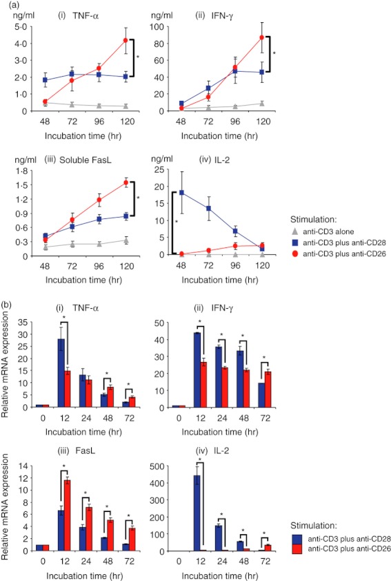
Figure 4.

CD26-mediated co-stimulation of CD8+ T cells induces greater levels of tumour necrosis factor-α (TNF-α), interferon-γ (IFN-γ), and soluble Fas ligand (sFasL) than CD28-mediated co-stimulation. Purified CD8+ T cells were stimulated with anti-CD3 monoclonal antibody (mAb) alone, anti-CD3 plus anti-CD28 mAbs, or anti-CD3 plus anti-CD26 mAbs for the indicated times. (a) Concentrations of TNF-α (i), IFN-γ (ii), sFasL (iii) and interleukin-2 (IL-2) (iv) were examined by ELISA. (b) mRNA expression of TNF-α (i), IFN-γ (ii), FasL (iii) and IL-2 (iv) was quantified by real-time RT-PCR. Each expression was normalized to HPRT1 and relative expression levels compared with resting CD8+ T cells (0 hr) were shown. Data are shown as mean ± SE of triplicate wells from six independent donors (a) and shown as mean ± SD of triplicate samples (b), comparing values in anti-CD3 plus anti-CD26 to that in anti-CD3 plus anti-CD28 (*P < 0·01).
