Background: Roles of zebrafish scube1 in primitive hematopoiesis remain unknown.
Results: scube1 knockdown by morpholino-oligonucleotides reduced the expression of marker genes associated with primitive hematopoiesis.
Conclusion: scube1 is critical for and acts at the top of the regulatory hierarchy of primitive hematopoiesis by modulating BMP signaling.
Significance: Scube1 is a key regulator for primitive hematopoiesis in vertebrates.
Keywords: Bone Morphogenetic Protein (BMP), Cell Surface Protein, Hematopoiesis, Signal Transduction, Zebrafish, Primitive Hematopoiesis
Abstract
scube1 (signal peptide-CUB (complement protein C1r/C1s, Uegf, and Bmp1)-EGF domain-containing protein 1), the founding member of a novel secreted and cell surface SCUBE protein family, is expressed predominantly in various developing tissues in mice. However, its function in primitive hematopoiesis remains unknown. In this study, we identified and characterized zebrafish scube1 and analyzed its function by injecting antisense morpholino-oligonucleotide into embryos. Whole-mount in situ hybridization revealed that zebrafish scube1 mRNA is maternally expressed and widely distributed during early embryonic development. Knockdown of scube1 by morpholino-oligonucleotide down-regulated the expression of marker genes associated with early primitive hematopoietic precursors (scl) and erythroid (gata1 and hbbe1), as well as early (pu.1) and late (mpo and l-plastin) myelomonocytic lineages. However, the expression of an early endothelial marker fli1a and vascular morphogenesis appeared normal in scube1 morphants. Overexpression of bone morphogenetic protein (bmp) rescued the expression of scl in the posterior lateral mesoderm during early primitive hematopoiesis in scube1 morphants. Biochemical and molecular analysis revealed that Scube1 could be a BMP co-receptor to augment BMP signaling. Our results suggest that scube1 is critical for and functions at the top of the regulatory hierarchy of primitive hematopoiesis by modulating BMP activity during zebrafish embryogenesis.
Introduction
SCUBE1 (signal peptide-CUB-EGF (epidermal growth factor) domain-containing protein 1) is the founding member of the newly identified, secreted, cell membrane-associated SCUBE family (1–7). This protein family is composed of three distinct members evolutionarily conserved in vertebrates (1, 2, 6–10). The proteins are organized in a modular fashion and share at least five protein domains: (i) an NH2-terminal signal peptide sequence, (ii) nine copies of EGF-like repeats, (iii) a spacer region, (iv) three cysteine-rich motifs, and (v) one CUB domain at the COOH terminus. In mice and humans, SCUBE genes are all expressed in the endothelium but have unique expression profiles during development. For example, mouse Scube1 is highly expressed in developing gonads, the central nervous system, somites, limb buds, and surface ectoderm (1). Scube2 is expressed in embryonic neuroectoderm, developing face, heart, and several sites of the endochondral skeleton (2). Scube3 transcript was localized to the neural tube, bronchial arches, the fronto-nasal region, somites, limb buds, developing teeth, and hair follicles (11). Thus, Scube genes may play important roles during early embryonic development.
However, we lack direct functional studies of these Scube genes by gene-targeting approaches to generate the null allele during mouse development, probably because of their large and complex genomic structures. Zebrafish have emerged as a powerful vertebrate model to knock down genes of interest to functionally assess genetic regulatory pathways (12). Indeed, forward genetic screens or knockdown experiments have revealed that the zebrafish scube genes are essential for proper Hedgehog signaling in development of slow muscle and ventral spinal cord fates (8–10, 13).
All vertebrates, including zebrafish, have two successive waves of hematopoiesis: primitive and definitive (14, 15). In mammalian and avian species, primitive hematopoiesis occurs in the extraembryonic yolk sac, but in zebrafish, it is at two intraembryonic locations: the anterior lateral mesoderm (ALM)2 and the posterior lateral mesoderm (PLM), which later fuses at the midline to form the intermediate cell mass (ICM). Primitive myelopoiesis is in the ALM, whereas primitive erythropoiesis initiates in the PLM/ICM region (14, 15).
The development of hematopoiesis precursor cells is regulated by both intrinsic and extrinsic factors. We have good documentation of the regulatory mechanisms underlying various intrinsic transcription factors (TFs) required for the formation of hematopoietic stems (e.g. scl, a basic helix-loop-helix TF) and the differentiation of stem cells along the myeloid (e.g. pu.1, an ETS-domain TF) or erythroid lineage (e.g. gata1, a zinc finger TF) (16, 17). However, the signaling regulation of extrinsic factors, such as secreted bone morphogenetic proteins (e.g. BMP4), upon induction of blood cells is less described (18–21). A recent molecular and genetic study showed that BMP acts upstream via Wnt signaling to activate the cdx-hox pathway, which directs the expression of scl, a key transcription factor specifying primitive hematopoietic progenitors (19).
In this study, we isolated and characterized the zebrafish scube1 gene and investigated its role during zebrafish embryonic development by using an antisense morpholino-oligonucleotide (MO) knockdown approach. We found that zebrafish scube1 plays an upstream role in primitive hematopoiesis by acting as a BMP co-receptor to increase its signal activity during early zebrafish embryonic development.
EXPERIMENTAL PROCEDURES
Ethics Statement
Animal handling protocols used in this study were reviewed and approved by the Institutional Animal Care and Utilization Committee, Academia Sinica (Protocol RMiIBMYR2010063).
Zebrafish Maintenance
Wild-type AB strain, Tg(kdr:EGFP) (22), and Tg(gata1:dsRed) (23) transgenic zebrafish were maintained in a 14-h light/10-h dark cycle at 28.5 °C. Zebrafish embryos were collected by natural spawning and incubated in 0.3× Danieau's buffer (1× Danieau's buffer: 58 mm NaCl, 0.7 mm KCl, 0.4 mm MgSO4, 0.6 mm Ca(NO3)2, and 5 mm HEPES (pH 7.6) with double-distilled water) until observation or fixation. The definition of embryo stage was as described (24), and the stages are indicated by hours postfertilization (hpf). An amount of 0.2 mm N-phenylthiourea in 0.3× Danieau's buffer was used to suppress melanin formation after bud stage. Dechorionation of embryos was achieved by protease (0.01 g/ml) digestion.
Identification of scube1 Full-length cDNA in Zebrafish
A TBLASTN search of the public database with mammalian SCUBE1 protein sequences identified one expressed sequence tag clone (GenBankTM accession number EB778403) containing the 5′-most coding sequences of zebrafish scube1. This cDNA clone was then purchased from Open Biosystems (Lafayette, CO). After complete sequencing of this clone, we identified a full-length cDNA sequence of zebrafish scube1 (3,842 bp) composed of a 122-bp 5′-UTR, a 3,075-bp protein-coding sequence, and a 645-bp 3′-UTR. This sequence was deposited in the public database with GenBankTM accession number JQ740109.
Whole-mount in Situ Hybridization (ISH)
Whole-mount ISH was performed essentially as described (25). A 640-bp cDNA in the 3′-UTR of zebrafish scube1 was used to synthesize the antisense RNA riboprobe. All other probes were synthesized as described in the following papers: scl (26), gata1 (27), pu.1 (28), hbbe1 (29), l-plastin (30), mpo (31) and fli1a (32). Two-color ISH was performed as described (33).
Morpholino-oligonucleotide and mRNA Microinjection
Two scube1 antisense translation-blocking MOs (tMOs) were designed with the primers tMO1 (5′-AGG CTG TGA GTG AAT GGG TCT CTC C-3′) and tMO2 (5′-CCA GGA GAA GCA GGC AGG ACC CAT G-3′) (Gene Tools, Philomath, OR). The scube1 tMO1 targets the 5′-UTR, and scube1 tMO2 blocks the AUG translation start site of zebrafish scube1. The scube1 tMOs diluted in Danieau's buffer (12) were injected at the one- or two-cell stage. Increasing amounts of each tMO were injected to titrate for the proper dose by monitoring the overall embryonic morphology or the expression of a mesoderm marker myod1 to rule out the nonspecific phenotypic effects (supplemental Fig. 1). As a control for nonspecific effects, each experiment was performed in parallel with a randomized control MO oligonucleotide. For knockdown experiments, we injected 0.625 ng per embryo of scube1 tMO1 or tMO2. Sense mRNA encoding full-length scube1 was transcribed in vitro from the pCS2+-scube1 plasmid by use of the mMESSAGE mMACHINE kit (Ambion).
Construction of MO Testing Plasmid and the Expression Plasmids
To construct the scube1-GFP testing plasmid, we cloned cDNA containing the region targeted by scube1 tMOs (−31 to +24) into the EcoRI-XbaI sites of pCS2-EGFP vector. The scube1 cDNA fragment was obtained by use of the following complementary oligonucleotides: 5′-aat tcG GAG AGA CCC ATT CAC TCA CAG CCT CTA ACC ATG GGT CCT GCC TGC TTC TCC TGG t-3′ and 5′-cta gaC CAG GAG AAG CAG GCA GGA CCC ATG GTT AGA GGC TGT GAG TGA ATG GGT CTC TCC g-3′. The FLAG or Myc epitope-tagged version of zebrafish Scube1 expression plasmid was constructed in the pFLAG-CMV1 (Sigma) or pSecTag2 (Invitrogen) as described (4). The expression plasmids coding for Myc-tagged BMP2 or BMP4 were prepared as described (34).
ο-Dianisidine Staining of Erythrocytes
Hemoglobin of erythrocytes was stained as described (35). Zebrafish embryos collected at the proper stage were incubated in fresh prepared ο-dianisidine solution (40% ethanol, 0.01 m sodium acetate, 0.65% H2O2, and 0.6 mg/ml ο-dianisidine, dissolved in double-distilled water) for 20 min in the dark.
Cell Culture and Transfection
Human embryonic kidney (HEK)-293T cells were maintained as described previously (5) and were transfected by use of Lipofectamine 2000 (Invitrogen).
Immunoprecipitation and Western Blot Analysis
Two days after transfection, cell lysates were clarified by centrifugation at 10,000 × g for 20 min at 4 °C. Samples were incubated with 1 μg of the indicated antibody and 20 μl of 50% (v/v) Protein A-agarose (Pierce) for 2 h with gentle rocking. After three washes with lysis buffer, precipitated complexes were solubilized by boiling in Laemmli sample buffer, fractionated by SDS-PAGE, and transferred onto PVDF membranes. The membranes were blocked with phosphate-buffered saline (pH 7.5) containing 0.1% gelatin and 0.05% Tween 20 and blotted with the indicated antibodies. After two washes, the blots were incubated with horseradish peroxidase-conjugated goat anti-mouse IgG (Jackson ImmunoResearch Laboratories) for 1 h. After washing the membranes, the reactive bands were visualized by use of the VisGlowTM chemiluminescent substrate, horseradish peroxidase system (Visual Protein).
Luciferase Activity Assay
Human HepG2 cells (3 × 10⁵ cells/well) were seeded into 24-well plates and transfected with 0.3 μg of the BMP-responsive luciferase reporter I-BRE-Luc, 0.01 μg of the Renilla luciferase reporter used as an internal control, and either empty vector or the zebrafish scube1 expression plasmid. Transfected cells were maintained in low serum medium (0.1% FBS) for 18 h. Luciferase activity was measured after a 24-h treatment with recombinant human BMP2 protein.
RESULTS
Identification and Embryonic Expression Pattern of Zebrafish scube1
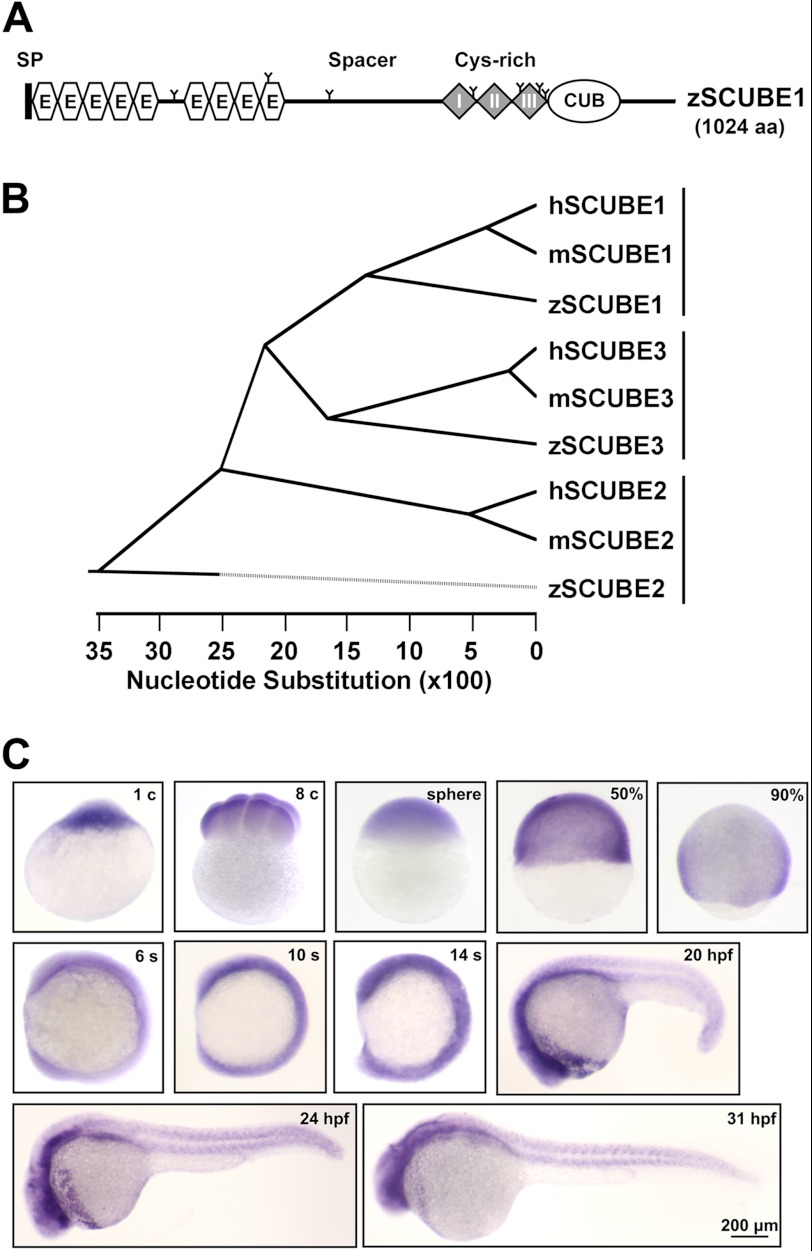
A homology search of the public database identified a full-length scube1 cDNA clone coding for a polypeptide of 1,024 amino acids sharing a conserved protein domain organization (Fig. 1A and supplemental Fig. 2) and overall 76 or 77% sequence identity with its mouse or human orthologue, respectively (supplemental Figs. 3 and 4). Furthermore, phylogenetic analysis showed that zebrafish Scube1 clustered closely with mouse and human SCUBE1 and separate from groups of SCUBE2 or SCUBE3 protein sequences (Fig. 1B). These results suggest that zebrafish scube1 is an authentic orthologue of mammalian SCUBE1.
FIGURE 1.
Protein domain organization, phylogenetic analysis, and embryonic expression patterns of zebrafish scube1. A, graphic illustration of the domain structure of zebrafish Scube1 protein. SP, signal peptide sequence; E, EGF-like domain; Cys-rich, cysteine-rich repeats. B, phylogenetic tree of the SCUBE protein family. Similarity of human (h), mouse (m), and zebrafish (z) SCUBE protein sequences was analyzed by use of the Lasergene MEGALIGN program (ClustalW algorithm). Branch order indicates structural relative, and branch length reflects sequence identity. C, whole-mount in situ hybridization of zebrafish embryos at the indicated stages with antisense scube1 probe. Corresponding control sense probes detected no apparent signal (not shown). c, cell stage; s, somite stage; hpf, hours postfertilization.
We then examined the spatiotemporal expression pattern of scube1 during embryonic development by whole-mount ISH. Maternal scube1 transcripts were distributed evenly in blastomeres (Fig. 1C). Likewise, zygotic scube1 was globally expressed up to the 14-somite stage. From 20 to 31 hpf, strong scube1 expression was detected in the brain and somitic region (Fig. 1C). The maternal and ubiquitous expression patterns of scube1 during early embryonic stages implied that scube1 may play critical roles during early embryogenesis.
Zebrafish Scube1 Is a Secreted and Surface-associated Glycoprotein
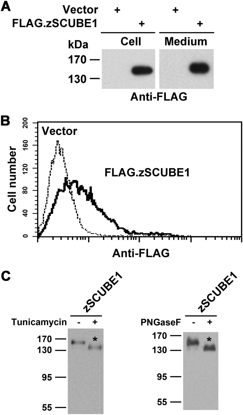
Because heterologous expression of human SCUBE1 resulted in secreted glycoprotein that could tether on the cell surface (7), we then examined whether overexpressed zebrafish Scube1 protein might also behave in a similar manner. The zebrafish Scube1 protein was heterologously expressed by transient transfection in HEK-293T cells. Overexpressed Scube1 (tagged with FLAG epitope) could be effectively secreted into the conditioned medium, targeted onto the cell surface, and post-translationally modified with N-linked oligosaccharides (Fig. 2).
FIGURE 2.
Overexpressed zebrafish Scube1 is a secreted, surface-anchored glycoprotein in HEK-293T cells. A, secretion of zebrafish Scube1 into the conditioned medium. HEK-293T cells were transfected with empty vector (Vector) or expression plasmid encoding FLAG-tagged zebrafish Scube1 (FLAG.zSCUBE1). Forty-eight hours post-transfection, the serum-free conditioned medium was collected, and cells were detached with phosphate-buffered saline/EDTA. Samples from cell lysate (Cell) or the serum-free conditioned culture medium (Medium) were separated by SDS-PAGE and transferred to polyvinylidene difluoride membranes. Recombinant FLAG.zSCUBE1 protein was detected by Western blot analysis with anti-FLAG antibody. B, cell surface expression of zebrafish Scube1 protein. HEK-293T cells were transfected with empty vector (Vector) or expression plasmid encoding FLAG.zSCUBE1 protein. Forty-eight hours post-transfection, cells were detached and probed with anti-FLAG antibody and subjected to flow cytometry analysis. C, N-linked glycosylation of zebrafish SCUBE1. HEK-293T cells were transfected with expression vector encoding FLAG.zSCUBE1 protein. Transfected cells were cultured in the absence (−) or presence (+) of tunicamycin (an inhibitor of N-glycosylation) for 24 h (left). Alternatively, cell lysates were left untreated (−) or treated (+) with peptide N-glycosidase F (PNGaseF) to remove the N-linked oligosaccharide chain (right). Cell lysates derived from each treatment were examined by Western blot analysis with anti-FLAG antibody.
Zebrafish scube1 Is Essential for Primitive Hematopoiesis
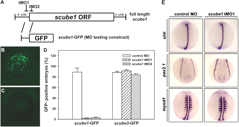
To further investigate the role of scube1 during early embryogenesis in zebrafish, we then designed two non-overlapping antisense tMOs targeting the 5′-UTR or the start codon sequence of scube1 to block translation of scube1 (named scube1 tMO1 or tMO2; Fig. 3A). A random sequence of antisense MO with minimal effect on embryonic development was used as a control (cMO). Both tMOs could specifically and effectively block translation of scube1, whereas cMO had no effect (Fig. 3D).
FIGURE 3.
Translational blocking efficiency of two scube1 tMOs and no effect of scube1 knockdown on the expression of various mesoderm genes. A, diagram showing the targeting sites of two scube1 tMOs and the scube1-GFP test construct. tMO1 targets the 5′-UTR, and tMO2 blocks at the AUG translation start site. The scube1-GFP plasmid contains pCS2-EGFP fused with a fragment of scube1 gene sequence from −31 to +24. Embryos at the one-cell stage were injected with 250 pg of scube1-GFP or scube3-GFP testing plasmid without or with 0.625 ng of scube1 tMO1 or tMO2, respectively. The expression of GFP was examined at 8 hpf and photographed by epifluorescent microscopy. Images are representative of embryos injected with scube1-GFP plasmid (B) or scube1 tMO1 (C). D, summary of translational blocking efficiency of two scube1 tMOs. The lack of suppression effect on the scube3-GFP testing construct confirms their specificity in blocking the translation of scube1 protein. Error bars, S.E. E, whole-mount ISH of shh (the axial mesoderm marker), pax2.1 (the lateral mesoderm marker), and myod1 (the somitic mesoderm marker) in cMO- or scube1 tMO1-injected embryos (0.625 ng/embryo) at the 6-somite stage (dorsal view).
We then injected embryos with scube1 tMOs (referred as morphants) and examined their effects on embryonic development. Zebrafish embryos injected with a low dose of cMO or scube1 tMOs (0.625 ng/embryo) produced no gross lethality or morphological defect because the expression of shh (the axial mesoderm marker), pax2.1 (the lateral mesoderm marker), and myod1 (the somitic mesoderm marker) was not affected (Fig. 3E). However, the scube1 morphants showed a markedly reduced number of circulating blood cells as compared with cMO-injected embryos at 30 hpf (supplemental Movies 1 and 2).
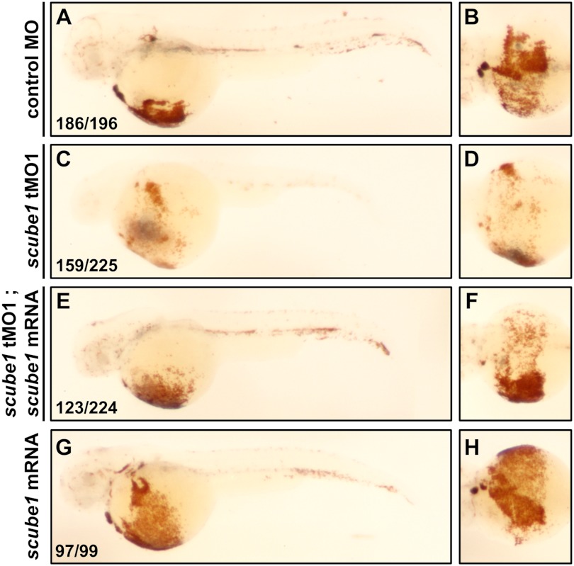
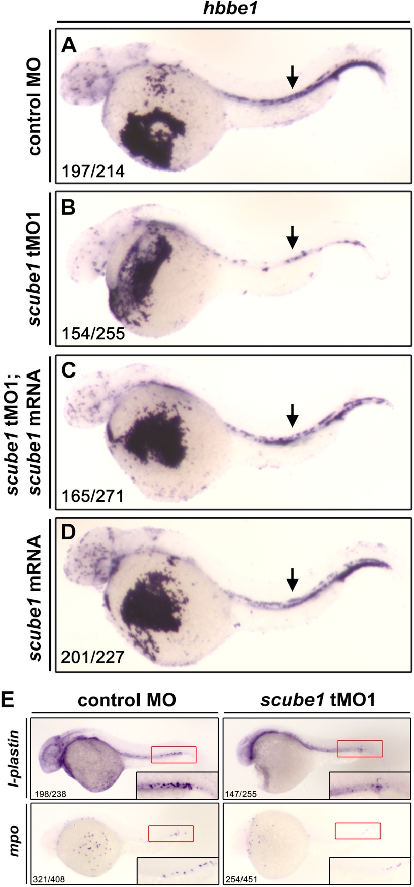
Because the first wave of blood production, primitive hematopoiesis, predominantly produced erythrocytes and some primitive macrophages in zebrafish (14), we then used ο-dianisidine staining to detect hemoglobin-containing erythrocytes. The scube1 morphant embryos showed severe reduction in proportion of differentiated erythrocytes in 48-hpf embryos (Fig. 4). Likewise, the expression of an erythroid-specific gene, β1-embryonic hemoglobin (hbbe1), was significantly reduced over the PLM at the 10-somite stage (supplemental Fig. 5) or in the ICM region at 31 hpf (Fig. 5B). Importantly, the loss of ο-dianisidine staining and hbbe1 expression could be rescued by the co-injection of scube1 mRNA in scube1 morphant embryos (Figs. 4 (E and F) and 5C), which confirmed the specificity of scube1 tMO targeting. Note that injection of scube1 mRNA alone produced normal expression of myod1 or hbbe1 (supplemental Fig. 6 and Fig. 5D). To investigate whether scube1 also plays an important role in regulating primitive myelopoiesis, we examined the expression of two myeloid markers, l-plastin (an actin-binding protein) and mpo (myeloperoxidase), which marks pan-myeloid cells and differentiated granulocytes, respectively (36). As shown in Fig. 5E, scube1 knockdown significantly decreased the expression of these two myeloid-specific genes.
FIGURE 4.
Effect of scube1 knockdown and scube1 mRNA rescue on the production of erythrocytes. A–D, detection of hemoglobin in erythrocytes stained with ο-dianisidine in control MO or tMO1-injected embryos at 48 hpf. E–H, forced expression of scube1 mRNA alone had little phenotypic effect (G and H) but could partially rescue the anemic defect in the scube1 tMO1-injected embryos (E and F). The proportion of erythrocytes was restored by co-injection of 50 pg of scube1 mRNA in scube1 morphants at 48 hpf. Embryos are shown in lateral (A, C, E, and G) and ventral view (B, D, F, and H). The numbers indicated in the bottom left-hand corners of photographs are the number of affected embryos with phenotype similar to what is shown in the picture (left) and the total number of observed embryos (right).
FIGURE 5.
Effect of scube1 knockdown on the expression of erythrocyte and myeloid-specific markers. A and B, expression of the erythrocyte marker hbbe1 in control MO- or tMO1-injected embryos at 31 hpf (lateral view). C and D, overexpression of scube1 mRNA produced little phenotypic effect (D) but could partially rescue hbbe1 expression in scube1 morphants (C). The expression of hbbe1 was restored by co-injection of 50 pg of scube1 mRNA in scube1 morphants at 31 hpf. The arrows indicate the hbbe1-expressing erythrocytes in the ICM region. E, expression of the granulocyte marker l-plastin and macrophage marker mpo in cMO- or tMO1-injected embryos at 31 hpf (lateral view). The ICM regions are boxed and shown at higher magnification in the bottom right-hand corner of each photograph. The numbers indicated in the bottom left-hand corners of photographs are the number of affected embryos with phenotype similar to what is shown in the picture (left) and the total number of observed embryos (right).
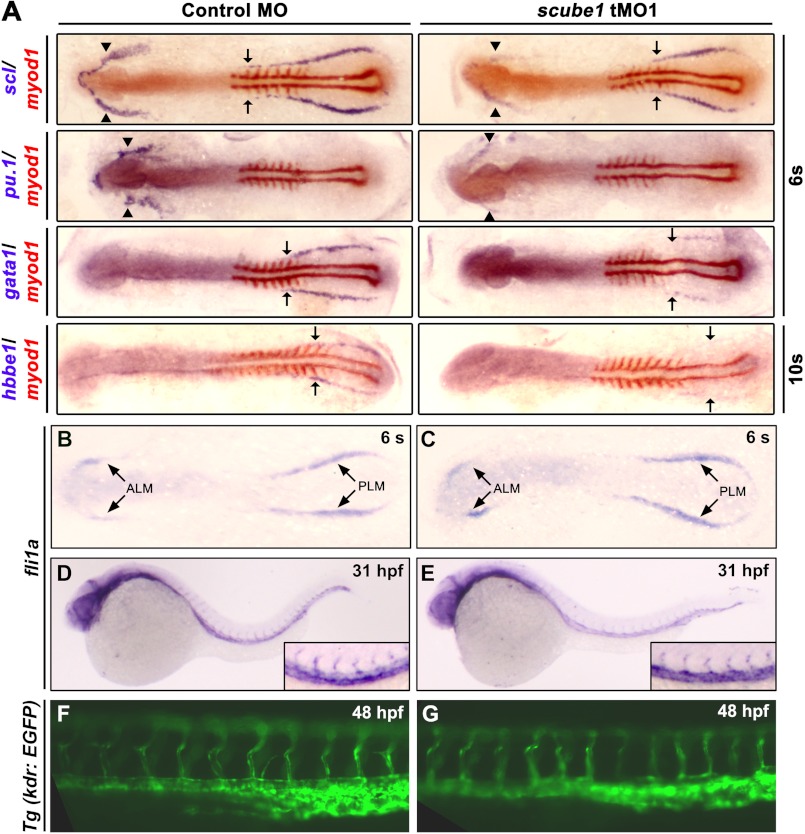
To further dissect whether scube1 is critical in these early primitive hematopoietic processes, we then examined the effect of scube1 tMO1 on gene expression associated with these early hematopoietic lineages. Whole-mount ISH at the 6-somite stage (12 hpf) showed that scube1 knockdown resulted in reduced expression of marker genes associated with primitive hematopoietic progenitors (scl) or primitive myelopoiesis (pu.1) and primitive erythropoiesis (hbbe1 and gata1) in the ALM or PLM (Fig. 6A and supplemental Fig. 5). Similar phenotypes were observed in tMO2-injected embryos (data not shown).
FIGURE 6.
Effect of scube1 knockdown on the expression of early hematopoiesis precursor markers, a vascular endothelial marker, and blood vessel morphogenesis. A, one-celled embryos were injected with cMO or scube1 tMO1 (1.25 ng/embryo) and examined for expression of the indicated markers at the 6- or 10-somite stage by two-color RNA ISH. Embryos are shown in dorsal view with anterior to the left. Although the expression of myod1 (red) remained relatively unaltered, the expression level of scl or pu.1 at the ALM (arrowheads) and the expression margins of scl, gata1, and hbbe1 at the PLM (arrows) were reduced in the scube1 morphants as compared with the control embryos. s, somite. B–G, expression of a vascular endothelial marker, fli1a, in cMO- or scube1 tMO1-injected embryos at the 6-somite stage (B and C, 6 s, dorsal view with anterior to the left) and 31 hpf (D and E, lateral view with anterior to the left). Shown are fluorescent images of Tg(kdr:EGFP) embryos at the trunk region injected with cMO (F) or scube1 tMO1 (G) at 48 hpf.
Zebrafish scube1 Is Not Involved in the Fate Determination of Vascular Endothelial Progenitors or Blood Vessel Formation
Because primitive hematopoietic and vascular endothelial precursors may be derived from common hemangioblasts in zebrafish (37), we then examined whether scube1 also plays a role in the fate determination of endothelial precursors and subsequent blood vessel morphogenesis. We examined the expression of a vascular endothelium-specific transcription factor, fli1a, in scube1 morphants and injected Tg(kdr:EGFP) (22) transgenic embryos with the scube1 tMO1. Expression of fli1a in the ALM and PLM at early 6-somite stage or in endothelial cells at 31 hpf was virtually unaffected (Fig. 6, B–E). In agreement with this finding, the scube1 tMO-injected Tg(kdr:EGFP) embryos showed a morphologically intact vasculature up to 48 hpf (Fig. 6, F and G). Thus, scube1 may not be needed for specification of angioblasts at early stages or angiogenesis at late stages of embryogenesis.
Forced Expression of scl Rescues the Primitive Hematopoietic Defects in scube Morphants
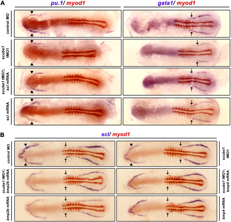
To establish the epistatic relationships between scube1 and genes involved in early primitive hematopoiesis, we further determined whether overexpression of scl could rescue the pu.1 or gata1 expression defects in scube1 morphants. Forced expression of scl by mRNA microinjection in scube1 morphants rescued the expression of both pu.1 and gata1 (Fig. 7A), which suggests that scl acts downstream of scube1 to specify early primitive myelopoietic and erythropoietic precursor formation.
FIGURE 7.
scube1 acts upstream of scl during primitive hematopoiesis, and overexpression of bmp rescues scl expression in scube1 morphants. A, injection of 50 pg of scl mRNA rescued pu.1 and gata1 expression in scube1 morphants at the 6-somite stage. Embryos are shown in dorsal view with anterior to the left. Expression of pu.1 or gata1 in ALM or PLM was marked by arrowheads or arrows, respectively. Note that the expression level of pu.1 in ALM or the expression margins of gata1 in PLM were reduced in the scube1 morphants that can be partially rescued by scl overexpression. B, injection of 50 pg of bmp2b and bmp4 mRNA restored scl expression in scube1 tMO1-injected embryos at the 6-somite stage. Embryos are shown in dorsal view with anterior to the left. Note that the margins of scl expression in the anterior region of PLM (arrows) were reduced in the scube1 morphants, which can be restored by bmp overexpression.
Overexpression of bmp2b or bmp4 Rescues the Expression of scl in scube1 Morphants
Because human SCUBE1 or SCUBE2 have been implicated in regulating the long range signaling of BMP (3, 34) and because BMP signaling is important for mesoderm patterning and specifying the blood fate (19, 21, 38), we then investigated the genetic relationship between scube1 and bmp genes by co-injection of bmp2b or bmp4 mRNA with scube1 tMO1 in embryos. Overexpression of bmp2b or bmp4 restored scl expression in scube1 morphants (Fig. 7B), which suggests that bmp lies downstream or parallel to scube1 for the initiation of zebrafish primitive hematopoiesis.
Zebrafish Scube1 Can Interact with BMP Ligands or Receptors and Enhance BMP Signaling
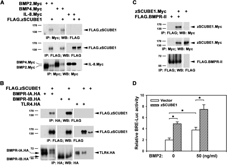
Because we observed a genetic interaction between scube1 and bmp genes, we then examined whether Scube1 could biochemically interact with BMP ligands and their receptors and, if so, whether zebrafish Scube1 could affect BMP signal activity. HEK-293T cells were transfected with the expression plasmid encoding Myc-tagged BMP2 or BMP4 alone or with the FLAG.zSCUBE1 expression construct. Two days after transfection, cell lysates underwent immunoprecipitation with the anti-Myc monoclonal antibody, and precipitates were analyzed by immunoblotting with anti-FLAG monoclonal antibody to determine protein interaction. Immunoprecipitation with anti-Myc antibody resulted in a specific coprecipitation of the Scube1 protein by BMP2/4 but not IL-8 (Fig. 8A). Thus, Scube1 may indeed form a complex with BMP2 or BMP4.
FIGURE 8.
Scube1 forms a complex with BMP ligands and receptors and augments BMP signal activity. The expression plasmid encoding the Myc-tagged BMP2/BMP4 or IL-8 (A), HA-tagged BMPR-IA/IB or TLR4 (B), or FLAG-tagged BMPR-II (C) was transfected alone or with the FLAG-tagged SCUBE1 or Myc-tagged SCUBE1 in HEK-293T cells. After 2 days, cell lysates underwent immunoprecipitation (IP) and then Western blot analysis (WB) of expression of the indicated proteins to determine protein-protein interactions. D, SCUBE1 enhances BMP signaling. HepG2 cells were transfected with BRE-Luc and pRL-TK alone or with the expression plasmid encoding SCUBE1. Transfected cells were then incubated for 24 h with or without 50 ng/ml BMP2, and luciferase activity was measured. Relative luciferase activity represents firefly luciferase values normalized to Renilla activity. Data are means ± S.E. (error bars). *, p < 0.05.
BMP signaling is initiated when BMP protein binds to its receptor complex, composed of two type I (BMPR-I) and two type II (BMPR-II) receptors (38, 39). Because Scube1 is also expressed on the cell surface (Fig. 2B), we next investigated whether Scube1 could interact with BMPR-I or -II. HEK-293T cells were transfected with BMPR-IA, -IB, or -II expression plasmids alone (tagged with the HA or FLAG epitope for easy detection) or with FLAG-tagged Scube1 (Fig. 8B) or Myc-tagged Scube1 (Fig. 8C) expression constructs, respectively. At 2 days after transfection, the BMPR-I or -II protein was immunoprecipitated with its corresponding anti-epitope antibody (anti-HA or anti-FLAG), and individual immunoprecipitates were analyzed by immunoblotting for the presence of Scube1 (anti-FLAG or anti-Myc). Scube1 protein co-immunoprecipitated with BMPR-IA, -IB, or -II protein (Fig. 8, B and C), which suggests that Scube1 can also interact with BMPRs on the plasma membrane. The interaction between Scube1 and BMPRs appears to be selective because Scube1 is incapable of forming a complex with Toll-like receptor 4 (TLR4) (Fig. 8B).
Because of the biochemical interactions between Scube1 and BMP ligands or receptors, we then evaluated whether Scube1 could regulate the BMP signal activity. HepG2 cells were transfected with a BMP-responsive luciferase reporter construct, BRE-Luc (40), alone or with plasmid encoding Scube1 (Fig. 8D). Transfected cells were then stimulated with or without BMP2 for 24 h before measurement of luciferase activity. In the absence of Scube1, treatment with BMP2 increased the relative luciferase activity as compared with untreated cells (Fig. 8D). Co-transfection with Scube1 similarly increased BRE luciferase activity even in the absence of exogenous BMP2 stimulation, probably because of Scube1-mediated cross-linking of BMPR-I and -II proteins (Fig. 8D). Most interestingly, Scube1 markedly enhanced signaling with exogenous BMP2 (Fig. 8D). As a control, co-transfection with Scube1 did not increase transforming growth factor (TGF)-β- or NF-κB-responsive luciferase activity above base line (data not shown). Furthermore, knockdown of scube1, not scube2, significantly decreased the BMP-responsive Smad activation (phosphorylated Smad1/5/8 protein level) (supplemental Fig. 7), which demonstrates the specific role of Scube1 in modulating BMP signaling. Consistently, whole-mount confocal immunofluorescent analysis also showed a reduction of p-Smad1/5/8 level in hematopoietic progenitors in the ICM region (supplemental Fig. 8). Thus, Scube1 may act as a BMP co-receptor and augment BMP-mediated signal activity.
DISCUSSION
In this study, we isolated the zebrafish scube1 gene and characterized its expression and function in primitive hematopoiesis. Although Scube1 is highly expressed in developing gonads, nervous system, somites, surface ectoderm, and limb buds in mice (1, 7), its involvement in primitive hematopoiesis in vertebrates has not yet been described. Our results demonstrate that scube1 is critical for primitive hematopoiesis in zebrafish embryos by positively regulating the expression of scl, a master gene in hematopoiesis. Epistasis experiments revealed an ordered scube1-bmp-scl pathway. Consistent with this finding, our biochemical and molecular analyses suggested that Scube1 may modulate BMP signal transduction during receipt of the BMP signal at the plasma membrane. This modulation may occur through the action of Scube1 as a BMP co-receptor in enhancing cellular responses to BMP for specifying the earliest hematopoietic precursor cells at the onset of primitive hematopoiesis. Thus, our findings position scube1 at the top of the regulatory hierarchy of primitive hematopoiesis in zebrafish.
Several lines of evidence indicate that scube1 is critical for the initiation of primitive hematopoiesis. First, knockdown of scube1 reduced the expression of the hematopoietic precursor marker scl during the early segmentation stage (Fig. 6A), and as a result, the expression of the erythroid marker gata1, hemoglobin gene hbbe1, and myeloid markers pu.1, mpo, and l-plastin was blocked at later stages (Figs. 5E and 6A). Second, the effect of scube1 knockdown could be rescued by coinjection of scube1 mRNA (Figs. 4 and 5). Third, forced expression of bmp2b or bmp4 could overcome the suppressive effect of scube1 knockdown on primitive hematopoiesis (Fig. 7B). Finally, we also validated hematopoietic phenotypes of scube1 knockdown by two independent splice-blocking MOs that target the exon2-intron2 or intron3-exon4 boundary sequences (data not shown).
BMPs belong to the TGF-β superfamily, initially essential for the patterning of ventral mesoderm from where the primitive hematopoietic stem cells arise. However, recent gain- or loss-of-function manipulation of the BMP signal pathway during middle or late gastrulation or even post-gastrulation suggested that BMP signaling also functions in blood fate specification (19, 21, 41–45). Overall, our genetic data suggest that scube1 may regulate the BMP signaling for its later role during somitogenesis in the specification of blood progenitors. Furthermore, biochemical and molecular experiments revealed that Scube1 expressed on the cell surface may act as a BMP co-receptor through its interactions with BMP ligands and receptors to facilitate the presentation of BMP ligands to its receptor complexes, thus promoting BMP signal transduction in the responding cells (Fig. 8). Our data suggest an interaction between SCUBE1 and BMP receptor protein when overexpressed in HEK-293T cells; however, it remains to be confirmed whether or not such an interaction indeed occurs in vivo.
In addition, we cannot exclude the possibility that a secreted extracellular form of Scube1 may be involved in transporting and/or protecting the BMP ligands against degradation in the extracellular environment. Alternatively, the secreted Scube1 protein may deposit in the extracellular milieu, where it cooperates with yet unknown regulatory molecules to modulate other cell growth signalings and establish a competent stem cell niche or microenvironment suitable for hematopoietic differentiation. We further performed RT-PCR analysis to examine the expression of scube1 in isolated hematopoietic progenitor cells from transgenic Tg(gata1:dsRed) embryos (23). Our results showed that scube1 is also expressed in hematopoietic progenitors, suggesting that Scube1 may act cell-autonomously in primitive hematopoiesis (supplemental Fig. 9). However, further cell transplantation experiments are needed to verify the cell-autonomous or non-cell-autonomous role of scube1.
During development, blood and endothelial cells differentiate in close proximity, which suggests that these cells may derive from common bipotential precursor cells, termed the hemangioblasts. A recent fate-mapping study provided clear evidence of the existence of hemangioblasts within zebrafish embryos (37). However, the number of such bipotential progenitors is limited in vivo, which suggests that not all endothelial and blood cells arise from a common progenitor. Instead, juxtaposed but distinct populations of angioblasts and hematopoietic precursors may develop independently from the posterior mesoderm and further differentiate into vascular endothelial cells or blood cells (46, 47). Consistent with this notion, our data show that scube1 is solely involved in the BMP regulatory pathway required for specifying hematopoietic fate and completely separate from the vascular endothelial lineage. Likewise, in Xenopus embryos, expression of the blood gene SCL but not endothelial gene Xfli1 depends on BMP signaling (48). However, additional studies are needed to understand the molecular mechanism underlying the context-dependent BMP regulatory function of scube1 in specifying hematopoietic progenitor cells.
Although the Scube protein family contains three members with similar domain structure and high protein sequence homology, knockdown of two other scube genes (scube2 and scube3) produced no apparent defects in primitive hematopoiesis.3 This observation can be simply due to a distinctive, non-overlapping spatiotemporal expression pattern and thus non-redundant functions for each scube gene during early zebrafish embryogenesis. For example, zebrafish scube2 has been implicated in the development of slow muscle fibers and the ventral neural tube by modulating the hedgehog signal transduction pathway (8–10). Furthermore, because BMP signaling is also involved in definitive hematopoiesis (42, 43, 49), we are interested in determining whether scube1 or other scube genes are also involved in definitive hematopoiesis.
Because the genetic program that drives hematopoiesis is evolutionarily conserved among vertebrates, whether SCUBE1 is also involved in primitive hematopoiesis in mammals is of interest. Because of the large and complex genomic structure of the mouse Scube1 gene, designing a targeting strategy for generating a null allele is difficult. Indeed, our previous targeting study disrupted only the COOH-terminal CUB domain of mouse Scube1 (mutant scube1Δcub allele), which resulted in brain malformation and perinatal lethality (34). Although this gene-targeting study clearly demonstrated the role of Scube1 during early stages of central nervous system development, its function in primitive hematopoiesis was not obvious and not thoroughly examined (34). Given the essential role of Scube1 in primitive hematopoiesis in zebrafish, we are interested in producing a conditional null allele to further explore the biological functions of Scube1 in mammalian hematopoiesis or other developmental processes.
In summary, our data demonstrate that zebrafish scube1 plays a critical role in the specification of blood cells by regulating the upstream, extrinsic Bmp signal activity during early somitogenesis. Given the wide spectrum expression of Scube1 in various developing tissues (1, 50), further investigation is needed to fully elucidate its stage- and tissue-specific functions during embryonic development.
Acknowledgments
We thank the Zebrafish International Resource Center (ZIRC), Taiwan Zebrafish Core Facility at Academia Sinica (supported by grant 101-2321-B-001-026 from the National Science Council), and Taiwan Zebrafish Core Facility at NTHU/NHRI (supported by grant 101-2321-B-400-014 from the National Science Council) for providing AB, Tg(kdr:EGFP), Tg(gata1:dsRed) fish lines, and some ISH probes used in this study.
This work was supported by National Science Council Grant NSC 99-2311-B-001-022-MY3 (to R.-B. Y.) and by National Science Council Grant NSC-98-2311-B-002-006-MY3 and National Taiwan University Grants NTU CESRP-10R70602A5 and NTU ERP-10R80600 (to S.-J. L.).

This article contains supplemental Figs. 1–9 and Movies 1 and 2.
The nucleotide sequence(s) reported in this paper has been submitted to the GenBankTM/EBI Data Bank with accession number(s) JQ740109.
K.-C. Tsao, C.-F. Tu, S.-J. Lee, and R.-B. Yang, unpublished data.
- ALM
- anterior lateral mesoderm
- PLM
- posterior lateral mesoderm
- ICM
- intermediate cell mass
- TF
- transcription factor
- BMP
- bone morphogenetic protein
- BMPR
- BMP receptor
- MO
- morpholino-oligonucleotide
- tMO
- translation-blocking MO
- cMO
- control MO
- ISH
- in situ hybridization
- HEK
- human embryonic kidney
- hpf
- hours postfertilization.
REFERENCES
- 1. Grimmond S., Larder R., Van Hateren N., Siggers P., Hulsebos T. J., Arkell R., Greenfield A. (2000) Cloning, mapping, and expression analysis of a gene encoding a novel mammalian EGF-related protein (SCUBE1). Genomics 70, 74–81 [DOI] [PubMed] [Google Scholar]
- 2. Grimmond S., Larder R., Van Hateren N., Siggers P., Morse S., Hacker T., Arkell R., Greenfield A. (2001) Expression of a novel mammalian epidermal growth factor-related gene during mouse neural development. Mech. Dev. 102, 209–211 [DOI] [PubMed] [Google Scholar]
- 3. Cheng C. J., Lin Y. C., Tsai M. T., Chen C. S., Hsieh M. C., Chen C. L., Yang R. B. (2009) SCUBE2 suppresses breast tumor cell proliferation and confers a favorable prognosis in invasive breast cancer. Cancer Res. 69, 3634–3641 [DOI] [PubMed] [Google Scholar]
- 4. Lin Y. C., Chen C. C., Cheng C. J., Yang R. B. (2011) Domain and functional analysis of a novel breast tumor suppressor protein, SCUBE2. J. Biol. Chem. 286, 27039–27047 [DOI] [PMC free article] [PubMed] [Google Scholar]
- 5. Tsai M. T., Cheng C. J., Lin Y. C., Chen C. C., Wu A. R., Wu M. T., Hsu C. C., Yang R. B. (2009) Isolation and characterization of a secreted, cell-surface glycoprotein SCUBE2 from humans. Biochem. J. 422, 119–128 [DOI] [PubMed] [Google Scholar]
- 6. Wu B. T., Su Y. H., Tsai M. T., Wasserman S. M., Topper J. N., Yang R. B. (2004) A novel secreted, cell-surface glycoprotein containing multiple epidermal growth factor-like repeats and one CUB domain is highly expressed in primary osteoblasts and bones. J. Biol. Chem. 279, 37485–37490 [DOI] [PubMed] [Google Scholar]
- 7. Yang R. B., Ng C. K., Wasserman S. M., Colman S. D., Shenoy S., Mehraban F., Komuves L. G., Tomlinson J. E., Topper J. N. (2002) Identification of a novel family of cell-surface proteins expressed in human vascular endothelium. J. Biol. Chem. 277, 46364–46373 [DOI] [PubMed] [Google Scholar]
- 8. Hollway G. E., Maule J., Gautier P., Evans T. M., Keenan D. G., Lohs C., Fischer D., Wicking C., Currie P. D. (2006) Scube2 mediates Hedgehog signalling in the zebrafish embryo. Dev. Biol. 294, 104–118 [DOI] [PubMed] [Google Scholar]
- 9. Kawakami A., Nojima Y., Toyoda A., Takahoko M., Satoh M., Tanaka H., Wada H., Masai I., Terasaki H., Sakaki Y., Takeda H., Okamoto H. (2005) The zebrafish-secreted matrix protein you/scube2 is implicated in long-range regulation of hedgehog signaling. Curr. Biol. 15, 480–488 [DOI] [PubMed] [Google Scholar]
- 10. Woods I. G., Talbot W. S. (2005) The you gene encodes an EGF-CUB protein essential for Hedgehog signaling in zebrafish. PLoS Biol. 3, e66. [DOI] [PMC free article] [PubMed] [Google Scholar]
- 11. Xavier G. M., Economou A., Senna Guimarães A. L., Sharpe P. T., Cobourne M. T. (2010) Characterization of a mouse Scube3 reporter line. Genesis 48, 684–692 [DOI] [PubMed] [Google Scholar]
- 12. Nasevicius A., Ekker S. C. (2000) Effective targeted gene “knockdown” in zebrafish. Nat. Genet. 26, 216–220 [DOI] [PubMed] [Google Scholar]
- 13. Johnson J. L., Hall T. E., Dyson J. M., Sonntag C., Ayers K., Berger S., Gautier P., Mitchell C., Hollway G. E., Currie P. D. (2012) Scube activity is necessary for Hedgehog signal transduction in vivo. Dev. Biol. 368, 193–202 [DOI] [PubMed] [Google Scholar]
- 14. de Jong J. L., Zon L. I. (2005) Use of the zebrafish system to study primitive and definitive hematopoiesis. Annu. Rev. Genet. 39, 481–501 [DOI] [PubMed] [Google Scholar]
- 15. Chen A. T., Zon L. I. (2009) Zebrafish blood stem cells. J. Cell. Biochem. 108, 35–42 [DOI] [PubMed] [Google Scholar]
- 16. Jing L., Zon L. I. (2011) Zebrafish as a model for normal and malignant hematopoiesis. Dis. Model Mech. 4, 433–438 [DOI] [PMC free article] [PubMed] [Google Scholar]
- 17. Paik E. J., Zon L. I. (2010) Hematopoietic development in the zebrafish. Int. J. Dev. Biol. 54, 1127–1137 [DOI] [PubMed] [Google Scholar]
- 18. Blank U., Karlsson S. (2011) The role of Smad signaling in hematopoiesis and translational hematology. Leukemia 25, 1379–1388 [DOI] [PubMed] [Google Scholar]
- 19. Lengerke C., Schmitt S., Bowman T. V., Jang I. H., Maouche-Chretien L., McKinney-Freeman S., Davidson A. J., Hammerschmidt M., Rentzsch F., Green J. B., Zon L. I., Daley G. Q. (2008) BMP and Wnt specify hematopoietic fate by activation of the Cdx-Hox pathway. Cell Stem Cell 2, 72–82 [DOI] [PubMed] [Google Scholar]
- 20. Ciau-Uitz A., Liu F., Patient R. (2010) Genetic control of hematopoietic development in Xenopus and zebrafish. Int. J. Dev. Biol. 54, 1139–1149 [DOI] [PubMed] [Google Scholar]
- 21. Hogan B. M., Layton J. E., Pyati U. J., Nutt S. L., Hayman J. W., Varma S., Heath J. K., Kimelman D., Lieschke G. J. (2006) Specification of the primitive myeloid precursor pool requires signaling through Alk8 in zebrafish. Curr. Biol. 16, 506–511 [DOI] [PubMed] [Google Scholar]
- 22. Jin S. W., Beis D., Mitchell T., Chen J. N., Stainier D. Y. (2005) Cellular and molecular analyses of vascular tube and lumen formation in zebrafish. Development 132, 5199–5209 [DOI] [PubMed] [Google Scholar]
- 23. Traver D., Paw B. H., Poss K. D., Penberthy W. T., Lin S., Zon L. I. (2003) Transplantation and in vivo imaging of multilineage engraftment in zebrafish bloodless mutants. Nat. Immunol. 4, 1238–1246 [DOI] [PubMed] [Google Scholar]
- 24. Kimmel C. B., Ballard W. W., Kimmel S. R., Ullmann B., Schilling T. F. (1995) Stages of embryonic development of the zebrafish. Dev. Dyn. 203, 253–310 [DOI] [PubMed] [Google Scholar]
- 25. Thisse C., Thisse B. (2008) High-resolution in situ hybridization to whole-mount zebrafish embryos. Nat. Protoc. 3, 59–69 [DOI] [PubMed] [Google Scholar]
- 26. Liao E. C., Paw B. H., Oates A. C., Pratt S. J., Postlethwait J. H., Zon L. I. (1998) SCL/Tal-1 transcription factor acts downstream of cloche to specify hematopoietic and vascular progenitors in zebrafish. Genes Dev. 12, 621–626 [DOI] [PMC free article] [PubMed] [Google Scholar]
- 27. Detrich H. W., 3rd, Kieran M. W., Chan F. Y., Barone L. M., Yee K., Rundstadler J. A., Pratt S., Ransom D., Zon L. I. (1995) Intraembryonic hematopoietic cell migration during vertebrate development. Proc. Natl. Acad. Sci. U.S.A. 92, 10713–10717 [DOI] [PMC free article] [PubMed] [Google Scholar]
- 28. Lieschke G. J., Oates A. C., Paw B. H., Thompson M. A., Hall N. E., Ward A. C., Ho R. K., Zon L. I., Layton J. E. (2002) Zebrafish SPI-1 (PU.1) marks a site of myeloid development independent of primitive erythropoiesis. Implications for axial patterning. Dev. Biol. 246, 274–295 [DOI] [PubMed] [Google Scholar]
- 29. Quinkertz A., Campos-Ortega J. A. (1999) A new β-globin gene from the zebrafish, βE1, and its pattern of transcription during embryogenesis. Dev. Genes Evol. 209, 126–131 [DOI] [PubMed] [Google Scholar]
- 30. Herbomel P., Thisse B., Thisse C. (1999) Ontogeny and behaviour of early macrophages in the zebrafish embryo. Development 126, 3735–3745 [DOI] [PubMed] [Google Scholar]
- 31. Bennett C. M., Kanki J. P., Rhodes J., Liu T. X., Paw B. H., Kieran M. W., Langenau D. M., Delahaye-Brown A., Zon L. I., Fleming M. D., Look A. T. (2001) Myelopoiesis in the zebrafish, Danio rerio. Blood 98, 643–651 [DOI] [PubMed] [Google Scholar]
- 32. Thompson M. A., Ransom D. G., Pratt S. J., MacLennan H., Kieran M. W., Detrich H. W., 3rd, Vail B., Huber T. L., Paw B., Brownlie A. J., Oates A. C., Fritz A., Gates M. A., Amores A., Bahary N., Talbot W. S., Her H., Beier D. R., Postlethwait J. H., Zon L. I. (1998) The cloche and spadetail genes differentially affect hematopoiesis and vasculogenesis. Dev. Biol. 197, 248–269 [DOI] [PubMed] [Google Scholar]
- 33. Prince V. E., Moens C. B., Kimmel C. B., Ho R. K. (1998) Zebrafish hox genes. Expression in the hindbrain region of wild-type and mutants of the segmentation gene, valentino. Development 125, 393–406 [DOI] [PubMed] [Google Scholar]
- 34. Tu C. F., Yan Y. T., Wu S. Y., Djoko B., Tsai M. T., Cheng C. J., Yang R. B. (2008) Domain and functional analysis of a novel platelet-endothelial cell surface protein, SCUBE1. J. Biol. Chem. 283, 12478–12488 [DOI] [PubMed] [Google Scholar]
- 35. Iuchi I., Yamamoto M. (1983) Erythropoiesis in the developing rainbow trout, Salmo gairdneri irideus. histochemical and immunochemical detection of erythropoietic organs. J. Exp. Zool. 226, 409–417 [DOI] [PubMed] [Google Scholar]
- 36. Meijer A. H., van der Sar A. M., Cunha C., Lamers G. E., Laplante M. A., Kikuta H., Bitter W., Becker T. S., Spaink H. P. (2008) Identification and real-time imaging of a myc-expressing neutrophil population involved in inflammation and mycobacterial granuloma formation in zebrafish. Dev. Comp. Immunol. 32, 36–49 [DOI] [PubMed] [Google Scholar]
- 37. Vogeli K. M., Jin S. W., Martin G. R., Stainier D. Y. (2006) A common progenitor for haematopoietic and endothelial lineages in the zebrafish gastrula. Nature 443, 337–339 [DOI] [PubMed] [Google Scholar]
- 38. Hogan B. L. (1996) Bone morphogenetic proteins. Multifunctional regulators of vertebrate development. Genes Dev. 10, 1580–1594 [DOI] [PubMed] [Google Scholar]
- 39. ten Dijke P., Korchynskyi O., Valdimarsdottir G., Goumans M. J. (2003) Controlling cell fate by bone morphogenetic protein receptors. Mol. Cell Endocrinol. 211, 105–113 [DOI] [PubMed] [Google Scholar]
- 40. Benchabane H., Wrana J. L. (2003) GATA- and Smad1-dependent enhancers in the Smad7 gene differentially interpret bone morphogenetic protein concentrations. Mol. Cell Biol. 23, 6646–6661 [DOI] [PMC free article] [PubMed] [Google Scholar]
- 41. Marom K., Levy V., Pillemer G., Fainsod A. (2005) Temporal analysis of the early BMP functions identifies distinct anti-organizer and mesoderm patterning phases. Dev. Biol. 282, 442–454 [DOI] [PubMed] [Google Scholar]
- 42. McReynolds L. J., Tucker J., Mullins M. C., Evans T. (2008) Regulation of hematopoiesis by the BMP signaling pathway in adult zebrafish. Exp. Hematol. 36, 1604–1615 [DOI] [PMC free article] [PubMed] [Google Scholar]
- 43. McReynolds L. J., Gupta S., Figueroa M. E., Mullins M. C., Evans T. (2007) Smad1 and Smad5 differentially regulate embryonic hematopoiesis. Blood 110, 3881–3890 [DOI] [PMC free article] [PubMed] [Google Scholar]
- 44. Schmerer M., Evans T. (2003) Primitive erythropoiesis is regulated by Smad-dependent signaling in postgastrulation mesoderm. Blood 102, 3196–3205 [DOI] [PubMed] [Google Scholar]
- 45. Gupta S., Zhu H., Zon L. I., Evans T. (2006) BMP signaling restricts hemato-vascular development from lateral mesoderm during somitogenesis. Development 133, 2177–2187 [DOI] [PubMed] [Google Scholar]
- 46. Davidson A. J., Ernst P., Wang Y., Dekens M. P., Kingsley P. D., Palis J., Korsmeyer S. J., Daley G. Q., Zon L. I. (2003) cdx4 mutants fail to specify blood progenitors and can be rescued by multiple hox genes. Nature 425, 300–306 [DOI] [PubMed] [Google Scholar]
- 47. Sumanas S., Lin S. (2006) Ets1-related protein is a key regulator of vasculogenesis in zebrafish. PLoS Biol. 4, e10. [DOI] [PMC free article] [PubMed] [Google Scholar]
- 48. Walmsley M., Ciau-Uitz A., Patient R. (2002) Adult and embryonic blood and endothelium derive from distinct precursor populations which are differentially programmed by BMP in Xenopus. Development 129, 5683–5695 [DOI] [PubMed] [Google Scholar]
- 49. Chiang P. M., Wong P. C. (2011) Differentiation of an embryonic stem cell to hemogenic endothelium by defined factors. Essential role of bone morphogenetic protein 4. Development 138, 2833–2843 [DOI] [PMC free article] [PubMed] [Google Scholar]
- 50. Xavier G. M., Sharpe P. T., Cobourne M. T. (2009) Scube1 is expressed during facial development in the mouse. J. Exp. Zool. B Mol. Dev. Evol. 312B, 518–524 [DOI] [PubMed] [Google Scholar]


