INTRODUCTION
It is generally acknowledged that inanimate objects can carry microorganisms originating from the surrounding environment. These attached microorganisms pose a biotransfer potential, that is the ability to be transferred to another substratum where growth is possible—for example on food, or on the human body.
Antibiotic-resistant strains of bacteria have been identified on some inanimate objects (1, 2), leading to concern regarding cross-contamination and infection, especially in hospitals. Many inanimate objects are rarely cleaned, yet are used frequently.
Most of the general public now carry mobile communication devices, as do many health-care workers. The most common of these devices is the mobile phone. These can be contaminated with bacteria known to cause disease (1, 2), as well as with a range of environmental microorganisms.
This tip describes a simple laboratory exercise to assess the microbial contamination of mobile phones, and suggests extension work that enables additional exploration of the topic. At its most basic, it is suitable for the school classroom; more advanced development of the suggested activities are suitable for undergraduate project work.
PROCEDURE
Students are asked to bring their mobile phones to the laboratory. The phone is pressed firmly onto a large nutrient agar plate (140 mm TV Petri Dish, code PET3007, SLS Ltd. Nottingham, UK) for 5 sec, and then removed. Tryptone soy is a more nutritious medium than nutrient agar, thus colonies are larger, and potentially a wider range of microorganisms might grow. However, any medium could be used for this initial sampling, depending on whether a particular (group of) microorganism(s) is of interest. The phone is wiped with a commercially available antimicrobial wipe (from supermarkets or drugstores: some mobile phone brands also sell antimicrobial wipes), and is then re-applied to a second agar plate. The phone is wiped again before being returned to its usual storage location. It is important that students wipe the phone a second time, so that there is no carryover of growth medium from plate to phone. It is also useful to check that the phone wipes used are actually antimicrobial, otherwise the wipes merely spread around what microorganisms are present and may appear to increase contamination; note the formulations as well as the key active ingredients. Also, if the phones are not dried after using the wipes, colonies on impression plates tend to merge.
Plates are incubated at 25°C for 48 h, after which time they are inspected for contamination. The effect of the wipe on contamination can be examined.
Typical results
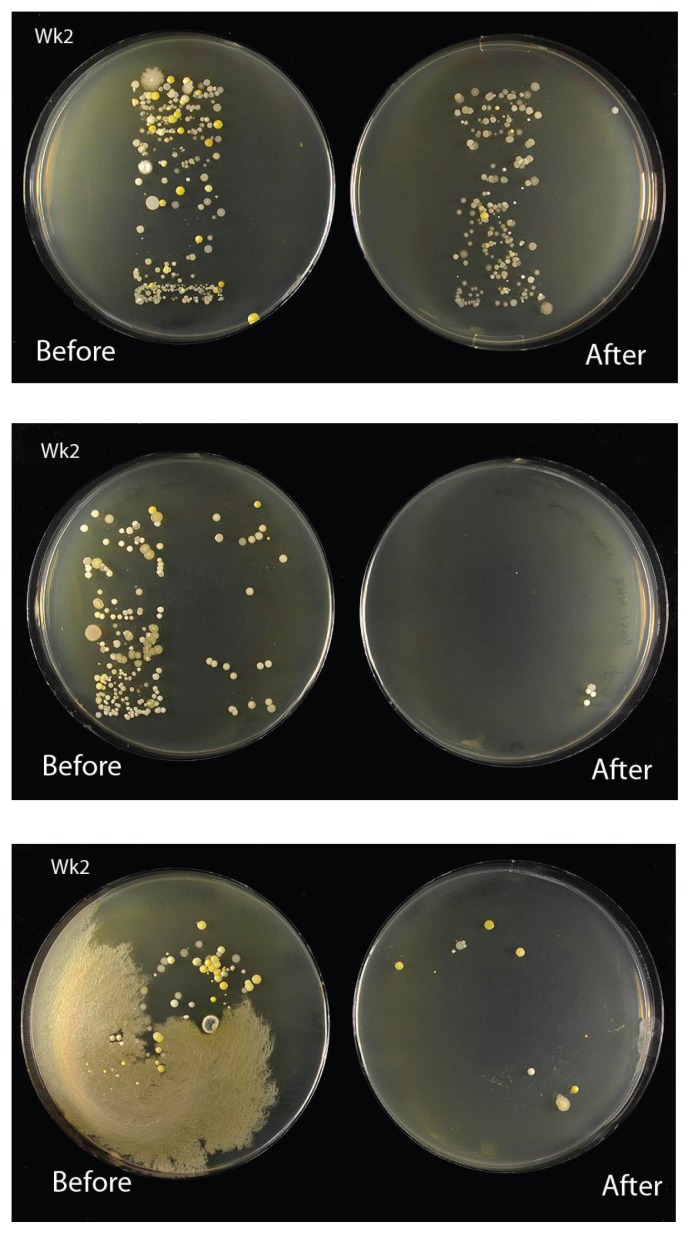
Plates usually present a ‘print’ of the mobile phone (Fig. 1), essentially a snapshot of the contamination of the phone at the time of sampling, with colonies predominant around any topographic features (raised areas, edges). It is easy to photograph these plates. The students can use their own phone cameras, or a stand with a digital camera can be set up in the laboratory and students queue to get photos taken. All images with student names attached are posted on the Intranet for use in reports, and a library of images is then acquired for the faculty.
FIGURE 1.
The contamination of three different mobile phones before and after wiping with an antimicrobial phone wipe containing benzalkonium chloride as the major active ingredient, and showing the range of contaminants and the varying effectiveness of the wipe.
Sometimes it is possible to count colonies and compare numbers before and after cleaning. On occasion, spreading colonies prevent counts from being made. Predominant colony morphologies can be described and compared. It is important that students learn how to describe colonies and to record the frequencies of different colony morphologies. By numbering their different colonies, students should be able to follow these isolates through any identification process. (In addition, students often confuse cells with colonies, thus asking for typical colony morphologies is important).
Gram staining of colonies tends to reveal a predominance of Gram-positive cocci, and occasional spore-forming rods. Typically there are many Micrococcus colonies, several coagulase-negative Staphylococci, and some Bacillus species.
Interpretation/extension
A range of questions can be asked that would enable the students to consider the significance of their findings, and/or design additional brief investigations.
Relating to phone contamination
Are you surprised/alarmed about your results?
Where do the microorganisms come from?
Does the style of the device affect the amount of contamination?
How does contamination differ from person to person?
Relating to phone hygiene
Are your wipes antimicrobial? How can you show this?
Do antimicrobial wipes reduce contamination?
How reliable are colony counts in this experiment?
Does the cost of a phone wipe affect its antimicrobial effectiveness?
What do the different components of the wipe formulation do?
Should you clean your phone? If not, why not; if yes, how often, and why?
Implications of findings
What contaminants would be undesirable on a mobile phone used in a hospital setting?
What further tests might you used to look for particular microorganisms?
Whose phone would be of most concern to you?
Is it dangerous to use a mobile phone in a hospital?
What other inert devices might pose a biotransfer potential?
Additional laboratory exercises
How does contamination of the same phone differ from week to week?
Does contamination build up over time (days)?
Isolate and identify staphylococci from the phone.
Compare identity and antibiotic sensitivity of Staphylococcus strains from the nares with those obtained from the mobile phone.
Comment on the variability of the skin/nasal community compared to the contamination of the inert mobile phone.
CONCLUSION
This very simple laboratory exercise can be used to illustrate a range of phenomena, from a simple demonstration of the contamination of inert devices and the importance of hygiene, to a consideration of the variability of results obtained and difficulties in interpreting findings. There is ample opportunity for extension activity and project work, alongside exploration and critical evaluation of the relevant scientific literature (and the popular press).
The 2009 H1N1 influenza pandemic focused on the importance of fomites in cross-contamination and the spread of infection. This laboratory activity can be used to illustrate the source of contamination and ways of controlling spread. Mobile phones and other communication devices are essential to the everyday life of today’s student, and the class provides an example of microbial contamination that is relevant to them.
SUPPLEMENTAL MATERIALS
Appendix 1: Poster - Investigations Into the Microbial Contamination of Mobile Phones
ACKNOWLEDGMENTS
The poster was presented at ASMCUE 2008. The author declares that there are no conflicts of interest.
Footnotes
Supplementary materials available at http://jmbe.asm.org
REFERENCES
- 1.Brady RRB, Wasson A, Stirling I, McAllister C, Damani NN. Is your phone bugged? The incidence of bacteria known to cause nosocomial infection on health care workers’ mobile phones. J. Hosp. Infect. 2006;62:112–125. doi: 10.1016/j.jhin.2005.05.005. [DOI] [PubMed] [Google Scholar]
- 2.Brady RRB, Verran J, Damani NN, Gibb AC. Review of mobile communication devices as potential reservoirs of nosocomial pathogens. J. Hosp. Infect. 2009;71:295–307. doi: 10.1016/j.jhin.2008.12.009. [DOI] [PubMed] [Google Scholar]
Associated Data
This section collects any data citations, data availability statements, or supplementary materials included in this article.
Supplementary Materials
Appendix 1: Poster - Investigations Into the Microbial Contamination of Mobile Phones


