Abstract
The central complex of Drosophila melanogaster plays important functions in various behaviors, such as visual and olfactory memory, visual orientation, sleep, and movement control. However little is known about the genes regulating the development of the central complex. Here we report that a mutant gene affecting central complex morphology, cbd (central brain defect), was mapped to ten-a, a type II trans-membrane protein coding gene. Down-regulation of ten-a in pan-neural cells contributed to abnormal morphology of central complex. Over-expression of ten-a by C767-Gal4 was able to partially restore the abnormal central complex morphology in the cbd mutant. Tracking the development of FB primordia revealed that C767-Gal4 labeled interhemispheric junction that separated fan-shaped body precursors at larval stage withdrew to allow the fusion of the precursors. While the C767-Gal4 labeled structure did not withdraw properly and detached from FB primordia, the two fan-shaped body precursors failed to fuse in the cbd mutant. We propose that the withdrawal of C767-Gal4 labeled structure is related to the formation of the fan-shaped body. Our result revealed the function of ten-a in central brain development, and possible cellular mechanism underlying Drosophila fan-shaped body formation.
Introduction
The central complex is an interconnected neuropil structure across and along the sagittal mid-section of the fly brain and includes the protocerebral bridge (PB), the fan-shaped body (FB), the paired nodule (NO), and the ellipsoid body (EB). It is involved in multi-modal behavioral control, such as locomotion [1]–[3], visual pattern memory [4], [5] and spatial orientation [6], [7]. The development of the central complex can be traced back to the larval stage [8], [9]. Lineage analysis has revealed the neurons that contribute to the central complex [8], [9], [10], but the molecular and cellular mechanism of central complex formation is not fully understood.
In the 1980s, Martin Heisenberg and coworkers generated a series of structural mutants, in which the morphology of adult central brain structures like mushroom bodies and the central complex were destroyed [1], [11], [12]. Among these mutants, mbm (mushroom body miniature), ceb (central brain deranged) and nob (no-bridge) have been identified. mbm was found to be a transcription factor, a nucleic acid-binding zinc finger protein [13], while ceb was reported to encode Neuroglian, a cell adhesion molecule that is crucial for axonal development, synapse formation and female receptivity [14]–[17]. As to nob, it interacted with drl at the interhemispheric junction to affect the formation of protocerebral bridge [18]. Another mutant type is central body defect (cbd), of which the most typical phenotype is that the fan-shaped body and the ellipsoid body are fragmented in the middle, or some fusion of the fan-shaped body and the ellipsoid body. So far, the molecular basis of most structural mutants is unclear.
Ten-a belongs to a large protein family, Teneurin, which contains an N-terminal intracellular domain, a single transmembrane domain, eight EGF-like domains, a 6-blade β-propeller TolB-like domain, and 26 YD repeats [19], [20]. From invertebrates to vertebrates, Teneurins function as signaling molecules at the cell surface as type II transmembrane receptors, while the intracellular domain cleaved from membrane works as a transcription regulator [21]–[23] and carboxyl terminus functions as a bioactive peptide [24]–[27]. The Teneurin family members are thought to be important for establishment and maintenance of neuronal connections, neurite outgrowth and axon guidance [20]. Recent reports showed that two Drosophila Teneurin members, Ten-a and Ten-m, are crucial for proper synaptic matching and the maintenance of neuromuscular junction [28], [29]. Although Teneurin may play a role in mammalian brain function [20], [30], detailed study is still largely lacking.
Here, we report that the Drosophila structural mutant gene cbd, the most typical phenotype of which is the fragmented fan-shaped body and ellipsoid body, is ten-a. The cbd mutation disrupts the formation of the FB, by preventing the merging of the two FB parts. This defect was rescued by over-expression of ten-a in a C767-Gal4 labeled structure which separated the FB parts but later disappeared to allow the merging of the two FB primordia. Our results might reveal the molecular and cellular mechanism of Drosophila central complex development.
Materials and Methods
Fly strains
Flies were cultured on standard cornmeal food at 25°C with a 12 h light : 12 h dark cycle at 60% humidity [31]. Wild-type flies Berlin (WTB) and w1118 were used in our study. The cbd lines were generated by EMS mutagenesis of WTB [32]. The deficiency lines (Df(1)ED7161, Df(1)ED7153, Df(1)KA10, Df(1)RC29, Df(1)ED7147), and flyC31 (y1M{vas-int.Dm}ZH-2Aw*; M{3xP3-RFP.attP}ZH-86Fb) were obtained from the Bloomington Drosophila Stock Center (Indiana University). The ten-a RNAi line (w1118; P{GD3330}v8322) was obtained from the Vienna Drosophila RNAi Center (Vienna, Austria). NP6510 was obtained from Drosophila Genetic Resource Center (Kyoto Institute of Technology, Japan). The ten-a imprecise jump out line ten-a#900 was a gift from Dr. Ron Wides.
DNA Sequencing
To determine whether there was a mutation in genes between 11A5 and 11A7, we sequenced the coding regions of CG1924, CG32655, and ten-a in wild-type flies (WTB) and cbd flies. The genome fragments were PCR amplified using Pfu polymerase (Promega). The gene-specific primers were designed according to the FlyBase Release 5.45 genome sequence (www.flybase.org). The primers for amplification also were used for sequencing for genes ten-a and CG32655. For gene CG1924, besides primer30 for amplifying and sequencing, other sequencing primers (S1, S2) were used.
Primers for ten-a:
Primer1: 5′-GGATCGTGGGCATCGGCGGTG-3′ and 5′-TTTACAAATTAGTTGAC-3′;
Primer2: 5′-GGAAGTTGGGTTCCATAGCA-3′ and 5′-GGCATCTATTTCCAGCCTGA-3′;
Primer3: 5′-CCCAACTGAGCGAGGAAATA-3′ and 5′-ACAATGTGGAGGTTCCAACG-3′;
Primer4: 5′-CAACAGACTGTTAGGCAAGAGA-3′ and 5′-TTGCACGCTTTTTCCCTATC-3′;
Primer5: 5′-GATAGGGATTTCGACGCAGA-3′ and 5′-AAGTGCATCGAGTGCATTATTTA-3′;
Primer6: 5′-TTCGAGTGCATCCCAAAAAT-3′ and 5′-CCCATATTCCGCATCTCCTA-3′;
Primer7: 5′-CCCACCCCCTTTTTGTTAAT-3′ and 5′-TTCTTAGCTGGCCGAAGTGT-3′;
Primer8: 5′-CGGCAGATAAGATGAAACAACA-3′ and 5′-GCCTCGTTGAACTCCTTCAG-3′;
Primer9: 5′-GTGGCATAATGAATGGTGGA-3′ and 5′-CTGCAGCAGGGATACATTCA-3′;
Primer10: 5′- GATACGGCCAAACAGCATCT-3′; and 5′-CGGGATTCCCCGTTATATTC-3′
Primer11: 5′-AAAACCACCAAATGCTGACC-3′ and 5′-AGCTCGTGATTTCCAGTTCC-3′;
Primer12: 5′-TAAATGCGCACAATGGAAAA-3′ and 5′-ACCGCAATGTTGCTGTTGTA-3′;
Primer13: 5′-CAATTCGATTGCGTGTCAAG-3′ and 5′-GAATCCCTGCGCACTAAGAG-3′;
Primer14: 5′-GCAAAAACTCGAACGCAAAT-3′ and 5′-GCAAAAACTCGAACGCAAAT-3′;
Primer15: 5′-ATTTCTCATGCCACCCACTC-3′ and 5′-TGGTAAATGAGGGGCACTTT-3′;
Primer16: 5′-CTGTCACCTGAGACCGATGA-3′ and 5′-ATTGCAGTAATCCGGACAGG-3′;
Primer17: 5′-GGAGTATCCGAGAACCGTCA-3′ and 5′-GGATCTTCATGTCCGAGGTG-3′;
Primer18: 5′-CATGGCCATCACAATCACTG-3′ and 5′-TAGCAGCGAACCTAATCGAA-3′;
Primer19: 5′-TCTAACACGCATTTCCCTCA-3′ and 5′-AAAATCCCACGAAAAACGTC-3′;
Primer20: 5′-CAGGTCAAATAGTGCGAATGC-3′ and 5′-CCCATGGTGACACTTTGATG-3′;
Primer21: 5′-CCCTAGTGATTCATGGCGATA-3′ and 5′-GAAGTCCCATCGGTACTCCA-3′;
Primer22: 5′-CTGCTCCAGACGATCCTACC-3′ and 5′-GCTCTTCTCTGGCATTTTGG-3′;
Primer23: 5′-GCCCAGGACAGGATTGTAAA-3′ and 5′-CGGCAAAGTCCTCTGGATAG-3′;
Primer24: 5′-CGTGTTGCCGAGAGGATTAT-3′ and 5′-GCTGGTCCACTACCCACAGT-3′;
Primer25: 5′-GCAGGACTCGTTCTTCTTCG-3′ and 5′-CTGTTCTTCGGTTTTCACTGC-3′;
Primer26: 5′-CGCAGATCCACCGATCTAAT-3′ and 5′-GTTGCCGTTCAATTGGTTTT-3′;
Primer27: 5′-TTTATGGGAATGGGCGTATC-3′ and 5′-GCATTGAGCTGAGTTCGAGA-3′;
primers for CG32655
Primer28: 5′-GAGCGACTGAGGATTCCCTA-3′ and 5′-TGGGTCTTTCGCTAGTCGTT-3′;
Primer29: 5′- CAGTGCTAGTTCCGTCGTGA-3′ and 5′-TGAAAAGGCTGGCTAGTTGG-3′;
primers for CG1924
Primer30: 5′-CCGGGAAAACTGTTGAAAAA-3′ and 5′-TATGAATGCCCGCTTACTCC-3′;
S1: 5′-TTCAAGTCGGAGAAGCCACT-3′
S2: 5′-ATCATCCGCAATCCCAACTA-3′
Construction of transgenic flies UAS-ten-a
The first strand cDNA of ten-a was synthesized from fly head mRNA using the SuperScript III first-strand synthesis system (Invitrogen). PCR reactions were performed by Pfu polymerase (Promega) with 5′ primer 5′-GCAGTCAGATCTATGACTATGAAATCGATGAAG-3′ and 3′ primer 5′-ATTACTGACGTCGGTACCCTAACAGTCGGCTTCGCG-3′. BglII and KpnI adapters were added to the 5′ and 3′ primers, respectively. The 9042 bp product was inserted into the BglII and KpnI sites of pUASTattB (a gift from Konrad Basler) after the sequence was confirmed. The purified construct was introduced to FlyC31 strains.
Immunohistochemistry
Flies were allowed to lay eggs on standard fly food for 30 min and brain dissection was performed at different time points of development: larvae, 48–72 h, 72–96 h and 96–120 h after egg laying; pupa, 0–2 h and 8–9 h after puparium formation; adult fly, 3∼5 days after eclosion. Immunostaining was performed as described previously [33] with modifications. Briefly, dissection was performed in a dish covered with cold PBS (phosphate-buffered saline, pH 7.4). The samples were fixed in freshly prepared paraformaldehyde (4% in PBS) for 3 h on ice. Brains were then washed in PBT (PBS+0.5% Triton X-100) for 3×15 min, followed incubation for 1 h in PNT (PBT+10% normal goat serum) at room temperature. Subsequently, rat monoclonal antibody to DN-cadherin (DSHB, DN-EX #8, dilution 1∶100) was used to track the development of the central complex in the larval and pupal stage. Nc82 antibody (DSHB; mAb nc82, dilution 1∶100) against Brp protein was the primary antibody used to stain the neuropil in adult flies. Rabbit anti-GFP antibody (Invitrogen Inc, dilution 1∶1000) was used to check the expression pattern of Gal4 lines. After incubating overnight at 4°C in primary antibody, samples were washed in PBT 3×15 min at room temperature, and then incubated overnight in secondary antibody. Goat anti-Rat antibody (TRITC-conjugated, Jackson Laboratories, dilution 1∶200), goat anti-Mouse antibody (TRITC-conjugated, Jackson Laboratories, dilution 1∶200), and goat anti-rabbit antibodies (FITC-conjugated, Invitrogen Inc, dilution 1∶200) were used as secondary antibodies. In the following day, after being rinsed in PBT 3×15 min, brains were mounted in Vectashield Fluorescent Mounting Media (Vector Laboratories, Burlingame) and observed.
Imaging
Mounted brains were scanned with a confocal microscope (Leica TCS SP5). Each brain stack z-resolution was 0.8 µm or 1.0 µm for adult fly brains and 0.5 µm for larval or pupal brains, at pixel resolution of 1024×1024. The images were then processed with ImageJ (National Institutes of Health, rsbweb.nih.gov/ij/).
Statistics
A two-tailed Fisher exact test was used to evaluate the efficiency of rescue experiments, and two sample t-test was carried out for the numbers of F1 neurons or F5 neurons. Statistical significance was assigned, *P<0.05, **P<0.01, ***P<0.001.
Results
cbd is mapped to ten-a
Although most structural mutants generated by Heisenberg and colleagues are still uncharacterized, some of them have been preliminarily mapped [13], [15], [18], [32]. The structural mutant gene cbd, which contributed to the disrupted fan-shaped body and ellipsoid body, was localized to 11A1–A7 on the X chromosome [1], [32]. To further localize cbd, we adopted a traditional method of deficiency mapping using complementation tests (Fig. 1). cbd KS171, a representative cbd mutant, was crossed with a series of deficiency lines around the 11A region and the brain morphology of trans-heterozygous female offspring was checked. Three large deficiencies, Df(1)ED7161, Df(1)ED7153, Df(1)KA10, which respectively remove 10D1-11B14, 11A1-11B1 and 11A1–11A7, failed to restore the deranged central complex in cbd KS171 mutants, confirming that the cbd gene lies within 11A1–11A7 (Fig. 1B–I). Next we used deficiencies with a breakpoint between 11A1 and 11A7 for fine mapping. Both deficiency lines Df(1)RC29 and Df(1)ED7147, that remove 11A1–11A5 and 10D6-11A1 respectively, could complement the cbd KS171 mutation, suggesting that cbd lies outside of 11A1–11A5 and locates in 11A5∼11A7 (Fig. 1J–M). The complementation tests for another two cbd mutants, cbd KS96 and cbd 2254 confirmed that cbd was located in 11A5–11A7 (Fig. 1N–Y).
Figure 1. Morphology of the fan-shaped body (FB) and the ellipsoid body (EB) in the complementation test.
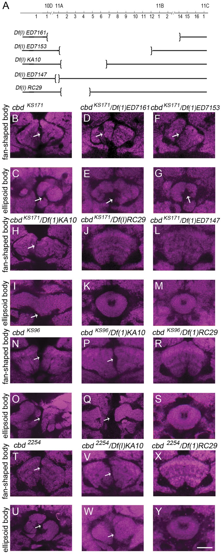
(A) Schematic drawing of deleting regions of five deficiency lines on the X chromosome. (B, C) cbd KS171 mutant showed a defect in FB and EB. (D–I) complementation test cbd KS171 and Df(1)ED7161 (D,E) or Df(1)ED7153 (F,G) or Df(1)KA10 (H,I) showed a defect in FB and EB. (J–M) complementation test cbd KS171 and Df(1)RC29 or Df(1)ED7147 showed a normal FB and EB. (N, O) cbd KS96 mutant showed a defect in FB and EB. (P–S) complementation test between cbd KS96 and Df(1)KA10 showed a defect in FB (P) and EB (Q). cbd KS96 and Df(1)RC29 showed a normal FB (R) and EB (S). (T, U) cbd 2254 mutant showed a defect in FB and EB. (V–Y) complementation test between cbd 2254 and Df(1)KA10 showed a defect in FB (V) and EB (W) while that between cbd 2254 and Df(1)RC29 showed a normal FB (X) and EB (Y). Scale bar, 25 µm. Arrows indicate the central complex defect.
We then checked the annotated genes in 11A5–11A7. According to FlyBase annotation release 5.45, there are three genes in this region: CG1924, CG32655 and CG42338. We sequenced the coding DNA region of these three genes in the three cbd mutant lines cbd KS171, cbd 2254 and cbd KS96, and found no sequence change in CG1924 and CG32655 that could lead to changes in amino acids or splicing sites. Interestingly, we found significant DNA sequence changes in the coding region of CG42338, also named ten-a (Fig. 2A). For convenience of illustration, the mutation points were assigned to the transcript RE, which encodes the largest protein isoform (3378 aa) according to the FlyBase annotation. In cbd 2254, a TGG codon (Tryptophan, 1562) was changed to TGA (a premature stop codon). In cbd KS171, a GGC codon was changed to AGC (Glycine in 1723 changed to Serine). In cbd KS96, the CGT codon was changed to TGT (Arginine 2846 changed to Cysteine) (Fig. 2B, S1). It is worth noting that all three amino acid changes occur in the conserved domains of the Ten-a protein (Fig. S2). Besides these three mutations, there are several amino acid residues that are same in the three cbd lines but different from control flies (WTB), and all are in non-conserved domain of Teneurin (Fig. S3). All of these data strongly hint that cbd is ten-a.
Figure 2. Schematic drawing of genomic region containing ten-a.
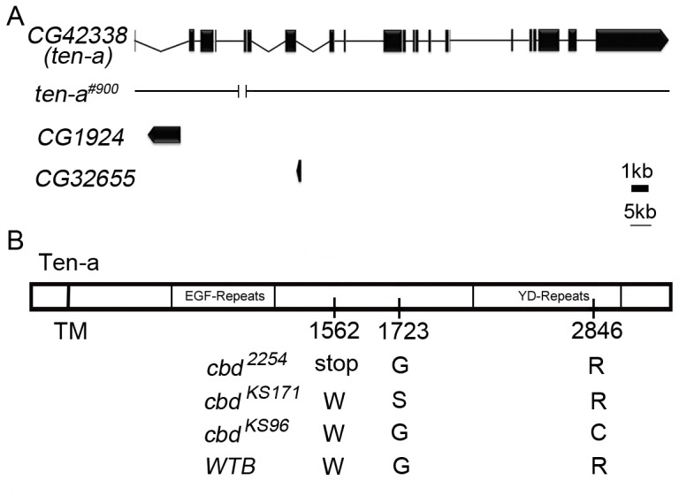
(A) Schematic drawing of CG42338 (ten-a), CG1924, CG32655, and ten-a deficiency line ten-a#900. (B) Schematic drawing of mutant site of Ten-a. In cbd 2254, Tryptophan at 1562 is changed to a premature stop codon. In cbd KS171, Glycine at 1723 is changed to Serine. In cbd KS96, Arginine at 2846 is changed to Cysteine.
ten-a is required for normal central complex morphology
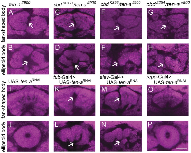
Since cbd is likely ten-a, we wondered if manipulating ten-a gene expression would affect morphology of the fly central complex, as cbd mutations do. We first made use of the ten-a deficiency line ten-a#900 which was generated by P element imprecise excision in ten-a. In ten-a#900, a 2219 bp region covering two ten-a exons was deleted (Fig. 2A). ten-a#900 is homozygous viable, but the central complex morphology is disrupted (Fig. 3A, B). Complementation tests between ten-a#900 and cbd mutants, cbd KS171, cbd KS96 and cbd 2254 showed that ten-a#900 was unable to complement the cbd mutant based on brain morphology (Fig. 3C–H). Thus, ten-a#900 is another cbd mutant allele. However, the question still remained that there might exist some other common mutation outside of ten-a in these mutant lines that generated the abnormal brain phenotype. We then specifically down-regulated ten-a expression by driving expression of UAS-ten-a RNAi with tub-Gal4. We noticed that morphology of both FB and EB was destroyed comparing with control flies (Fig. 3I–L). We further asked where ten-a functioned to affect fly brain morphology. By driving UAS-ten-a RNAi with pan-neuronal elav-Gal4 and pan-glial repo-Gal4, we found distinct results: down-regulation of ten-a in neurons produced strong brain derangement, while down-regulation of ten-a in glial cells had no effect on fly brain morphology (Fig. 3M–P). It suggests that ten-a is required in neurons for normal central complex morphology.
Figure 3. ten-a deficiency line ten-a#900 and down-regulated ten-a expression by RNAi caused FB and EB defects.
(A, B) ten-a#900 showed a FB and EB defect. (C–H) cbd KS171, cbd 2254 and cbd KS96 could not complement the FB and EB defect of ten-a#900. (I–L) Both the FB and the EB were destroyed when ten-a was down-regulated by driving expression of UAS-ten-a RNAi with tub-Gal4. (M–N) Both the FB and the EB were destroyed when ten-a was down-regulated by driving expression of UAS-ten-a RNAi with pan-neuronal elav-Gal4. (O–P) Both the FB and the EB were normal when ten-a was down-regulated by driving expression of UAS-ten-a RNAi with pan-glial repo-Gal4. Scale bar, 25 µm. Arrows indicate the central complex defect.
Over-expression of ten-a restored normal brain morphology in cbd mutant
Next, we undertook fine mapping of the cells in which Ten-a functions to affect the central complex morphology by attempting to rescue the cbd mutant phenotype via over-expressing ten-a with screening various Gal4 lines. During the screening, pan-neuronal Gal4 line, glia-specific Gal4 line, and some region specific Gal4 lines were chosen for rescue experiments. Finally only C767-Gal4, which labeled the EB as well as the median bundle in adult central brain, significantly rescued the cbd mutant phenotype (Fig. 4). Since the phenotype is the cleavage of FB, it is reasonable to postulate that it is the C767-Gal4 labeled structure at midline but not at other regions that affected the FB morphology. Interestingly, the percentage of rescue was a little higher when flies were cultured at a constant temperature of 18°C comparing with flies cultured at a constant temperature of 25°C (Fig. 4I). Thus, Ten-a in C767-Gal4 labeled cells contributed to normal central complex formation.
Figure 4. The cbd mutant phenotype could be significantly rescued when ten-a was driven by C767-Gal4.

(A, B) WTB showed normal FB and EB. (C–F) Both UAS-ten-a and C767-Gal4 in the cbd KS171 background showed defect in FB and EB. (G, H) FB and EB were restored to normal when UAS-ten-a was driven by C767-Gal4. (I) Percentage of normal FB and EB in controls (5.6% for cbd KS171;UAS-ten-a, n = 36; 10% for cbd KS171;C767-Gal4/+, n = 30) and in flies with UAS-ten-a driven by C767-Gal4 (36%, n = 25). Two tailed Fisher exact test. *P<0.05, **P<0.01, ***P<0.001. When ten-a over-expressing flies were kept at 18°C, 47% (n = 32) of flies showed a normal FB and EB, much higher than control flies (12.5% for cbd KS171;UAS-ten-a, n = 24; 11.1% for cbd KS171;C767-Gal4/+, n = 18). Scale bar, 25 µm. Arrows indicate the central complex defect.
Defective FB in cbd mutants is caused by the failure of FB merging
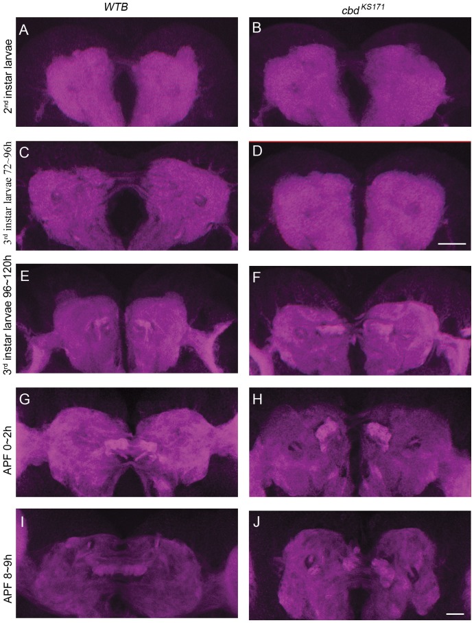
We then asked how ten-a mutation led to the deranged central complex morphology. Theoretically, the deranged FB in cbd mutants could result from either abnormal development, or from degeneration of a normal mature FB. We tracked the whole developmental process of the FB in both wild-type and cbd mutants from 2nd instar larva to pupa (Fig. 5), when the FB is supposed to form [8]. We could see several thin commissural axon tracts which was supposed as supraesophageal commissure [34] in 2nd and early 3rd instar larval brains in both wild-type and cbd mutants (Fig. 5A–D), which suggests that ten-a mutation doesn't affect the midline crossing of this structure, at least in these stages. In late 3rd instar larval brains, we found immature FB stained by DN-cadherin anterior to the supraesophageal commissure on the two sides of the midline (Fig. 5E, F). The FB precursors expand and merge at the midline by 8–9 h after pupa formation (APF) in wild-type flies (Fig. 5G, I). In cbd KS171 flies, the similar event happens until 0∼2 h APF. However, in cbd mutants the two strong DN-cadherin labeled precursors were unable to merge at 8∼9 h APF (Fig. 5H, J), even at later time points that we have checked (data not shown). Thus, the broken FB in cbd mutants was likely caused by a developmental defect in FB formation, but not by neuronal degeneration of the formed FB.
Figure 5. The development of the FB in both WTB and cbd KS171 from 2nd instar larva to pupa.
(A–D) DN-cadherin signal in the commissure in 2nd instar larval brain of WTB (A) or cbd KS171 mutant (B), and in early 3rd instar larval brain of WTB (C) or cbd KS171 mutant (D). Scale bar in (D) equals 25 µm and applies to (A–D). (E–J) FB precursors strongly labeled by DN-cadherin in the commissure in a late 3rd instar larval brain from WTB (E) or a cbd KS171 mutant (F), and in a 0–2 h APF pupal brain from WTB (G) or a cbd KS171 mutant (H), and in an 8–9 h APF pupal brain from WTB (I) or a cbd KS171 mutant (J). Scale bar in (J) equals 25 µm and applies to (E–J).
C767-Gal4 labeled structure at interhemispheric junction might be required for FB formation
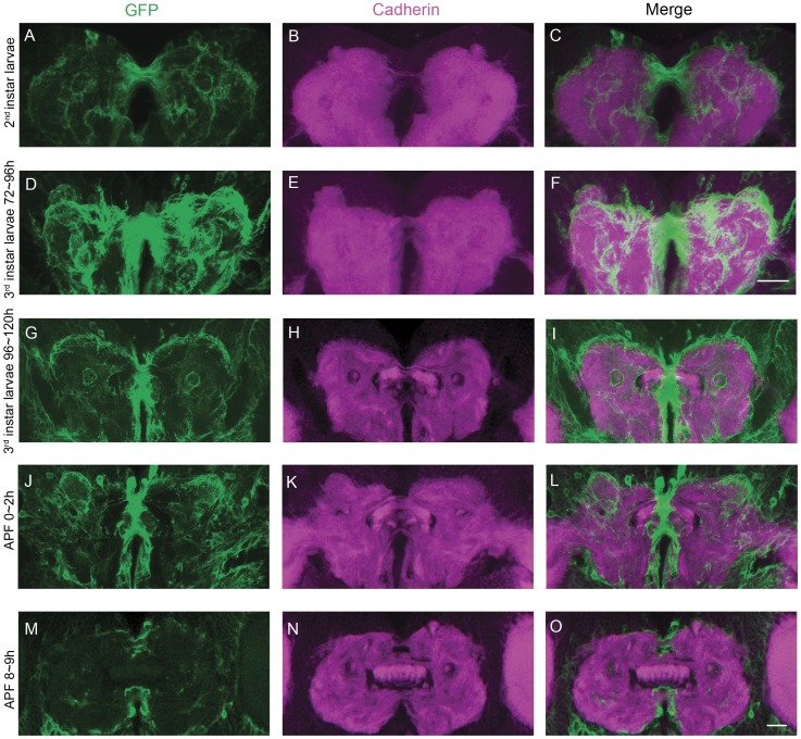
We then asked exactly how ten-a affected the process of FB development. As cbd mutant phenotype can be partially rescued by over-expressing ten-a in C767-Gal4 labeled structure, we supposed that the labeled cells are closely related to the event. The expression pattern of C767-Gal4 in the region of larval FB precursors was tracked during the process of FB formation in both wild-type flies and cbd mutants. At the 2nd instar larval stage, C767-Gal4 labeled universally in the central brain, but was significantly strong in midline regions joining the two hemispheres (Fig. 6A–C). It is worth noting that the two DN-cadherin stained FB patches were separated by a C767-Gal4 labeled structure at interhemispheric junction (Fig. 6G, J, M). Later in the 3rd instar larval stage, the interhemispheric junction was narrowed while the FB precursors expanded and invaded medially. In wild type background, the interhemispheric junction and the FB precursors are still tightly connected as if the withdrawal of C767-Gal4 labeled structure at interhemispheric junction was accompanied with the extending of the DN-cadherin signal (Fig. 6G). By the pupal stage, the C767-Gal4 labeled midline structure further shrank and disappeared completely to allow complete merging of the two FB primordium parts (Fig. 6J, M). Judging from the concurrency between the morphological changes in C767-Gal4 stained structure and DN-cadherin labeled FB primordia, we postulated that the merging of FB primordia was influenced by the C767-Gal4 labeled midline junction.
Figure 6. The expression pattern of C767-Gal4 and DN-cadherin signal during FB formation in wild type flies.
(A–C), The expression pattern of C767-Gal4 (green) and DN-cadherin signal (magenta) in a 2nd instar larval brain. (D–F) The expression pattern of C767-Gal4 (green) and DN-cadherin signal (magenta) in an early 3rd instar larval brain. Scale bar in (F) equals 25 µm and applies to (A–F). (G–I) The expression pattern of C767-Gal4 (green) and DN-cadherin signal (magenta) in a late 3rd instar larval brain. (J–L) The expression pattern of C767-Gal4 (green) and DN-cadherin signal (magenta) in a pupal brain at 0–2 h APF. (M–O) The expression pattern of C767-Gal4 (green) and DN-cadherin signal (magenta) in a pupal brain at 8–9 h APF. Scale bar in (O) equals 25 µm and applies to (G–O).
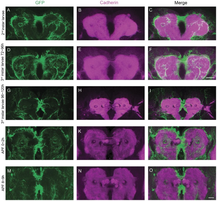
To confirm this hypothesis, we checked the development of C767-Gal4 labeled cells and FB morphology in cbd KS171 mutant (Fig. 7). In cbd KS171 mutant, the C767-Gal4 labeled midline structure that separated the two FB patches persisted during 0–2 h APF. But the structure is detached from the precursors and failed to guide the merging of FB parts and consequently the formation of a normal FB (Fig. 7). Based on this morphological observation, as well as the rescue results, we concluded that the Ten-a molecule in the C767-Gal4 labeled structure affected merging of FB primordia.
Figure 7. The expression pattern of C767-Gal4 and DN-cadherin signal during FB formation in cbd KS171 mutants.
(A–C) 2nd instar larval brain. (D–F) early 3rd instar larval brain. Scale bar in (F) equals 25 µm and applies to (A–F). (G–I) late 3rd instar larval brain. (J–L) pupal brain 0–2 h APF. (M–O) pupal brain 8–9 h APF. Scale bar in (O) equals 25 µm and applies to (G–O).
Defective FB in cbd mutants is not caused by abnormal axonal projections
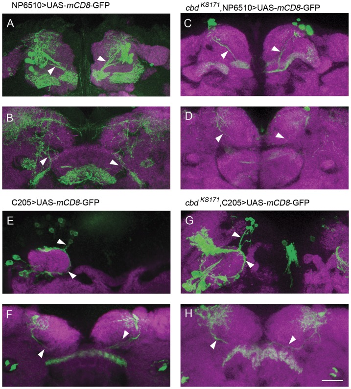
Since the deficiency is not due to the degeneration, we also wondered if the failure in FB part merging in cbd mutants was due to improper generation or specification of FB neurons. We counted the neurons labeled by NP6510-Gal4 and C205-Gal4 in both cbd KS171 mutant and wild type adult flies. The somata of F1 neurons were located anterior to the antennal lobes and the somata of F5 neurons were around the calyxes of mushroom bodies. The numbers of neurons varied even in each hemisphere in the same brain and there was no significant difference between wild type and cbd KS171 flies (Fig. 8A, C, E, G, Fig. S4). We also wondered if the abnormal morphology of FB was caused by abnormal axonal projections of FB neurons. The morphology of F1 neurons labeled by NP6510-Gal4 and F5 neurons labeled by C205-Gal4 were checked in adult cbd mutant flies. As shown in Figure 8, FB in cbd KS171 mutants were fragmented into pieces (Fig. 8D, H), but the projection tracts of both F1 neurons and F5 neurons appeared to be normal in cbd KS171 mutants (Fig. 8B, F). Thus, the mutant FB phenotype in cbd mutants is not caused by abnormalities in neuronal projection. Rather, the FB defect in cbd mutants was caused by abnormalities in growth of the FB midline-crossing arborizations, but terminal arborizations in the non-cleft region could still form in cbd mutants.
Figure 8. The projection tracts of both F1 neurons and F5 neurons appeared to be normal in cbd KS171 mutants.
(A–D) Neural projection and arborizations of F1 neurons in wild type flies and cbd KS171 mutants. (E–H) Neural projection and arborizations of F5 neurons in wild type flies and cbd KS171 mutants. Scale bar, 25 µm. Arrowheads indicate the normal neural projection.
Discussion
In this study, we found that the Drosophila central brain morphological mutant cbd is actually ten-a, a member of the teneurin family. Ten-a is required for fusion of the fan-shaped body precursors, before the formation of the complete normal FB. Mutation in ten-a leads to the failure of the two FB precursors to merge and consequently to the deranged fan-shaped body in adult flies.
Aside from the FB morphological defect itself, ten-a mutation might cause other abnormalities that contribute to the morphological defect. For example, Ten-a might affect the projections and contra-lateral crossing of FB neurons resulting from lineages of FBP1 and FBP2, which contribute to two staves of the fan-shaped body [10], consequently led to a cleaved fan-shaped body. Nevertheless, the generation and projection of large field ExFl neurons labeled by NP6510-Gal4 or C205-Gal4 are not affected when ten-a mutated (Fig. 8), which suggested that ten-a mainly produce the morphological defect by exerting its effect on FBP1 and FBP2 neuron arborizations. Actually, based on the morphological observation in cbd KS171 flies and the rescue results, we postulated that the interhemispheric structure C767-Gal4 labeled was related to FB primordial fusion. But it seemed to have no effect on axonal projections and terminal arborization of F1 and F5 neurons.
ten-a knockdown results showed that the neuronal ten-a was required for the central complex formation. Further rescue experiments suggested that neither neurons nor glial cells alone were sufficient for normal central complex formation. After screening, one Gal4 line was found finally. C767-Gal4 could be used to rescue the cbd mutant phenotype significantly. To identify the cell types labeled by C767-Gal4, we used neuron specific marker ELAV or glial cell specific marker REPO to co-stain with C767-Gal4 labeling cells. The results showed that nlsGFP driven by C767-Gal4 was co-localized with both neuronal and glial markers from larval to early pupal stages (Fig. S5). Since previous studies showed some adhesion molecules were expressed both in neurons and glia for mediating the fasciculation of axon bundles, axon guidance or targeting [35], we suggested that the rescue results by C767-Gal4 might just attribute to that the Gal4 expressed both in neurons and glial cells. That is to say, only when ten-a functions in certain neurons and glial cells together, the FB precursors could merge normally. However, we could not identify which neurons and glial cells were required for the partial rescue from current results. To solve this problem, more Gal4 lines which can rescue the cbd mutant phenotype are needed. Then, dependent on the expression patterns of these Gal4 lines, the neurons and glial cells which ten-a functions in may be identified.
If Ten-a functions in C767-Gal4 labeled cells to influence the merging of FB primordia, what is its working partner for the arborization of FB neurons? As a Drosophila homolog of vertebrate Teneurin, Ten-a has been reported to be involved in embryo development, especially in the central nervous system. Ten-a, as well as its homologue Ten-m, was recently found to be required for synaptic matching between olfactory receptor neurons and corresponding projection neurons [28]. Ten-a and Ten-m were also important for establishing the correct connection in the larval neuromuscular junction [29], [36]. In our work, lack of normal Ten-a function led to failure in merging of FB precursors. It is possible to assume that Ten-a itself mediates homophilic interaction between neurons and glial cells to regulate the fusion of the central complex, such as Nrg, which is expressed on both neurons and glial cells and interacts to control axonal sprouting and dendrite branching [37]. Meanwhile, Ten-a may interact with other molecules such as Ten-m, or other membrane proteins that function in heterophilic way at the cell surface. Further molecular and cellular experiments are needed to elaborate this important issue.
Vertebrate Teneurins have been suggested to be related to mental diseases and our discovery of Ten-a function in Drosophila brain development seems to support the hypothesis. Neuroglian (Nrg), whose vertebrate homologue L1-CAM has been implicated in neurological disorders [38], [39], is also required for development of normal brain morphology in Drosophila [40], [41]. Considering that both Nrg and Ten-a are type-II transmembrane proteins with extracellular EGF repeats and also function in glial cells for brain development [40], it is possible that Teneurins in vertebrates also affect brain development, and probably synapse formation, as vertebrate Nrg does.
In summary, our work elucidates the function of ten-a in development of the Drosophila central brain, and the cellular mechanism underlying FB formation.
Supporting Information
Sequence traces show the mutation in cbd lines. (A) Sequence trace shows the nucleotide change of G in control flies to A in cbd2254 leading to the nonsense mutation (W1562*). (B) Sequence trace in cbd KS171 shows G to A nucleotide change, leading to missense mutation (G1723S). (C) Sequence trace in cbd KS96 shows C to T nucleotide change, leading to missense mutation (R2846C). The underlines indicate the base substitution position.
(TIF)
Conservation analysis of amino acids which were mutated in cbd2254, cbd KS171, cbd KS96 , respectively. Multiple-sequence alignment for Teneurin homologues surrounding the coding changes (boxed) was done by MegAlign program. We found the three regions all are with high conservation (Cyan), especially the changed sites, W1562, G1723, R2846, which can be found in all 20 homologues.
(TIF)
Conservation analysis of amino acids that were seen in all three cbd lines, but not in control flies ( WTB ). (A) Schematic drawing of sites that are different between cbd and control flies. At position 168, P in WTB was changed to A in all three cbd lines. D328 was changed to G328. I691 was changed to M691. L3372 was changed to F3372. (B) Conservation analysis of the sites. From the alignment results, we can see these sites are in a region with low conservation (Cyan) and the changed sites (boxed) are not appeared in other homologues.
(TIF)
Average numbers of F1 and F5 neurons in control flies ( C205 -Gal4>UAS-GFP and NP6510 -Gal4>UAS-GFP) and cbd KS171 mutant flies. No significant difference of neuron numbers was observed between control flies (light grey) and cbd KS171 (dark grey), either for NP6510-Gal4 labeled F1 neurons (left) or for C205-Gal4 labeled F5 neurons (right). Two sample t-test, error bars represent the s.e.m.; n.s., not significant.
(TIF)
C767 -Gal4 labels both neurons and glial cells from the 3rd instar larval to early pupal stage. For easy illustration, multiple middle z-axis slices were stacked. GFP-labeled cell bodies (green) driven by C767-Gal4 co-localized with neurons (arrowheads) with a neural specific marker, ELAV, stained by anti-ELAV antibody (magenta) in 3rd instar larval brain (A), pupal brain 0∼2 h APF (B), and pupal brain 8∼9 h APF (C). GFP-labeled cell bodies driven by C767-Gal4 co-localized with glial cells (arrowheads) with a glial specific marker, REPO, stained by anti-REPO antibody (blue) in 3rd instar larval brain (D), pupal brain 0∼2 h APF (E), and pupal brain 8∼9 h APF (F). Schematics of distributions of neurons and glial cells in whole brains of 3rd instar larva (G), pupa 0∼2 h APF (H), and pupa 8∼9 h APF (I). Scale bars, 25 µm.
(TIF)
Acknowledgments
We are grateful to Martin Heisenberg and Ron Wides for providing the flies, Konrad Basler for providing pUASTattB. We thank Haiyun Gong, Mo Han, and Jianwen Wu for technical assistance. We also thank the Bloomington Drosophila Stock Centers, the Vienna Drosophila RNAi Center, and Drosophila Genetic Resource Center at Kyoto Institute of Technology for fly stocks.
Funding Statement
This work was supported by the ‘973 Program’ (2009CB918702, 2012CB825504), the National Natural Sciences Foundation of China (31030037, 30921064 and 31070944), and the External Cooperation Program of the Chinese Academy of Sciences (GJHZ1005). The funders had no role in study design, data collection and analysis, decision to publish, or preparation of the manuscript.
References
- 1. Strauss R, Heisenberg M (1993) A higher control center of locomotor behavior in the Drosophila brain. J Neurosci 13: 1852–1861. [DOI] [PMC free article] [PubMed] [Google Scholar]
- 2. Martin JR, Raabe T, Heisenberg M (1999) Central complex substructures are required for the maintenance of locomotor activity in Drosophila melanogaster. J Comp Physiol [A] 185: 277–288. [DOI] [PubMed] [Google Scholar]
- 3. Strauss R (2002) The central complex and the genetic dissection of locomotor behaviour. Curr Opin Neurobiol 12: 633–638. [DOI] [PubMed] [Google Scholar]
- 4. Liu G, Seiler H, Wen A, Zars T, Ito K, et al. (2006) Distinct memory traces for two visual features in the Drosophila brain. Nature 439: 551–556. [DOI] [PubMed] [Google Scholar]
- 5. Pan Y, Zhou Y, Guo C, Gong H, Gong Z, et al. (2009) Differential roles of the fan-shaped body and the ellipsoid body in Drosophila visual pattern memory. Learn Mem 16: 289–295. [DOI] [PubMed] [Google Scholar]
- 6. Neuser K, Triphan T, Mronz M, Poeck B, Strauss R (2008) Analysis of a spatial orientation memory in Drosophila . Nature 453: 1244–1247. [DOI] [PubMed] [Google Scholar]
- 7. Xiong Y, Lv H, Gong Z, Liu L (2010) Fixation and locomotor activity are impaired by inducing tetanus toxin expression in adult Drosophila brain. Fly (Austin) 4: 194–203. [DOI] [PubMed] [Google Scholar]
- 8. Young JM, Armstrong JD (2010) Building the central complex in Drosophila: the generation and development of distinct neural subsets. J Comp Neurol 518: 1525–1541. [DOI] [PubMed] [Google Scholar]
- 9. Boyan GS, Reichert H (2011) Mechanisms for complexity in the brain: generating the insect central complex. Trends Neurosci 34: 247–257. [DOI] [PubMed] [Google Scholar]
- 10. Ito K, Awasaki T (2008) Clonal unit architecture of the adult fly brain. Adv Exp Med Biol 628: 137–158. [DOI] [PubMed] [Google Scholar]
- 11. Hanesch U, Fischbach K, Heisenberg M (1989) Neuronal architecture of the central complex in Drosophila melanogaster . Cell Tissue Res 257: 343–366. [Google Scholar]
- 12. Heisenberg M, Borst A, Wagner S, Byers D (1985) Drosophila mushroom body mutants are deficient in olfactory learning. J Neurogenet 2: 1–30. [DOI] [PubMed] [Google Scholar]
- 13. Raabe T, Clemens-Richter S, Twardzik T, Ebert A, Gramlich G, et al. (2004) Identification of mushroom body miniature, a zinc-finger protein implicated in brain development of Drosophila . Proc Natl Acad Sci U S A 101: 14276–14281. [DOI] [PMC free article] [PubMed] [Google Scholar]
- 14. Callaerts P, Sidhu N, Hiesinger PR, Islam R, Hortsch M, et al. (2003) Neuroglian/Central brain deranged signals through small GTPase during axon growth, guidance, and branching in the developing mushroom bodies. Dev Biol 259: 445–603. [Google Scholar]
- 15. Carhan A, Allen F, Armstrong JD, Hortsch M, Goodwin SF, et al. (2005) Female receptivity phenotype of icebox mutants caused by a mutation in the L1-type cell adhesion molecule neuroglian. Genes Brain Behav 4: 449–465. [DOI] [PubMed] [Google Scholar]
- 16. Godenschwege TA, Kristiansen LV, Uthaman SB, Hortsch M, Murphey RK (2006) A conserved role for Drosophila Neuroglian and human L1-CAM in central-synapse formation. Curr Biol 16: 12–23. [DOI] [PubMed] [Google Scholar]
- 17. Goossens T, Kang YY, Wuytens G, Zimmermann P, Callaerts-Végh Z, et al. (2011) The Drosophila L1CAM homolog Neuroglian signals through distinct pathways to control different aspects of mushroom body axon development. Development 138: 1595–1605. [DOI] [PMC free article] [PubMed] [Google Scholar]
- 18. Hitier R, Simon AF, Savarit F, Preat T (2000) no-bridge and linotte act jointly at the interhemispheric junction to build up the adult central brain of Drosophila melanogaster . Mech Dev 99: 93–100. [DOI] [PubMed] [Google Scholar]
- 19. Minet AD, Chiquet-Ehrismann R (2000) Phylogenetic analysis of teneurin genes and comparison to the rearrangement hot spot elements of E. coli . Gene 257: 87–97. [DOI] [PubMed] [Google Scholar]
- 20. Kenzelmann D, Chiquet-Ehrismann R, Tucker RP (2007) Teneurins, a transmembrane protein family involved in cell communication during neuronal development. Cell Mol Life Sci 64: 1452–1456. [DOI] [PMC free article] [PubMed] [Google Scholar]
- 21. Bagutti C, Forro G, Ferralli J, Rubin B, Chiquet-Ehrismann R (2003) The intracellular domain of teneurin-2 has a nuclear function and represses zic-1-mediated transcription. J Cell Sci 116: 2957–2966. [DOI] [PubMed] [Google Scholar]
- 22. Baumgartner S, Chiquet-Ehrismann R (1993) Tena, a Drosophila gene related to tenascin, shows selective transcript localization. Mech Dev 40: 165–176. [DOI] [PubMed] [Google Scholar]
- 23. Nunes SM, Ferralli J, Choi K, Brown-Luedi M, Minet AD, et al. (2005) The intracellular domain of teneurin-1 interacts with MBD1 and CAP/ponsin resulting in subcellular codistribution and translocation to the nuclear matrix. Exp Cell Res 305: 122–132. [DOI] [PubMed] [Google Scholar]
- 24. Qian X, Barsyte-Lovejoy D, Wang L, Chewpoy B, Gautam N, et al. (2004) Cloning and characterization of teneurin C-terminus associated peptide (TCAP)-3 from the hypothalamus of an adult rainbow trout (Oncorhynchus mykiss). Gen Comp Endocrinol 137: 205–216. [DOI] [PubMed] [Google Scholar]
- 25. Wang L, Rotzinger S, Al Chawaf A, Elias CF, Barsyte-Lovejoy D, et al. (2005) Teneurin proteins possess a carboxy terminal sequence with neuromodulatory activity. Brain Res Mol Brain Res 133: 253–265. [DOI] [PubMed] [Google Scholar]
- 26. Lovejoy D, AI Chawaf A, Cadinouche MZ (2006) Teneurin C-terminal associated peptides: an enigmatic family of neuropeptides with structural similarity to the corticotropin-releasing factor and calcitonin families of peptides. Gen Comp Endocrinol 148: 299–305. [DOI] [PubMed] [Google Scholar]
- 27. Chand D, Song L, Delannoy L, Barsyte-Lovejoy D, Ackloo S, et al. (2012) C-terminal region of teneurin-1 co-localizes with dystroglycan and modulates cytoskeletal organization through an extracellular signal-regulated kinase-dependent stathmin- and filamin A-mediated mechanism in hippocampal cells. Neuroscience 219: 255–270. [DOI] [PubMed] [Google Scholar]
- 28. Hong W, Mosca TJ, Luo L (2012) Teneurins instruct synaptic partner matching in an olfactory map. Nature 484: 201–207. [DOI] [PMC free article] [PubMed] [Google Scholar]
- 29. Mosca TJ, Hong W, Dani VS, Favaloro V, Luo L (2012) Trans-synaptic Teneurin signaling in neuromuscular synapse organization and target choice. Nature 484: 237–241. [DOI] [PMC free article] [PubMed] [Google Scholar]
- 30. Leamey CA, Merlin S, Lattouf P, Sawatari A, Zhou X, et al. (2007) Ten_m3 regulates eye-specific patterning in the mammalian visual pathway and is required for binocular vision. PLoS Biol 5: e241. [DOI] [PMC free article] [PubMed] [Google Scholar]
- 31. Guo A, Li L, Xia SZ, Feng CH, Wolf R, et al. (1996) Conditioned visual flight orientation in Drosophila: dependence on age, practice, and diet. Learn Mem 3: 49–59. [DOI] [PubMed] [Google Scholar]
- 32.Hanesch U (1987) Der Zentralkomplex von Drosophila melanogaster. Ph. D. Thesis, Universiaet Wuerzburg, Germany.
- 33. Li W, Pan Y, Wang Z, Gong H, Gong Z, et al. (2009) Morphological characterization of single fan-shaped body neurons in Drosophila melanogaster . Cell Tissue Res 336: 509–519. [DOI] [PubMed] [Google Scholar]
- 34. Nassif C, Noveen A, Hartenstein V (2003) Early development of the Drosophila brain: III. The pattern of neuropile founder tracts during the larval period. J Comp Neurol 455: 417–434. [DOI] [PubMed] [Google Scholar]
- 35. Silies M, Klambt C (2011) Adhesion and signaling between neurons and glial cells in Drosophila . Curr Opin Neurobiol 21: 11–16. [DOI] [PubMed] [Google Scholar]
- 36. Kurusu M, Cording A, Taniguchi M, Menon K, Suzuki E, et al. (2008) A screen of cell-surface molecules identifies leucine-rich repeat proteins as key mediators of synaptic target selection. Neuron 59: 972–985. [DOI] [PMC free article] [PubMed] [Google Scholar]
- 37. Yamamoto M, Ueda R, Takahashi K, Saigo K, Uemura T (2006) Control of axonal sprouting and dendrite branching by the Nrg-Ank complex at the neuron-glia interface. Curr Biol 16: 1678–1683. [DOI] [PubMed] [Google Scholar]
- 38. Engle EC (2010) Human genetic disorders of axon guidance. Cold Spring Harb Perspect Biol 2: a001784. [DOI] [PMC free article] [PubMed] [Google Scholar]
- 39. Kurumaji A, Nomoto H, Okano T, Toru M (2001) An association study between polymorphism of L1CAM gene and schizophrenia in a Japanese sample. Am J Med Genet 105: 99–104. [PubMed] [Google Scholar]
- 40. Chen W, Hing H (2008) The L1-CAM, Neuroglian, functions in glial cells for Drosophila antennal lobe development. Dev Neurobiol 68: 1029–45. [DOI] [PubMed] [Google Scholar]
- 41. Godenschwege TA, Murphey RK (2008) Genetic interaction of Neuroglian and Semaphorin1a during guidance and synapse formation. J Neurogenet 23: 147–55. [DOI] [PMC free article] [PubMed] [Google Scholar]
Associated Data
This section collects any data citations, data availability statements, or supplementary materials included in this article.
Supplementary Materials
Sequence traces show the mutation in cbd lines. (A) Sequence trace shows the nucleotide change of G in control flies to A in cbd2254 leading to the nonsense mutation (W1562*). (B) Sequence trace in cbd KS171 shows G to A nucleotide change, leading to missense mutation (G1723S). (C) Sequence trace in cbd KS96 shows C to T nucleotide change, leading to missense mutation (R2846C). The underlines indicate the base substitution position.
(TIF)
Conservation analysis of amino acids which were mutated in cbd2254, cbd KS171, cbd KS96 , respectively. Multiple-sequence alignment for Teneurin homologues surrounding the coding changes (boxed) was done by MegAlign program. We found the three regions all are with high conservation (Cyan), especially the changed sites, W1562, G1723, R2846, which can be found in all 20 homologues.
(TIF)
Conservation analysis of amino acids that were seen in all three cbd lines, but not in control flies ( WTB ). (A) Schematic drawing of sites that are different between cbd and control flies. At position 168, P in WTB was changed to A in all three cbd lines. D328 was changed to G328. I691 was changed to M691. L3372 was changed to F3372. (B) Conservation analysis of the sites. From the alignment results, we can see these sites are in a region with low conservation (Cyan) and the changed sites (boxed) are not appeared in other homologues.
(TIF)
Average numbers of F1 and F5 neurons in control flies ( C205 -Gal4>UAS-GFP and NP6510 -Gal4>UAS-GFP) and cbd KS171 mutant flies. No significant difference of neuron numbers was observed between control flies (light grey) and cbd KS171 (dark grey), either for NP6510-Gal4 labeled F1 neurons (left) or for C205-Gal4 labeled F5 neurons (right). Two sample t-test, error bars represent the s.e.m.; n.s., not significant.
(TIF)
C767 -Gal4 labels both neurons and glial cells from the 3rd instar larval to early pupal stage. For easy illustration, multiple middle z-axis slices were stacked. GFP-labeled cell bodies (green) driven by C767-Gal4 co-localized with neurons (arrowheads) with a neural specific marker, ELAV, stained by anti-ELAV antibody (magenta) in 3rd instar larval brain (A), pupal brain 0∼2 h APF (B), and pupal brain 8∼9 h APF (C). GFP-labeled cell bodies driven by C767-Gal4 co-localized with glial cells (arrowheads) with a glial specific marker, REPO, stained by anti-REPO antibody (blue) in 3rd instar larval brain (D), pupal brain 0∼2 h APF (E), and pupal brain 8∼9 h APF (F). Schematics of distributions of neurons and glial cells in whole brains of 3rd instar larva (G), pupa 0∼2 h APF (H), and pupa 8∼9 h APF (I). Scale bars, 25 µm.
(TIF)


