Abstract
Technology of somatic cell nuclear transfer (SCNT) has been adapted worldwide to generate transgenic animals, although the traditional procedure relies largely on instrumental micromanipulation. In this study, we used the modified handmade cloning (HMC) established in cattle and pig to produce transgenic sheep with elevated levels of omega-3 (n−3) fatty acids. Codon-optimized nematode mfat-1 was inserted into a eukaryotic expression vector and was transferred into the genome of primary ovine fibroblast cells from a male Chinese merino sheep. Reverse transcriptase PCR, gas chromatography, and chromosome analyses were performed to select nuclear donor cells capable of converting omega-6 (n−6) into n−3 fatty acids. Blastocysts developed after 7 days of in vitro culture were surgically transplanted into the uterus of female ovine recipients of a local sheep breed in Xinjiang. For the HMC, approximately 8.9% (n = 925) of reconstructed embryos developed to the blastocyst stage. Four recipients became pregnant after 53 blastocysts were transplanted into 29 naturally cycling females, and a total of 3 live transgenic lambs were produced. Detailed analyses on one of the transgenic lambs revealed a single integration of the modified nematode mfat-1 gene at sheep chromosome 5. The transgenic sheep expressed functional n−3 fatty acid desaturase, accompanied by more than 2-folds reduction of n−6/n−3 ratio in the muscle (p<0.01) and other major organs/tissues (p<0.05). To our knowledge, this is the first report of transgenic sheep produced by the HMC. Compared to the traditional SCNT method, HMC showed an equivalent efficiency but proved cheaper and easier in operation.
Introduction
Sheep is one of the most important domestic animal species for human consumption of meat protein and milk. With new knowledge and understanding that a number of human diseases can be effectively prevented via improved and balanced nutrition, the nutritional value of sheep meat and milk could be further increased by elevated levels of polyunsaturated fatty acids (omega-3 or n−3 PUFAs). Omega-3 is an essential nutrient for human and has been demonstrated to have preventive and therapeutic effects on certain diseases of cardiovascular nature, arthritis, cancer, as well as neuropathic problems [1]–[4]. Unfortunately, supply of omega-3 in human is totally dependent on dietary intake due to our body’s inability to synthesize the essential nutrients. While the exact reason on why human and most other mammals have lost their abilities to synthesize omega-3 during the evolution remains unclear [5], certain primary organisms like C. elegans can efficiently covert n−6 into n−3 PUFAs [6]–[8]. It would be ideal to produce sufficient amount of omega-3 in domestic animals via genetic engineering, which would be much better than capturing and killing of deep-sea fishes for the same essential nutrient. In this regard, transgenic pigs and cattle rich in n−3 fatty acids were generated to explore the possibility [2], [9].
Technology of somatic cell nuclear transfer (SCNT) has been adapted worldwide since the successful generation of the sheep “Dolly” [10]. However, the traditional SCNT procedures rely on specialized instruments of micromanipulation, which are expensive and technologically demanding. An alternative technique termed handmade cloning (HMC), established previously [11], has proved to be efficient for animal cloning in several domestic species [12]–[17]. For the procedure of HMC, in contrast to traditional SCNT, the nucleus of an oocyte is removed manually by slicing off a portion of the zona-free oocyte with a sharp microblade, and a somatic nucleus is introduced by fusion of two enucleated oocytes with the somatic cell under an ordinary light microscope.
In this study, we aimed to establish a reliable and robust HMC procedure to generate transgenic sheep with increased levels of omega-3. We report here that a single copy of nematode mfat-1 gene was successfully integrated into the sheep Cep120 genomic locus on chromosome 5. Both of the introduced mfat-1 and the host gene Cep120 were functional, and three live births of the transgenic sheep were produced by the HMC. Tissue examination of one of the three new born transgenic lambs showed that the introduced mfat-1 effectively lowered the n−6/n−3 ratio in the muscle and other major organs/tissues. If the nematode mfat-1 gene is finally proved to be heritable and functionally stable in the host genome, the transgenic animals would potentially contribute better to human health for evaluated levels of omega-3 in meat and milk.
Results and Discussion
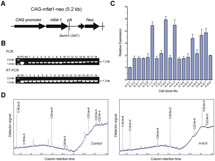
Following the HMC procedure, we successfully generated 3 transgenic sheep that carry and express the nematode mfat-1 gene. The coding sequence of nematode fat-1 (1209 bps) was modified for optimal expression in mammalian system (mfat-1) prior integrating to the primary fibroblast cells of the Chinese merino sheep (Figure 1 A, B). Recombinant cells were screened by PCR and RT-qPCR for the presence and level of mfat-1 mRNA expression (Figure 1C), and the cellular fatty acids composition was assessed by gas chromatography for mfat-1 function (Figure 1D). In the recombinant clonal cells expressing mfat-1, n−6 fatty acids (18∶2n−6, 20∶4n−6, and 22∶4n−6) were successfully converted to the corresponding n−3 (18∶3n−3, 20∶5n−3, and 22∶5n−3), and the ratio of n−6/n−3 were reduced from 8.60∶1 to 0.45∶1, a change of more than 19-folds of reduction compared to the non-transfected cells (Figure 1D, Table 1). The candidate clones selected (F-1-1, F-6-5, and H-6-6) showed the normal karyotype (data not shown). We eventually selected the clone H-6-6 for the subsequent HMC procedure; partially due to a relatively higher ratio of n−6/n−3 conversion was observed in that clone (Table 1).
Figure 1. Establishment and analysis of transgenic clonal donor cells.
(A) Schematic representation of n−3 fatty acid desaturase gene with linearized expression vectors. (B) Detection of the mfat-1 gene in Geneticin-resistant cell clones by PCR and RT-qPCR. mfat-1 expression vector was used as the template for positive control (PC) and untransfected syngenic cells was used as the negative control (NC). (C) Quantitative PCR analysis of mfat-1 expression in positive cell clones. cDNA representing mfat-1 was amplified with sequence specific primers. The beta-actin was used as internal control and the expression level observed in the transgenic donor cell was normalized to the value of A-3-1. (D) Partial gas chromatograph traces showing the polyunsaturated fatty acid profiles of total cellular lipids from the H-6-6 cells and the control cells. Note the level of n-6 polyunsaturated acids are lower whereas n−3 fatty acids are abundant in the mfat-1 cells (right) as compared with the control cells (left), in which there is very little n−3 fatty acid.
Table 1. Composition and ratio of n−6 and n−3 PUFA in the syngenic control and the transgenic cells expressing mfat-1 gene.
| Fatty acids | Control Cells | mfat-1 Cells | ||
| F-1-1 | F-6-5 | H-6-6 | ||
| LA (18∶2n−6) | 6.35±0.48 | 4.85±0.03** | 5.01±0.13** | 4.57±0.19** |
| ALA (18∶3n−3) | 0.06±0.05 | 1.61±0.13** | 1.32±0.16** | 3.92±0.45** |
| AA (20∶4n−6) | 5.90±1.06 | 4.06±0.50* | 5.62±0.14 | 2.64±0.38** |
| EPA (20∶5n−3) | 0.00 | 6.24±0.06** | 4.24±0.06** | 10.54±0.18** |
| ADA (22∶4n−6) | 2.47±0.39 | 1.86±0.36 | 1.83±0.30* | 1.73±0.29* |
| DPA (22∶5n−3) | 1.11±0.18 | 3.61±0.16** | 1.55±0.18* | 4.72±0.26** |
| DHA (22∶6n−3) | 0.54±0.12 | 0.76±0.06* | 0.43±0.22 | 0.71±0.06 |
| Total n−6 | 14.72±1.90 | 10.76±0.84* | 12.49±0.65 | 8.95±0.53** |
| Total n−3 | 1.71±0.08 | 12.22±0.35** | 7.54±0.55** | 19.94±0.65** |
| n−6/n−3 ratio | 8.60±1.50 | 0.88±0.09** | 1.65±0.16** | 0.45±0.04** |
Fatty acids composition is presented as a percentage of the total cellular lipids from the control cells and mfat-1 cells. Each value represented the mean ± standard deviation from three cell samples in each group with two independent measurements for each sample.
Statistical analysis using the two tailed student t-test. Significant differences between the syngenic control and mfat-1 cells were marked (*P<0.05; **P<0.01).
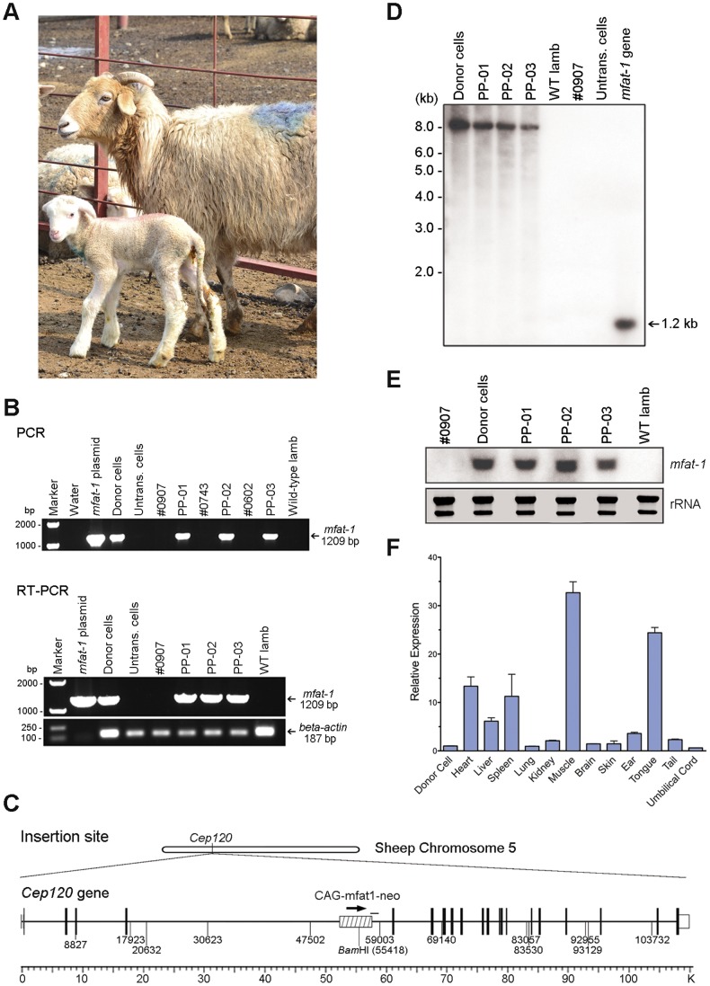
For the HMC procedure, approximately 3,740 (68%) out of 5,465 collected ovine oocytes were selected and utilized in the subsequent experiments. According to the established protocol, every two enucleated oocytes and a nuclear donor cell were combined by fusion to form one reconstructed embryo for the subsequent in vitro development. As a consequence, a total of 925 reconstructed embryos were produced and 82 (∼8.9%) of them successfully developed to the blastocyst stage after 7 days of in vitro culture (Table 2). A total of 53 blastocysts were surgically transplanted to uterus of 29 naturally cycling female recipients, with 1 to 2 blastocysts per recipient. Four pregnancies were detected and 3 live lambs were born either by caesarean section or natural delivery (Figure 2A and Table 3).
Table 2. Nuclear transfer efficiencies of the handmade cloning.
| Donor cells | No. of reconstructed embryos | No. and (%) of cloned embryos developed to blastocysts | No. of blastocysts transferred | No. of recipients | No. and (%) ofpregnant | No. and (%) of lambs born | |
| H-6-6 | Passage 8 | 196 | 27 (13.8%) | 13 | 6 | 0 | 0 |
| Passage 9 | 178 | 12 (6.7%) | 9 | 5 | 2 (40.0%) | 2 (22.2%) | |
| Passage 10 | 229 | 15 (6.6%) | 11 | 6 | 0 | 0 | |
| Passage 11 | 210 | 18 (8.6%) | 13 | 7 | 1 (14.3%) | 0 | |
| Passage 12 | 112 | 10 (8.9%) | 7 | 5 | 1 (20%) | 1 (14.3%) | |
| Total | 925 | 82 (8.9±2.9%) | 53 | 29 | 4 (13.8%) | 3 (5.7%) | |
Figure 2. Production of transgenic lambs by handmade cloning.
(A) The recipient #0907 and the transgenic lamb (PP-01). (B) Detection of the mfat-1 gene in umbilical cord samples of three cloned lambs by PCR and RT-qPCR. (C) Insertion site of mfat-1 vector in the sheep genome. Arrows indicate the mfat-1 transcriptional direction, which is identical with the endogenous putative sheep Cep120 gene. PCR fragment obtained in this study are shown by black bars. (D) Southern blot using the 32P-labled mfat-1 specific sequence as a probe to hybridize the genomic DNA from the transgenic donor cells and the cloned lambs. The genomic DNA was digested with BamHI before the gel electrophoresis. (E) Northern blot analysis. Total RNAs were loaded on each lane (15 µg per sample) and the coding region of mfat-1 was used as a probe. Shown below is the gel electrophoresis of rRNA as control. (F) Quantitative PCR analysis of mfat-1 expression in major tissues from the transgenic lambs (PP-02). Compared with the mRNA expression level normalized to the donor cell, the highest level of mfat-1 expression was observed in transgenic muscle sample.
Table 3. Production of transgenic cloned lambs by HMC.
| Lamb name | Donor cell | Recipient no. | Type of delivery (length of gestation) | Birth weight (kg) | Status |
| PP-01 | H-6-6, Passage 9 | #0907 | Caesarean section (152 day) | 5.74 | Live (>7 months) |
| PP-02 | H-6-6, Passage 9 | #0743 | Caesarean section (151 day) | 3.86 | Dead (2 days) |
| PP-03 | H-6-6, Passage 12 | #0602 | Naturally (149 day) | 5.24 | Live (>7 months) |
Genotype analysis using 13 ovine microsatellite markers demonstrated that all 3 transgenic sheep shared the same genotype to that of the nuclear donor cells (Table 4). The presence and function of mfat-1 gene in the transgenic sheep were confirmed by PCR, RT-qPCR, Southern, Northern, and DNA sequencing (Figure 2 B, D, E). All 3 transgenic sheep are males as expected. The efficiency of nuclear transfer varied from 22.2% for the passage 9 to 14.3% for the passage 12 of the donor clonal cells, expressed as the number of live lambs per 100 blastocysts transplanted. The overall efficiency for production of live lambs (5.7%) was close to those reported previously [10], [18], [19]. The birth weight of the transgenic lambs ranged from 3.86 kg to 5.74 kg. The lambs PP-01 and PP-03 were healthy and behaved normally after the birth. The lamb PP-02, however, died within 48 hours after the birth with the lowest body weight among the three. The dead lamb was utilized as a representative sample to assess the levels of omega-3 in the muscle and other organs/tissues.
Table 4. Microsatellite analysis of three transgenic cloned lambs, donor cell, and recipient.
| Loci | Lamb PP-01 | Lamb PP-02 | Lamb PP-03 | Donor cell H-6-6 | Recipient #0907 | Recipient #0743 |
| BMS460 | 122 | 122 | 122 | 122 | 122 | 122 |
| 138 | 138 | 138 | 138 | 138 | 122 | |
| AE129 | 145 | 145 | 145 | 145 | 147 | 147 |
| 147 | 147 | 147 | 147 | 149 | 149 | |
| MB009 | 142 | 142 | 142 | 142 | 142 | 142 |
| 142 | 142 | 142 | 142 | 142 | 146 | |
| ETH3 | 103 | 103 | 103 | 103 | 95 | 103 |
| 103 | 103 | 103 | 103 | 103 | 103 | |
| TGLA53 | 132 | 132 | 132 | 132 | 126 | 132 |
| 132 | 132 | 132 | 132 | 126 | 132 | |
| INRA063 | 172 | 172 | 172 | 172 | 164 | 164 |
| 178 | 178 | 178 | 178 | 172 | 172 | |
| ADCYC | 246 | 246 | 246 | 246 | 246 | 244 |
| 246 | 246 | 246 | 246 | 246 | 244 | |
| TGLA126 | 116 | 116 | 116 | 116 | 122 | 116 |
| 116 | 116 | 116 | 116 | 126 | 116 | |
| MAF209 | 121 | 121 | 121 | 121 | 121 | 109 |
| 127 | 127 | 127 | 127 | 121 | 121 | |
| TGLA122 | 146 | 146 | 146 | 146 | 158 | 138 |
| 158 | 158 | 158 | 158 | 158 | 138 | |
| PZ963 | 253 | 253 | 253 | 253 | 227 | 209 |
| 253 | 253 | 253 | 253 | 227 | 209 | |
| BMS2079 | 109 | 109 | 109 | 109 | 109 | 109 |
| 109 | 109 | 109 | 109 | 115 | 109 | |
| BMS2104 | 147 | 147 | 147 | 147 | 147 | 147 |
| 147 | 147 | 147 | 147 | 153 | 147 |
Insertion site analysis demonstrated that a single copy of the mfat-1 was integrated into the sheep genome on chromosome 5, located within the 5th intron of a functional gene Centrosomal protein of 120 kDa (Cep120, Figure 2C). DNA sequencing of a 1.2 kb PCR fragment across the 3′-end of mfat-1 and the neighbor host genome showed a 99% sequence homology to exons 5 and 6 of Cep120 (Figure 2C). The transcriptional orientation of the transgene mfat-1 was identical to that of the host gene. We found that the insertion of the mfat-1 showed no significant interfering effect on the function of Cep120, which is involved in centriole assembly and is essential for cell division [20], [21]. The remaining 2 transgenic lambs showed no obvious sign of neurogenic or other defects up to the date.
Single-copy insertion of the mfat-1 to ovine genome was cross-verified by the BamHI restriction enzyme analysis. Southern blot revealed a single target band sized ∼8.0 kb in BamHI digested DNA only from the transgenic lambs (Figure 2D). This result was consistent with the previous analysis in that the DNA fragment digested with BamHI was expected to be 7916 bps in length (Figure 2C). The possibility of tandem insertion of multiple mfat-1 copy was excluded, because the digested BamHI band from the transgenic sheep failed to show a predictive 5.2 kb fragment, a band representing multiple copies of the mfat-1 with the maximum size of 5.2 kb.
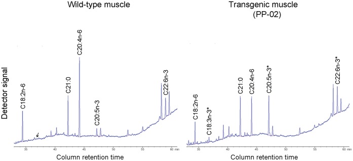
Analyses of fatty acid composition showed that the omega-3 (n−3) peaks in the umbilical cords of all three transgenic lambs were significantly higher than those in the age-matched wild type controls (data not shown). The results was cross-verified by systematic analysis of the fatty acid in major tissues of the lamb PP-02, which showed a two-fold reduction of n−6/n−3 ratio (2.63 to 1.24, p<0.01) in muscles compared to the wild-type lamb (Figure 3, Table 5). The lower ratio of n−6/n−3 was also found in other major tissues (p<0.05, Table 5). Consistent with these results, the level of mfat-1 mRNA varied among different tissues, with the highest expression found in the skeletal muscle (Figure 2F). More importantly, the total n−3 fatty acids (a-linolenic acid, eicosapentaenoic acid (EPA), and docosahexaenoic acid (DHA)) constituted approximately 6.2% of total muscle fat in the transgenic lamb, significantly higher than those in the control lambs, which was 3.5% on average (p<0.01). All of these indicated that the mfat-1 gene was functional in the transgenic lambs. Compared to the earlier studies of transgenic animals expressing the n−3 fatty acid desaturase gene [22], [23], our results help to provide additional evidence on the feasibility of transgenic animals.
Figure 3. Partial gas chromatograph of fatty acids in muscle sample of mfat-1 transgenic (PP-02) and the control lamb.
Fatty acid methyl esters were quantified using a fully automated 6890 Network GC System with an Agilent J&W fused-silica DB-23 capillary column. The peaks were identified by comparison with the internal fatty acid standards, and area percentage for all of resolved peaks was analyzed using GC ChemStation software. Compared with the wild-type control (left), the level of n−6 polyunsaturated acids in the transgenic muscle (right) are significantly lower, whereas n−3 fatty acids are abundant.
Table 5. Comparison of n−6/n−3 ratios between wild-type and mfat-1 transgenic lamb.
| Organs or Tissues | n−6/n−3 Ratioa | |
| Wild-type | Transgenic | |
| Heart | 2.28±0.01 | 0.93±0.02** |
| Liver | 1.56±0.05 | 0.66±0.04** |
| Spleen | 3.04±0.23 | 1.50±0.16** |
| Lung | 2.66±0.46 | 1.40±0.12* |
| Kidney | 2.45±0.05 | 1.36±0.15** |
| Brain | 0.42±0.01 | 0.32±0.01** |
| Ear | 2.89±0.02 | 1.18±0.25** |
| Tongue | 1.66±0.13 | 1.25±0.02* |
| Tail | 4.60±1.14 | 2.08±0.63* |
| Muscle | 2.63±0.05 | 1.24±0.06** |
Ratio of n−6/n−3 fatty acid was calculated from n−6 fatty acids [linoleic acid (LA, 18∶2n−6) and arachidonic acid (AA, 20∶4n−6)] versus n−3 fatty acids [α-linolenic acid (ALA, 18∶3n−3), eicosapentaenoic acid (EPA, 20∶5n−3), and docosahexaenoic acid (DHA, 22∶6n−3)]. Each value represents the mean ± standard deviation from three replicated sample measurements of each tissue.
Statistical analysis using the two tailed student t-test. Significant differences between the wild-type and the transgenic lamb samples were marked (*P<0.05; **P<0.01).
It was not escaped from our attention that, in contrast to the SCNT, the procedure of HMC introduced an obvious heteroplasmy into the transgenic animals, mitochondrial DNAs in particular. While the potential interactions and the long-term consequences of such heteroplasmy remain to be determined, it seems that the developmental reprogramming driven by the enucleated oocytes was not significantly affected. Although certain concern was raised regarding the three origins of mitochondrial DNAs in the developing fetus from the cells of different sheep, no evidence of the negative effect to the transgenic animal is currently available [24]. The two remaining HMC sheep in our study showed normal growth and development up to the date (Figure S1), consistent with those of the HMC pigs [3], [15].
Over the past decade the nematode fat-1 has been successfully introduced into mice [8], [25], pigs [2], [3] and cattle [9]. The fat-1 mice produced by pronuclei microinjection have possessed good value for the production of n−3 PUFAs [8], [25], particularly for DHA and docosapentaenoic acid (DPA) in transgenic sFat-1 mice [25]. In the transgenic pig, Lai et al. (2006) reported that there was a fivefold reduction of the n−6/n−3 ratio in tail tissues of hfat-1 transgenic piglets. The other tissues from transgenic piglets also showed a substantially lower n−6/n−3 ratio [2]. Wu et al. successfully cloned transgenic cow expressing fat-1 gene by traditional SCNT, with increased levels of n−3 PUFAs in the ear tissue, and reduced ratio of n−6/n−3 in the milk [9]. Our previous results also showed that nematode mfat-1 could effectively lower the n−6/n−3 ratio in muscle and other major organs of the transgenic pig [3].
In summary, we used the modified HMC procedure to generate a group of transgenic sheep carrying a functional nematode mfat-1 gene, and the resulting sheep were rich in omega-3 fatty acids in the muscles and other organ/tissues. Our study demonstrated once again that the alternative HMC is helpful to accelerate the speed of transgenic animal research with cost efficiency and easy in operation. With an increasing demand for dietary intake of omega-3 for a better quality of our life via disease prevention, generation of transgenic animals rich in omega-3 could be an attractive alternative not only for human health, also for environmental conservation.
Materials and Methods
All the chemical reagents used in this research were obtained from Sigma-Aldrich Chemical (St Louis, USA) except for otherwise indicated.
Ethics Statement
This study was carried out following the recommendations in the Guide for the Care and Use of Laboratory Animals of the Chinese Academy of Sciences. Procedures for all of the animal experiments were approved in advance by the Animal Care and Ethics Committee of the Institute of Genetics and Developmental Biology, Chinese Academy of Sciences.
Ovine Primary Fibroblasts Cell Culture
Punch collected ear tissue from a Chinese merino male sheep was washed twice with Ca2+- and Mg2+-free PBS (DPBS), minced with a surgical blade on a 10-cm culture dish (Becton Dickinson, Lincoln Park, USA) and then dissociated with 0.25% (v/v) trypsin-EDTA (GIBCO, New York, USA) containing Dulbecco’s modified Eagle’s medium (DMEM, GIBCO) at 37°C. Cell pellets after centrifuging were seeded onto a 35 mm culture dish and cultured for 8–10 days in DMEM supplemented with 10% (v/v) fetal bovine serum (FBS, HyClone, Logan, USA) at 37°C and 5% CO2. The construction of mfat-1 expression vector CAG-mfat1-neo and the transfection were performed as described previously [3], [26]. Cells in each well were trypsinized and seed onto a 10-cm culture dish containing 450 µg/mL Geneticin (Invitrogen, USA) for 24 hrs after the transfection. Drug selection was conducted for 2 weeks with a medium change per 3 days and the Geneticin-resistant colonies were isolated. Each colony was transferred to a 6-well plate for expansion. The resulting clones were frozen at −80°C overnight and stored in liquid nitrogen.
Analysis of mfat-1 in the Ovine Cell Clones
Geneticin selected cells were analyzed by PCR and RT-PCR as described previously [3]. Quantitative PCR (qPCR) was performed with sequence-specific primer pairs for mfat-1 and beta-actin, respectively. Reagents of Power SYBR® Green PCR Master Mix were used for the PCR amplification in triplicates under the following thermal program: 10 min at 95°C, 40 cycles of 95°C for 15 sec, 60°C for 25 sec, and 72°C for 20 sec.
The mfat-1 positive cells were cultured in medium supplemented with 10 µM 18∶2n−6 and 15 µM 20∶4n−6 prior to fatty acid analysis. Cells were collected in a glass methylation tube for fatty acid analysis at 80% confluence. The fatty acid compositions of total cellular lipids were analyzed for both of the transgenic and control clones using the gas chromatography as previously described [27]. Fatty acid methyl esters were quantified using a fully automated 6890 Network GC System (Agilent Technologies, Palo Alto, USA) with an Agilent J&W fused-silica DB-23 capillary column. The injector and detector were maintained at 260°C and 270°C, respectively. The oven program was maintained initially at 180°C for 10 min, then ramped to 200°C at 4°C/min and held for 15 min, finally ramped to 230°C at 10°C/min and maintained for 6 min. Carrier gas-flow rate was maintained at a constant rate of 2.0 mL/min throughput. The peaks were identified by comparison with the internal fatty acid standards, and area percentage for all of resolved peaks was analyzed using the GC ChemStation software (Agilent Technologies, USA).
Procedure Outline for the Handmade Cloning
Sheep ovaries were collected at a local slaughterhouse of Xinjiang Hualing Industry and Trade (Group) Co. Ltd., with the authorization from the business administration for the collection and usage of ovary tissues on the transgenic research. The fresh ovary tissues were transferred to a portable incubator for processing within 120 min and the oocytes were isolated by the ovarian slicing method described previously [28]. Prior to slicing, ovaries were washed in phosphate buffered saline (PBS), and then placed in a Petri dish and covered with Hepes-buffered TCM-199 (GIBCO, USA) containing 60 IU/mL heparin. The compact cumulus-oocytes complexes (COCs) were selected and washed twice in Hepes-buffered TCM-199. Each group of 50 COCs was matured in 400 µL bicarbonate-buffered TCM-199 supplemented with 10% (V:V) fetal bovine serum (FBS), 2 mM L-Glutamine, 0.3 mM sodium pyruvate, 0.1 mM cysteine, 5 µg/mL follicle stimulating hormone, 5 µg/mL luteinizing hormone, 1 µg/mL β-Estradiol in an incubator maintained at 38.6°C with humidified air and 5% CO2.
The HMC was performed essentially as previously described [13], [15], with minor modifications to adapt for the ovine nuclear transferring. Briefly, at 21–22 h after the starting point of maturation, the cumulus cells were removed by repeated pipetting in 1 mg/mL hyaluronidase in Hepes-buffered TCM-199. All the remaining manipulations were performed on a hotplate adjusted to 30°C. Zonae pellucidae were partially digested with 8 mg/mL pronase solution dissolved in T33 (T for Hepes-buffered TCM 199 medium, and the number for the percentage of calf serum supplement), then washed quickly in T2 and T20 drops. The oocytes with softened zonae pellucidae were lined up in T20 drops supplemented with 2.5 µg/mL cytochalasin B (CB), and were enucleated by oriented bisection with an ultra sharp microblade (AB Technology, Pullman, USA) under a stereomicroscope. Approximately 1/3−1/2 cytoplasm close to the polar body was removed manually, and the remaining putative cytoplast were washed twice in T2 drops and collected in a T10 drop. The nuclear donor cells were trypsinized and kept in T2. Fusion was performed in two steps where the second step included the initiation of activation. For the first step, half of the available cytoplasts were transferred into 1 mg/mL of phytohaemagglutinin (PHA; ICN Pharmaceuticals, Australia) dissolved in T0, and then each one was quickly dropped over a single fibroblast. After attachment, cytoplast-fibroblast pairs were equilibrated in a fusion medium (0.3 M mannitol and 0.01% polyvinyl alcohol). Using an alternative current (AC) of 4 V (CF-150/B fusion machine; BLS, Budapest, Hungary), cell pairs were aligned to the wire of a fusion chamber (BTX microslide 0.5 mm fusion chamber, model 450; BTX, SanDiego, USA) with the somatic cells farthest from the wire, then fused with a single direct current (DC) of 1.3 kV/cm for 10 µsec. After the pulse, cytoplast-fibroblast pairs were incubated in T10 drops to examine whether or not the fusion had occurred. Approximately 1 h after the first fusion, each pair was fused with the second cytoplasts and activated simultaneously in activation medium (0.3 M mannitol, 0.1 mM MgSO4, 0.1 mM CaCl2 and 0.01% polyvinyl alcohol) with a double DC pulse of 0.9 kV/cm, each pulse for 15 µsec and 1 sec apart. When fusion had been observed in T10 drops, the reconstructed embryos were first incubated in 40 µL Hepes-buffered TCM 199 medium containing 5 µM Ionomycin for 5 min at 38.6°C. After subsequent washing in culture medium 3–5 times, reconstructed embryos were incubated in culture medium containing 2 mM 6-dimethylamino-purine (6-DMAP) for 4 h at 38.6°C in 5% CO2 with maximum humidity, then washed thoroughly with IVC medium for culture.
The reconstructed embryos were in vitro cultured in wells of a Nunc 4-well dish in 400 µL SOFaaci medium [29] supplemented with 4 mg/mL BSA and covered with oil. Zona-free embryos produced from the HMC were cultured in modified WOWs system [30], [31] at 38.6°C in 5% CO2 with saturated humidity. Cleavage and blastocyst formation of the reconstructed embryos were monitored during the in vitro culture for 7 days.
Embryo Transfer and Pregnancy Diagnosis
The healthy ewes from a local Xinjiang sheep breed aged 12–18 months were selected as the embryo-transfer recipients. The blastocysts with clearly visible inner cell mass were surgically transplanted into the uterine horns of a naturally cycling ewe on 6 or 7 days of standing estrus by a certified veterinarian. Pregnancies were diagnosed by ultrasonography on day 45 after the surgical embryo transfer.
Screening of the Transgenic Sheep
Umbilical cord from each newborn lamb was collected and the genomic DNA was isolated for the genotyping and genetic screening. Presence of mfat-1 gene in each DNA sample was testified by SP-PCR and Southern blot. The expression of mfat-1 mRNA was evaluated by RT-PCR and Northern blot.
Comparative genotype analyses were performed for each of the newborn lambs, the recipients, and the donor fibroblasts using a set of microsatellite markers. Thirteen polymorphic microsatellite loci (BMS460, AE129, MB009, ETH3, TGLA53, INRA063, ADCYC, TGLA126, MAF209, TGLA122, PZ963, BMS2079, BMS2104) located on different ovine chromosomes were amplified by 3-color multiplex PCR and the products were analyzed on a 3130XL Genetic Analyzer (Applied Biosystems) with GeneMapper ID Software v3.2 (Applied Biosystems, USA).
For Southern blot analysis, approximately 20 µg of genomic DNA was digested with BamHI for agarose gel electrophoresis. The probes were 32P -labeled PCR fragments of mfat-1 sequence, generated using the Random Prime Labeling System Redi PrimeTMII (GE Healthcare, Piscataway, USA). Northern blot analysis was performed as described previously [32]. About 15 µg of total RNA were loaded per lane, and the coding region of mfat-1 was used as a probe.
To identify the insertion site, the genomic DNA flanking inserted mfat-1 was amplified by a thermal asymmetric interlaced PCR (TAIL-PCR) using Genome Walking Kit (TaKaRa, Japan). The specific primers (CAG4823-1F: 5′-cctcgtgctttacggtatcg-3′; CAG-4939-2F: 5′-caacctgccatcacgagatt-3′ and CAG5044-3F: 5′-tctcatgctggagttcttcg-3′) were designed with their melting temperatures sufficiently high to ensure a maximum thermal asymmetric priming and to ensure the cloning of genomic DNA flanking the mfat-1 gene. The tertiary TAIL-PCR products were purified, cloned into pMD18-T (TaKaRa, Japan), and sequenced. The BLAST homology searches and the sequence comparisons with the draft sheep genome assembly OARv2.0 (International Sheep Genomics Consortium) were performed to identify the site of mfat-1 insertion at the level of nucleotide sequences.
To evaluate the functional activity of desaturase encoded by nematode mfat-1 gene, the tissues from one of the transgenic lambs (PP-02) was utilized as representative samples for fatty acid analyses. The major organ/tissue samples (heart, liver, spleen, lung, kidney, tongue, ear, muscle, brain, and tail) were collected from both transgenic and age-matched wild-type lamb, and the fatty acid analysis was performed as described previously. Briefly, the oven program of the gas chromatography instrument was maintained initially at 100°C for 5 min, then ramped to 180°C at 6°C/min and held for 10 min, then ramped to 205°C at 2°C/min and held for 10 min, finally ramped to 225°C at 2°C/min and maintained for 7 min. The ratio of n-6/n-3 from each organ/tissue was used for comparison between the transgenic and the wild-type lambs.
Statistical Analysis
Statistical analysis was performed using a two-tailed Student’s t-test, and p-values <0.05 were considered statistically significant.
Supporting Information
Image of mfat-1 transgenic lambs of approximately 75 days (PP-01) and 100 days (PP-03) after the birth. The lamb (PP-02) was used for n−3 fatty acid composition analyses of major organ/tissues approximately 3 days after the birth, due to obvious weakness at the time of birth.
(TIF)
Acknowledgments
We gratefully thank Min Han for the gift of C. elegans culture stock; Mingjun Liu, Juncheng Huang and Bin Han for the animal surgery facility and technical assistance.
Funding Statement
This study was funded by the research grants from the China Ministry of Agriculture (2009ZX08008-005B) and the Chinese Academy of Sciences (KSZD-EW-005). The funders had no role in study design, data collection and analysis, decision to publish, or preparation of the manuscript.
References
- 1. Riediger ND, Othman RA, Suh M, Moghadasian MH (2009) A systemic review of the roles of n-3 fatty acids in health and disease. J Am Diet Assoc 109: 668–679. [DOI] [PubMed] [Google Scholar]
- 2. Lai L, Kang JX, Li R, Wang J, Witt WT, et al. (2006) Generation of cloned transgenic pigs rich in omega-3 fatty acids. Nat Biotechnol 24: 435–436. [DOI] [PMC free article] [PubMed] [Google Scholar]
- 3. Zhang P, Zhang Y, Dou H, Yin J, Chen Y, et al. (2012) Handmade cloned transgenic piglets expressing the nematode fat-1 gene. Cell Reprogram 14: 258–266. [DOI] [PMC free article] [PubMed] [Google Scholar]
- 4. Simopoulos AP (2000) Human requirement for N-3 polyunsaturated fatty acids. Poult Sci 79: 961–970. [DOI] [PubMed] [Google Scholar]
- 5. Kang JX (2005) From fat to fat-1: a tale of omega-3 fatty acids. J Membr Biol 206: 165–172. [DOI] [PubMed] [Google Scholar]
- 6. Spychalla JP, Kinney AJ, Browse J (1997) Identification of an animal omega-3 fatty acid desaturase by heterologous expression in Arabidopsis. Proc Natl Acad Sci U S A 94: 1142–1147. [DOI] [PMC free article] [PubMed] [Google Scholar]
- 7. Kang ZB, Ge Y, Chen Z, Cluette-Brown J, Laposata M, et al. (2001) Adenoviral gene transfer of Caenorhabditis elegans n–3 fatty acid desaturase optimizes fatty acid composition in mammalian cells. Proc Natl Acad Sci U S A 98: 4050–4054. [DOI] [PMC free article] [PubMed] [Google Scholar]
- 8. Kang JX, Wang J, Wu L, Kang ZB (2004) Transgenic mice: fat-1 mice convert n-6 to n-3 fatty acids. Nature 427: 504. [DOI] [PubMed] [Google Scholar]
- 9. Wu X, Ouyang H, Duan B, Pang D, Zhang L, et al. (2012) Production of cloned transgenic cow expressing omega-3 fatty acids. Transgenic Res 21: 537–543. [DOI] [PubMed] [Google Scholar]
- 10. Wilmut I, Schnieke AE, McWhir J, Kind AJ, Campbell KH (1997) Viable offspring derived from fetal and adult mammalian cells. Nature 385: 810–813. [DOI] [PubMed] [Google Scholar]
- 11. Vajta G, Lewis IM, Hyttel P, Thouas GA, Trounson AO (2001) Somatic cell cloning without micromanipulators. Cloning 3: 89–95. [DOI] [PubMed] [Google Scholar]
- 12. Vajta G, Lewis IM, Trounson AO, Purup S, Maddox-Hyttel P, et al. (2003) Handmade somatic cell cloning in cattle: analysis of factors contributing to high efficiency in vitro. Biol Reprod 68: 571–578. [DOI] [PubMed] [Google Scholar]
- 13. Vajta G, Bartels P, Joubert J, de la Rey M, Treadwell R, et al. (2004) Production of a healthy calf by somatic cell nuclear transfer without micromanipulators and carbon dioxide incubators using the Handmade Cloning (HMC) and the Submarine Incubation System (SIS). Theriogenology 62: 1465–1472. [DOI] [PubMed] [Google Scholar]
- 14. Kragh PM, Vajta G, Corydon TJ, Purup S, Bolund L, et al. (2004) Production of transgenic porcine blastocysts by hand-made cloning. Reprod Fertil Dev 16: 315–318. [DOI] [PubMed] [Google Scholar]
- 15. Du Y, Kragh PM, Zhang Y, Li J, Schmidt M, et al. (2007) Piglets born from handmade cloning, an innovative cloning method without micromanipulation. Theriogenology 68: 1104–1110. [DOI] [PubMed] [Google Scholar]
- 16. Kragh PM, Nielsen AL, Li J, Du Y, Lin L, et al. (2009) Hemizygous minipigs produced by random gene insertion and handmade cloning express the Alzheimer’s disease-causing dominant mutation APPsw. Transgenic Res 18: 545–558. [DOI] [PubMed] [Google Scholar]
- 17. Luo Y, Li J, Liu Y, Lin L, Du Y, et al. (2011) High efficiency of BRCA1 knockout using rAAV-mediated gene targeting: developing a pig model for breast cancer. Transgenic Res 20: 975–988. [DOI] [PubMed] [Google Scholar]
- 18. Schnieke AE, Kind AJ, Ritchie WA, Mycock K, Scott AR, et al. (1997) Human factor IX transgenic sheep produced by transfer of nuclei from transfected fetal fibroblasts. Science 278: 2130–2133. [DOI] [PubMed] [Google Scholar]
- 19. Lee JH, Peters A, Fisher P, Bowles EJ, St John JC, et al. (2010) Generation of mtDNA homoplasmic cloned lambs. Cell Reprogram 12: 347–355. [DOI] [PubMed] [Google Scholar]
- 20. Xie Z, Moy LY, Sanada K, Zhou Y, Buchman JJ, et al. (2007) Cep120 and TACCs control interkinetic nuclear migration and the neural progenitor pool. Neuron 56: 79–93. [DOI] [PMC free article] [PubMed] [Google Scholar]
- 21. Mahjoub MR, Xie Z, Stearns T (2010) Cep120 is asymmetrically localized to the daughter centriole and is essential for centriole assembly. J Cell Biol 191: 331–346. [DOI] [PMC free article] [PubMed] [Google Scholar]
- 22. Pan D, Zhang L, Zhou Y, Feng C, Long C, et al. (2010) Efficient production of omega-3 fatty acid desaturase (sFat-1)-transgenic pigs by somatic cell nuclear transfer. Sci China Life Sci 53: 517–523. [DOI] [PubMed] [Google Scholar]
- 23. Duan B, Cheng L, Gao Y, Yin FX, Su GH, et al. (2012) Silencing of fat-1 transgene expression in sheep may result from hypermethylation of its driven cytomegalovirus (CMV) promoter. Theriogenology 78: 793–802. [DOI] [PubMed] [Google Scholar]
- 24. Hiendleder S, Zakhartchenko V, Wolf E (2005) Mitochondria and the success of somatic cell nuclear transfer cloning: from nuclear-mitochondrial interactions to mitochondrial complementation and mitochondrial DNA recombination. Reprod Fertil Dev 17: 69–83. [DOI] [PubMed] [Google Scholar]
- 25. Zhu G, Chen H, Wu X, Zhou Y, Lu J, et al. (2008) A modified n-3 fatty acid desaturase gene from Caenorhabditis briggsae produced high proportion of DHA and DPA in transgenic mice. Transgenic Res 17: 717–725. [DOI] [PubMed] [Google Scholar]
- 26. Zhang P, Cao LX, Chen H, Ma RL (2007) Prokaryotic expression of the C. elegans fat-1 gene and preparation of antibody. J Northwest A & F Univ (Nat Sci Ed) 35: 24–28. [Google Scholar]
- 27. Kang JX, Wang J (2005) A simplified method for analysis of polyunsaturated fatty acids. BMC Biochem 6: 5. [DOI] [PMC free article] [PubMed] [Google Scholar]
- 28. Bhojwani S, Vajta G, Callesen H, Roschlau K, Kuwer A, et al. (2005) Developmental competence of HMC(TM) derived bovine cloned embryos obtained from somatic cell nuclear transfer of adult fibroblasts and granulosa cells. J Reprod Dev 51: 465–475. [DOI] [PubMed] [Google Scholar]
- 29. Holm P, Booth PJ, Schmidt MH, Greve T, Callesen H (1999) High bovine blastocyst development in a static in vitro production system using SOFaa medium supplemented with sodium citrate and myo-inositol with or without serum-proteins. Theriogenology 52: 683–700. [DOI] [PubMed] [Google Scholar]
- 30. Vajta G, Peura TT, Holm P, Paldi A, Greve T, et al. (2000) New method for culture of zona-included or zona-free embryos: the Well of the Well (WOW) system. Mol Reprod Dev 55: 256–264. [DOI] [PubMed] [Google Scholar]
- 31. Feltrin C, Forell F, dos Santos L, Rodrigues JL (2006) In vitro bovine embryo development after nuclear transfer by handmade cloning using a modified wow culture system. Reprod Fertil Dev 18: 126. [Google Scholar]
- 32. Chen L, Liu K, Zhao Z, Blair HT, Zhang P, et al. (2012) Identification of Sheep Ovary Genes Potentially Associated with Off-season Reproduction. J Genet Genomics 39: 181–190. [DOI] [PubMed] [Google Scholar]
Associated Data
This section collects any data citations, data availability statements, or supplementary materials included in this article.
Supplementary Materials
Image of mfat-1 transgenic lambs of approximately 75 days (PP-01) and 100 days (PP-03) after the birth. The lamb (PP-02) was used for n−3 fatty acid composition analyses of major organ/tissues approximately 3 days after the birth, due to obvious weakness at the time of birth.
(TIF)


