Abstract
Throughout the animal kingdom, Wnt-triggered signal transduction pathways play fundamental roles in embryonic development and tissue homeostasis. Wnt proteins are modified as glycolipoproteins and are secreted into the extracellular environment as morphogens. Recent studies on the intracellular trafficking of Wnt proteins demonstrate multiple layers of regulation along its secretory pathway. These findings have propelled a great deal of interest among researchers to further investigate the molecular mechanisms that control the release of Wnts and hence the level of Wnt signaling. This review is dedicated to Wntless, a putative G-protein coupled receptor that transports Wnts intracellularly for secretion. Here, we highlight the conclusions drawn from the most recent cellular, molecular and genetic studies that affirm the role of Wntless in the secretion of Wnt proteins.
Keywords: Wntless, Gpr177, Wnt, trafficking, secretion, exocytosis, exocytosi
Introduction
Wnts are acylated and glycosylated secretory proteins with established roles in embryonic development and tissue homeostasis (Logan and Nusse, 2004; Tang et al., 2011). Defects in Wnt signaling are associated with various human diseases including developmental abnormalities and cancers (Clevers, 2006). Extensive studies have been performed on Wnt downstream signaling events, e.g. the canonical and non-canonical pathways, which regulate target gene expression in signal-receiving cells (Logan and Nusse, 2004; Clevers, 2006; MacDonald et al., 2009). However, less is known about Wnt trafficking in ligand-producing cells, and a great amount of interest is now dedicated to dissect the Wnt secretory pathways completely.
In this review, we highlight the most recent studies on Wntless, a conserved transmembrane protein fundamental for transporting Wnts for secretion. Here, we integrate and discuss the conclusions made by various biochemical, cellular and genetic studies that contribute to the role of Wntless in Wnt secretion.
Wntless: biochemical and molecular characteristics
Discovered in 2006, Wntless (Wls or Evenness Interrupted/Sprinter in Drosophila, MOM-3/Mig-14 in C. elegans, and GPR177 in mammals) is an evolutionarily conserved trans-membrane protein and is indispensible for the secretion of multiple Wnt proteins (Bänziger et al., 2006; Bartscherer et al., 2006; Goodman et al., 2006; Belenkaya et al., 2008; Franch-Marro et al., 2008; Port et al., 2008; Yang et al., 2008; Silhankova et al., 2010). Studies till date show that depletion of Wntless in both vertebrates and invertebrates gives rise to Wnt loss-of-function phenotypes (Belenkaya et al., 2008; Franch-Marro et al., 2008; Port et al., 2008; Yang et al., 2008; Carpenter et al., 2010; Silhankova et al., 2010; Fu et al., 2011), thus illustrating a fundamentally conserved function of Wntless in Wnt secretion (MacDonald et al., 2009).
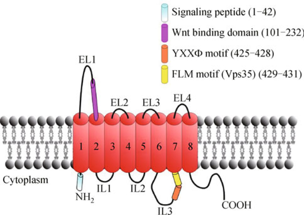
Structural details of Wntless protein have provided the first piece of evidence in support of its role in intracellular Wnt trafficking. Through amino acid sequence analysis Wntless is predicted to contain a long N-terminal region, seven or eight transmembrane segments and an intracellular C terminus similar to the member of G-protein coupled receptor (GPCR) superfamily (Fig. 1). Indeed, Wntless has been proposed as a putative orphan GPCR (Jin et al., 2010). However, there is a lack of evidence supporting whether or not Wntless may signal as a GPCR. As great structural and functional diversities lie within the GPCR superfamily, it is difficult to speculate function of Wntless purely based on our classic GPCR knowledge. In addition, Wntless contains almost no known motifs that are homologous to existing GPCRs, greatly impeding our understanding of its molecular function and regulation.
Figure 1.
The putative structure of Wntless protein with specific protein-interaction domains highlighted.
It is well known that most membrane associated secretory proteins, including GPCRs, are usually N-linked glycosylated. To determine whether Wntless is modified with N-linked oligosaccharide sides, Wntless protein has been treated with N-glycosidase, which leads to the occurrence of a fast-migrating band, indicating N-linked glycosylation ofWls (Jin et al., 2010). However, whether this glycosylation is essential for the interaction between Wntless and Wnts, or for the trafficking of Wntless itself, is not clear.
Through the detection of N-terminal Flag-tagged Wntless under non-permeabilized conditions, Jin et al. determined that the N terminus of Wntless is localized intracellularly, thereby suggesting presence of an even number of membrane-spanning segments in the protein (Jin et al., 2010). Using GST pull-down to investigate the interaction between Wntless and Wnts, a lipocalin-like domain in the N-terminal region near the second transmembrane domain of Wntless is found to be required for its association with Wnt1, Wnt3 and Wnt5a (Fu et al., 2009) (Fig. 1). However, little information is available to date regarding the role of other extracellular loops of Wntless in the interaction between Wntless and Wnts. In terms of the structural property of Wnts, a conserved serine residue within most of Wnts (e.g. Ser239 in Drosophila Wg) has been found to be indispensable for their interaction with Wntless (Herr and Basler, 2011). Substitution of this serine residue with alanine significantly affects the physical association of Wnts with Wntless. However, the exact mechanism underlying the recognition of Wnts by Wntless still remains elusive.
Similar to many known GPCRs, Wntless can be internalized from the cell membrane through endocytosis (Pan et al., 2008; Port et al., 2008; Harterink et al., 2011; Temkin et al., 2011). Indeed, a conserved YXXΦ endocytotic motif has been identified in the third intracellular loop of Wntless (Fig. 1). This motif serves as an endocytic signal for recognition by a clathrin adaptor protein 2 (AP2), and is critical for recycling Wntless from the plasma membrane (Gasnereau et al., 2011). Site-directed mutation of this conserved motif in Wntless results in its accumulation on the cell surface. However, sequestration of Wntless on the plasma membrane can be restored via the addition of the classic motif to its C-terminal tail. Interestingly, in C. elegans Wntless homolog, this key motif is missing from the third intracellular loop and is found in the second intracellular loop.
Two independent studies have identified the interaction between Wntless and Vps35, a core subunit of retromers responsible for retrograde transport of Wntless from Plasma membrane to Golgi (discussed in details below). These studies have promoted the discovery of a FLM tripeptide motif at the end of the third intracellular loop of Wntless, required for retromer-dependent transportation (Fig. 1). Immunofluorescence and co-immunoprecipitation studies indicate that Wntless localizes with yet another subunit of retromer, a cargo sorting protein SNX3 (Harterink et al., 2011; Zhang et al., 2011). Unfortunately, there is lack of studies to verify whether this interaction is direct.
Taken together, further structural analysis of Wntless is needed to understand whether this GPCR-like protein can indeed recruit heterotrimeric G-proteins and subsequently initiate related signaling pathways in the process of Wnt secretion.
Protein modifications of Wnts: Wntless-Wnts interaction
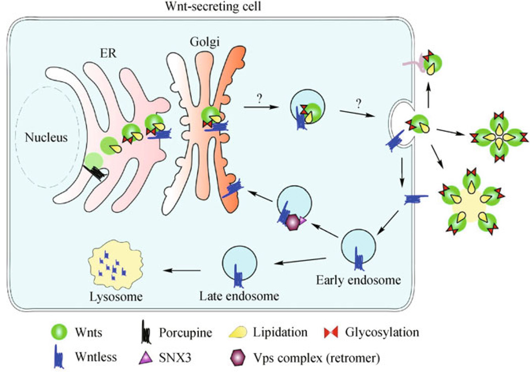
The posttranslational modifications of Wnts are critically coupled to their secretion and activity. Wnts are produced and then modified in ER by a membrane-bound acyltransferase, Porcupine (Fig. 2). Porcupine is believed to facilitate the lipidation of Wnt proteins on at least two distinct sites: the Nterminal cysteine rich residues (Tanaka et al., 2000, 2002) and a C-terminal serine 209 residue (Coombs et al., 2010). The conserved cysteine residues at the N terminus of Wnt proteins are subjected to the addition of palmitate groups (Takada et al., 2006), creating a binding site for porcupine (Tanaka et al., 2002). However, it is less clear if porcupine itself adds or facilitates addition of palmitate group to this locus, through other proteins.
Figure 2.
The putative role of Wntless in the transport of Wnts through the secretory pathway in the Wnt-producing cell.
Unlike the incomprehensible role of Porcupine in Wnt palmitation at the cysteine rich residues, Porcupine is strongly believed to add palmitoyl group to the Serine 209 (van den Heuvel et al., 1993; Zhai et al., 2004; Galli et al., 2007). Mutation causing loss of acyltransferase activity in Porcupine leads to impaired Wnt secretion, suggesting the necessity of these lipid modifications in the release of Wnts (van den Heuvel et al., 1993).
Wntless has been found to be localized in the entire Wnt secretory route including ER, Golgi, vesicles and Plasma membrane (Bänziger et al., 2006; Bartscherer et al., 2006; Fu et al., 2009; Coombs et al., 2010) (Fig. 2). Having a lipocalin-like structure allows Wntless capable to bind to hydrophobic regions like the palmitate groups in mature Wnts. The affinity of Wntless toward the hydrophobic regions in Wnts might facilitate the physical association of Wnts with Wntless. Rather than palmitation at the cysteine residues, palmitoylation at Ser209 in Wnt3a by Porcupine is required for its physical interaction with Wntless (Coombs et al., 2010) and its release from ER (Willert et al., 2003; Takada et al., 2006; Komekado et al., 2007). Consistently, reduced Ser209 palmitoylation of Wnt3a demonstrates ER retention (Takada et al., 2006), reflecting a defective Wnt secretion probably due to disrupted Wnt-Wntless association.
On the other hand, as impaired Wnt secretion also affects downstream Wnt signaling, different interpretations of the experimental findings exist on whether a specific Wnt lipidation promotes only its secretion, signaling, or both (Tang et al., 2011). For example, the absence of cysteine rich residues in Wnt1, Wnt3a and Wnt5a results in depleted Wnt signaling in vertebrates (Galli et al., 2007; Kurayoshi et al., 2007), whereas absence of palmitation at cysteine residue in Drosophila Wg causes ER retention (Franch-Marro et al., 2008). Whether it is the amino acid itself altering protein folding, or the lipid modification on the amino acids affecting Wnt secretion and its signaling is yet to be established. Furthermore, the Drosophila Wnt D is recently identified as the only Wnt protein that neither requires lipid modification nor an association with Wntless for its secretion (Ching et al., 2008; Herr and Basler, 2011).
Another posttranslational modification undergone by Wnts in the ER is N-terminal glycosylation (Fig. 2). In Drosophila, N-terminal glycosylation at Ser239 is stimulated by Porcupine, and this modification is indispensable for its interaction with Wntless (Tanaka et al., 2002). Overall, this suggests that the posttranslational modifications of Wnts contribute to their transport and secretion from ligand-producing cells. However, whether these specific modifications provide opportunities for precise regulation of Wnt trafficking and exocytosis via Wntless or other secretory pathway components awaits definitive studies (Fig. 2).
Wnt-Wntless dissociation
Apart from post translational modifications, an interesting physical parameter that seems to have a strong impact on Wnt secretion is the environmental pH. Endosomal pH gradient plays an important role in the secretory pathway of proteins. An acidic pH ~ 5.5 of secretory vesicle appears to promote dissociation of Wnt3a from Wntless and facilitates Wnt secretion out of the cell (Coombs et al., 2010). The precise role of the endosomal pH gradient in Wnt secretory pathway is yet to be explored.
Retrograde trafficking of Wntless
The fate of Wntless is distinct from Wnts after the Wnt-Wls protein complex reaches the plasma membrane. Wntless is recycled back into Wnt-producing cells and is reutilized for yet another round of Wnt secretion (Belenkaya et al., 2008; Franch-Marro et al., 2008; Port et al., 2008; Yang et al., 2008; Silhankova et al., 2010) (Fig. 2). Clathrin coated (Port et al., 2008; Harterink et al., 2011) and Rab5 (Rojas et al., 2008; Harterink et al., 2011) positive early endosomal vesicles retrieve Wntless from Plasma membrane. This transport is also demonstrated to be AP2 and dynamin dependent (Belenkaya et al., 2008; Franch-Marro et al., 2008; Pan et al., 2008; Port et al., 2008; Yang et al., 2008; Silhankova et al., 2010).
From early endosomes, Wntless is carried to the Golgi in a retromer-dependent manner in small vesicles (Belenkaya et al., 2008; Franch-Marro et al., 2008; Port et al., 2008; Silhankova et al., 2010; Yang et al., 2008) (Fig. 2). Retromer is a complex of different protein subcomplexes including a cargo recognition Vps26-Vps29-Vps35 trimer (Seaman, 2005; Attar and Cullen, 2010) and a cargo sorting, SNX-BAR (Sorting Nexin Protein with Carboxyl terminal Bin amphiphysin Rvs) hetero or homodimer (Carlton et al., 2004; Carlton et al., 2005; Wassmer et al., 2007). These SNX proteins contain BAR domains that tether them to specific regions of endosomal membrane, in this case phosphatidy-linositol 3-phosphate (PI3P) rich region (Seaman, 2005). Hence, functionality of SNX proteins is also dependent on the overall lipid composition of vesicular membranes and the activity of enzymes like PI3P phosphatases (Seaman, 2005). The Vps subcomplex identifies and binds to the cargo whereas SNX-BAR sorts out proteins into tubular structures which are directed constitutively to different cellular subcompartments (Belenkaya et al., 2008; Franch-Marro et al., 2008; Port et al., 2008; Yang et al., 2008; Silhankova et al., 2010; Harterink et al., 2011).
In contrast, it was recently found that SNX-BAR is dispensable for a retromer dependent Wntless recycling. Instead, Wntless retrieval requires SNX3, yet another nexin family protein without BAR domains (Harterink et al., 2011; Zhang et al., 2011) (Fig. 2). The absence of BAR domains in SNX3 facilitates formation of small endosomal vesicles rather than larger tubular structures. Probably, this is how Wntless traffic is sorted from other housekeeping proteins in a more regulated manner. SNX3 sorts Wntless traffic to the trans-Golgi network (TGN) by preventing its degradation in Lysosomes and promoting its stabilization. This sorting action is possibly achieved by SNX3 via recruitment of Vps26 to the endosomal membrane containing Wntless (Harterink et al., 2011).
Other experimental data from invertebrates illustrate the inevitability of Vps complex components, Vps26 and Vps35, in the retrograde trafficking of Wntless from the plasma membrane to the TGN (Belenkaya et al., 2008; Franch-Marro et al., 2008; Port et al., 2008; Yang et al., 2008; Silhankova et al., 2010). Overall, it is believed that the retromer complex maintains the stability of Wntless (Belenkaya et al., 2008; Franch-Marro et al., 2008; Harterink et al., 2011; Port et al., 2008; Yang et al., 2008; Silhankova et al., 2010). The molecular details involving recruitment of Wntless-recognition retromer component to the endosomal membrane by SNX3 and the precise underlying mechanism that prevents the degradation of Wntless by retrieval to the TGN are still unclear.
Synaptic transmission of Wnts via Wntless
Till date it is known that Wntless is associated with Wnt secretion as discussed above, and is believed to be present in all Wnt-producing cells. Nonetheless, the precise function of Wntless in Wnt-receiving cells is obscure. RNAi studies in Drosophila neuromuscular junction show that Wnt1 travels through the synapse via exocytic vesicles containing multi-vesicular bodies (Korkut et al., 2009). The transport of Wnt1 from the cell body to the presynaptic terminal and further trans-synaptic transport to the post-synaptic terminal are found to be Wntless dependent. These vesicles are endocytosed and the Wnt-Wntless complex is internalized by the post-synaptic neuron. Furthermore, Wntless appears to target a Wnt receptor interacting protein, dGRIP, to the post-synaptic plasma membrane (Korkut et al., 2009). This study demonstrates the role of Wntless in the post-synaptic terminal, which is comparable to Wnt-receiving non-neuronal cells. However, whether the same is true for vertebrates is yet to be established.
Gpr177 knockout mouse models: insights into role of Wntless in development
To investigate the role of Wntless in mammalian development, two independent groups have generated Gpr177 conditional knockout mouse models using different gene targeting strategies. Homozygous Gpr177 knockout mice in both studies exhibited embryonic lethality (Fu et al., 2009; Carpenter et al., 2010). Gpr177 knockout mice developed by Fu et al. survived through E10.5 while the mice generated by Carpenter et al. survived through E8.5. At E7.5–8.5 Gpr177−/− embryos lacked primitive streak and mesoderm, hence appearing as an egg cylinder (Fu et al., 2009; Carpenter et al., 2010).
Studies of E6.5–7.5 embryos demonstrate that Gpr177-knockout affects β-catenin activation in the canonical Wnt-signaling pathway. This advises three possibilities: Gpr177 affects Wnt production, Wnt secretion, or both. The first possibility is unlikely as Western blots for Wnt3 showed increased protein levels in E7.5 knockout embryos (Fu et al., 2009). This also suggests an accumulation of Wnt3 possibly due to a defective secretion. Alternatively, this Wnt3 accumulation may attribute to a regulatory mechanism that compensates for reduced extracellular Wnts, thus producing more Wnts within the cell. Absence of Gpr177 at E7.5 affects the accumulation of Wnt3 more severely than any other time point (E6.5 or E9.5) (Fu et al., 2009, 2011). In contrast, absence of Gpr177 at E9.5 does not affect Wnt1 or Wnt5a levels (Fu et al., 2011). These results advocate the presence of a narrow developmental window when individual Wnt proteins are actively secreted and hence are more sensitive to the availability of Gpr177. Defining this “window” for individual Wnts and assessment of their dependability on GPR177 requires further study.
Wnt3 signaling is accountable for formation of anterior-posterior embryonic axis, making it inevitable for early embryonic development (Liu et al., 1999). The Gpr177 knockout mouse phenotype resembles Wnt3 knockouts, β-catenin mutants (Brault et al., 2001), as well as Wnt1 and Wnt3 double knockouts (Ikeya et al., 1997). The overall phenotypes in Gpr177 knockouts appear more severe than the phenotypes observed by knocking out Wnt1 alone (McMahon and Bradley, 1990; Thomas and Capecchi, 1990). Together, these observations further affirm the role of Gpr177 in secreting multiple Wnts in mammals (Fu et al., 2009; Carpenter et al., 2010).
In situ hybridization and immunostaining analysis show that Gpr177 is expressed in some organs, such as the inner ear (Jin et al., 2010), the development of which depends on both canonical and non-canonical Wnts. Loss of Gpr177 in mice clearly affects the canonical Wnt signaling pathway as suggested by the depletion of active β-catenin (Fu et al., 2009). Mouse Gpr177 is also associated with non-canonical Wnts, such as Wnt5a, which is confirmed by co-immunoprecipitation with Gpr177 (Fu et al., 2009). Recently, it has been established that Gpr177 affects Wnt5a/11-regulated angiogenesis in retinal myeloid cells (Stefater et al., 2011). Hence, there is a possibility that Gpr177 has an effect on cell polarity formation and epithelial to mesenchymal transition.
Gpr177 heterozygous mice are fertile but demonstrate certain developmental defects suggesting an essential Gpr177 dosage dependence (Fu et al., 2009; Carpenter et al., 2010). Phenotypes, such as defects in the brain regions (i.e., telencephalon), observed by Carpenter et al. are more severe than those observed by Fu et al. The difference in mortality rate of embryos and severity of phenotypes between these models could be due to the difference in gene targeting strategies.
The animal studies performed by the above two groups have elucidated the role of Gpr177 in mouse embryonic axis formation, organogenesis and neural development. In contrast to the studies in C. elegans, where Wntless is responsible for the migration of Q descendent neural crest cells (Harterink et al., 2011), murine Gpr177 does not seem to play a role in this aspect. This suggests that some functions of Wntless during the development of neural crest may have evolved with time and differ between species.
Closing remarks
In terms of Wnt-Wntless trafficking, the question remains in whether the direct binding of Wnts to Wntless is required for the transport of Wnts through the entire secretory route within the ligand-producing cell. The nature of the signal that triggers the secretion of Wnt-Wntless complex from the ER and the Golgi is yet to be characterized (Fig. 2). Also, whether the physical association of Wnt and Wntless prompts a secretory signal remains obscure. It is tempting to speculate that the exit of Wnt-Wntless from the Golgi and the vesicular fusion to the plasma membrane may also be regulated to distinguish out Wnts from other constitutively secretory proteins (Fig. 2).
More importantly, the exact biochemical and cellular function of Wntless in Wnt secretion awaits resolution. Here lie many possibilities. Wntless could function as a chaperone-like protein, a cargo receptor, or a sorter. Wntless could also be involved in further modification of Wnts in Golgi. Furthermore, Wntless itself is a downstream effector of Wnt signaling (Fu et al., 2009), thus it is also present in signal-receiving cells. However, the exact function of Wntless inWnt-responsive cells is still mysterious. A possibility exists that the Wnt-secreting cells activate their Wntless expression through an autocrine mechanism to amplify Wnt signals. There is a need for further investigations to prove these predictions for the understanding of the Wnt signal transduction pathway. Genetic manipulations of Wntless in specific mouse tissue cell types, and in-depth structural-functional characterization of Wntless will certainly help establish a clearer picture of Wnt/Wntless function at cellular and molecular levels.
Acknowledgements
This work is supported by NIH/NIDDK (5K01DK085194); Charles and Johanna Busch Memorial Award 659160; Rutgers University Faculty Research Grant and a Biologic Sciences Departmental Fund to N.G. We sincerely apologize to researchers for any unintentional omissions of their works in this review.
References
- Attar N, Cullen PJ. The retromer complex. Adv Enzyme Regul. 2010;50(1):216–236. doi: 10.1016/j.advenzreg.2009.10.002. [DOI] [PubMed] [Google Scholar]
- Bänziger C, Soldini D, Schütt C, Zipperlen P, Hausmann G, Basler K. Wntless, a conserved membrane protein dedicated to the secretion of Wnt proteins from signaling cells. Cell. 2006;125(3):509–522. doi: 10.1016/j.cell.2006.02.049. [DOI] [PubMed] [Google Scholar]
- Bartscherer K, Pelte N, Ingelfinger D, Boutros M. Secretion of Wnt ligands requires Evi, a conserved transmembrane protein. Cell. 2006;125(3):523–533. doi: 10.1016/j.cell.2006.04.009. [DOI] [PubMed] [Google Scholar]
- Belenkaya TY, Wu Y, Tang X, Zhou B, Cheng L, Sharma YV, Yan D, Selva EM, Lin X. The retromer complex influences Wnt secretion by recycling wntless from endosomes to the trans-Golgi network. Dev Cell. 2008;14(1):120–131. doi: 10.1016/j.devcel.2007.12.003. [DOI] [PubMed] [Google Scholar]
- Brault V, Moore R, Kutsch S, Ishibashi M, Rowitch DH, McMahon AP, Sommer L, Boussadia O, Kemler R. Inactivation of the beta-catenin gene by Wnt1-Cre-mediated deletion results in dramatic brain malformation and failure of craniofacial development. Development. 2001;128(8):1253–1264. doi: 10.1242/dev.128.8.1253. [DOI] [PubMed] [Google Scholar]
- Carlton J, Bujny M, Peter BJ, Oorschot VM, Rutherford A, Mellor H, Klumperman J, McMahon HT, Cullen PJ. Sorting nexin-1 mediates tubular endosome-to-TGN transport through coincidence sensing of high- curvature membranes and 3-phosphoinositides. Curr Biol. 2004;14(20):1791–1800. doi: 10.1016/j.cub.2004.09.077. [DOI] [PubMed] [Google Scholar]
- Carlton JG, Bujny MV, Peter BJ, Oorschot VM, Rutherford A, Arkell RS, Klumperman J, McMahon HT, Cullen PJ. Sorting nexin-2 is associated with tubular elements of the early endosome, but is not essential for retromer-mediated endosome-to-TGN transport. J Cell Sci. 2005;118(19):4527–4539. doi: 10.1242/jcs.02568. [DOI] [PMC free article] [PubMed] [Google Scholar]
- Carpenter AC, Rao S, Wells JM, Campbell K, Lang RA. Generation of mice with a conditional null allele for Wntless. Genesis. 2010;48(9):554–558. doi: 10.1002/dvg.20651. [DOI] [PMC free article] [PubMed] [Google Scholar]
- Ching W, Hang HC, Nusse R. Lipid-independent secretion of a Drosophila Wnt protein. J Biol Chem. 2008;283(25):17092–17098. doi: 10.1074/jbc.M802059200. [DOI] [PMC free article] [PubMed] [Google Scholar]
- Clevers H. Wnt/beta-catenin signaling in development and disease. Cell. 2006;127(3):469–480. doi: 10.1016/j.cell.2006.10.018. [DOI] [PubMed] [Google Scholar]
- Coombs GS, Yu J, Canning CA, Veltri CA, Covey TM, Cheong JK, Utomo V, Banerjee N, Zhang ZH, Jadulco RC, Concepcion GP, Bugni TS, Harper MK, Mihalek I, Jones CM, Ireland CM, Virshup DM. WLS-dependent secretion of WNT3A requires Ser209 acylation and vacuolar acidification. J Cell Sci. 2010;123(19):3357–3367. doi: 10.1242/jcs.072132. [DOI] [PMC free article] [PubMed] [Google Scholar]
- Franch-Marro X, Wendler F, Guidato S, Griffith J, Baena-Lopez A, Itasaki N, Maurice MM, Vincent JP. Wingless secretion requires endosome-to-Golgi retrieval of Wntless/Evi/Sprinter by the retromer complex. Nat Cell Biol. 2008;10(2):170–177. doi: 10.1038/ncb1678. [DOI] [PMC free article] [PubMed] [Google Scholar]
- Fu J, Ivy Yu HM, Maruyama T, Mirando AJ, Hsu W. Gpr177/mouse Wntless is essential for Wnt-mediated craniofacial and brain development. Dev Dyn. 2011;240(2):365–371. doi: 10.1002/dvdy.22541. [DOI] [PMC free article] [PubMed] [Google Scholar]
- Fu J, Jiang M, Mirando AJ, Yu HM, Hsu W. Reciprocal regulation of Wnt and Gpr177/mouse Wntless is required for embryonic axis formation. Proc Natl Acad Sci USA. 2009;106(44):18598–18603. doi: 10.1073/pnas.0904894106. [DOI] [PMC free article] [PubMed] [Google Scholar]
- Galli LM, Barnes TL, Secrest SS, Kadowaki T, Burrus LW. Porcupine-mediated lipid-modification regulates the activity and distribution of Wnt proteins in the chick neural tube. Development. 2007;134(18):3339–3348. doi: 10.1242/dev.02881. [DOI] [PubMed] [Google Scholar]
- Gasnereau I, Herr P, Chia PZ, Basler K, Gleeson PA. Identification of an endocytosis motif in an intracellular loop of Wntless, essential for its recycling and the control of Wnt signalling. J Biol Chem. 2011;286:43324–43333. doi: 10.1074/jbc.M111.307231. [DOI] [PMC free article] [PubMed] [Google Scholar]
- Goodman RM, Thombre S, Firtina Z, Gray D, Betts D, Roebuck J, Spana EP, Selva EM. Sprinter: a novel transmembrane protein required for Wg secretion and signaling. Development. 2006;133(24):4901–4911. doi: 10.1242/dev.02674. [DOI] [PubMed] [Google Scholar]
- Harterink M, Port F, Lorenowicz MJ, McGough IJ, Silhankova M, Betist MC, van Weering JR, van Heesbeen RG, Middelkoop TC, Basler K, Cullen PJ, Korswagen HC. A SNX3-dependent retromer pathway mediates retrograde transport of the Wnt sorting receptor Wntless and is required for Wnt secretion. Nat Cell Biol. 2011;13(8):914–923. doi: 10.1038/ncb2281. [DOI] [PMC free article] [PubMed] [Google Scholar]
- Herr P, Basler K. Porcupine-mediated lipidation is required for Wnt recognition by Wls. Dev Biol. 2011;361(2):392–402. doi: 10.1016/j.ydbio.2011.11.003. [DOI] [PubMed] [Google Scholar]
- Ikeya M, Lee SM, Johnson JE, McMahon AP, Takada S. Wnt signalling required for expansion of neural crest and CNS progenitors. Nature. 1997;389(6654):966–970. doi: 10.1038/40146. [DOI] [PubMed] [Google Scholar]
- Jin J, Kittanakom S, Wong V, Reyes BA, Van Bockstaele EJ, Stagljar I, Berrettini W, Levenson R. Interaction of the mu-opioid receptor with GPR177 (Wntless) inhibits Wnt secretion: potential implications for opioid dependence. BMC Neurosci. 2010;11(1):33. doi: 10.1186/1471-2202-11-33. [DOI] [PMC free article] [PubMed] [Google Scholar]
- Komekado H, Yamamoto H, Chiba T, Kikuchi A. Glycosylation and palmitoylation of Wnt-3a are coupled to produce an active form of Wnt-3a. Genes Cells. 2007;12(4):521–534. doi: 10.1111/j.1365-2443.2007.01068.x. [DOI] [PubMed] [Google Scholar]
- Korkut C, Ataman B, Ramachandran P, Ashley J, Barria R, Gherbesi N, Budnik V. Trans-synaptic transmission of vesicular Wnt signals through Evi/Wntless. Cell. 2009;139(2):393–404. doi: 10.1016/j.cell.2009.07.051. [DOI] [PMC free article] [PubMed] [Google Scholar]
- Kurayoshi M, Yamamoto H, Izumi S, Kikuchi A. Posttranslational palmitoylation and glycosylation of Wnt-5a are necessary for its signalling. Biochem J. 2007;402(3):515–523. doi: 10.1042/BJ20061476. [DOI] [PMC free article] [PubMed] [Google Scholar]
- Liu P, Wakamiya M, Shea MJ, Albrecht U, Behringer RR, Bradley A. Requirement for Wnt3 in vertebrate axis formation. Nat Genet. 1999;22(4):361–365. doi: 10.1038/11932. [DOI] [PubMed] [Google Scholar]
- Logan CY, Nusse R. The Wnt signaling pathway in development and disease. Annu Rev Cell Dev Biol. 2004;20(1):781–810. doi: 10.1146/annurev.cellbio.20.010403.113126. [DOI] [PubMed] [Google Scholar]
- MacDonald BT, Tamai K, He X. Wnt/beta-catenin signaling: components, mechanisms, and diseases. Dev Cell. 2009;17(1):9–26. doi: 10.1016/j.devcel.2009.06.016. [DOI] [PMC free article] [PubMed] [Google Scholar]
- McMahon AP, Bradley A. The Wnt-1 (int-1) proto-oncogene is required for development of a large region of the mouse brain. Cell. 1990;62(6):1073–1085. doi: 10.1016/0092-8674(90)90385-r. [DOI] [PubMed] [Google Scholar]
- Pan CL, Baum PD, Gu M, Jorgensen EM, Clark SG, Garriga G. C. elegans AP-2 and retromer control Wnt signaling by regulating mig-14/Wntless. Dev Cell. 2008;14(1):132–139. doi: 10.1016/j.devcel.2007.12.001. [DOI] [PMC free article] [PubMed] [Google Scholar]
- Port F, Kuster M, Herr P, Furger E, Bänziger C, Hausmann G, Basler K. Wingless secretion promotes and requires retromer-dependent cycling of Wntless. Nat Cell Biol. 2008;10(2):178–185. doi: 10.1038/ncb1687. [DOI] [PubMed] [Google Scholar]
- Rojas R, van Vlijmen T, Mardones GA, Prabhu Y, Rojas AL, Mohammed S, Heck AJ, Raposo G, van der Sluijs P, Bonifacino JS. Regulation of retromer recruitment to endosomes by sequential action of Rab5 and Rab7. J Cell Biol. 2008;183(3):513–526. doi: 10.1083/jcb.200804048. [DOI] [PMC free article] [PubMed] [Google Scholar]
- Seaman MN. Recycle your receptors with retromer. Trends Cell Biol. 2005;15(2):68–75. doi: 10.1016/j.tcb.2004.12.004. [DOI] [PubMed] [Google Scholar]
- Silhankova M, Port F, Harterink M, Basler K, Korswagen HC. Wnt signalling requires MTM-6 and MTM-9 myotubularin lipidphosphatase function in Wnt-producing cells. EMBO J. 2010;29(24):4094–4105. doi: 10.1038/emboj.2010.278. [DOI] [PMC free article] [PubMed] [Google Scholar]
- Stefater JA, 3rd, Lewkowich I, Rao S, Mariggi G, Carpenter AC, Burr AR, Fan J, Ajima R, Molkentin JD, Williams BO, Wills-Karp M, Pollard JW, Yamaguchi T, Ferrara N, Gerhardt H, Lang RA. Regulation of angiogenesis by a non-canonical Wnt-Flt1 pathway in myeloid cells. Nature. 2011;474(7352):511–515. doi: 10.1038/nature10085. [DOI] [PMC free article] [PubMed] [Google Scholar]
- Takada R, Satomi Y, Kurata T, Ueno N, Norioka S, Kondoh H, Takao T, Takada S. Monounsaturated fatty acid modification of Wnt protein: its role in Wnt secretion. Dev Cell. 2006;11(6):791–801. doi: 10.1016/j.devcel.2006.10.003. [DOI] [PubMed] [Google Scholar]
- Tanaka K, Kitagawa Y, Kadowaki T. Drosophila segment polarity gene product porcupine stimulates the posttranslational N-glycosylation of wingless in the endoplasmic reticulum. J Biol Chem. 2002;277(15):12816–12823. doi: 10.1074/jbc.M200187200. [DOI] [PubMed] [Google Scholar]
- Tanaka K, Okabayashi K, Asashima M, Perrimon N, Kadowaki T. The evolutionarily conserved porcupine gene family is involved in the processing of the Wnt family. Eur J Biochem. 2000;267(13):4300–4311. doi: 10.1046/j.1432-1033.2000.01478.x. [DOI] [PubMed] [Google Scholar]
- Tang X, Fan X, Lin X. Regulation of Wnt Secretion and Distribution. Vol. 2011. Springer Science + Business Media, LLC; 2011. pp. 19–33. [Google Scholar]
- Temkin P, Lauffer B, Jäger S, Cimermancic P, Krogan NJ, von Zastrow M. SNX27 mediates retromer tubule entry and endosome-to-plasma membrane trafficking of signalling receptors. Nat Cell Biol. 2011;13(6):717–721. doi: 10.1038/ncb2252. [DOI] [PMC free article] [PubMed] [Google Scholar]
- Thomas KR, Capecchi MR. Targeted disruption of the murine int-1 proto-oncogene resulting in severe abnormalities in midbrain and cerebellar development. Nature. 1990;346(6287):847–850. doi: 10.1038/346847a0. [DOI] [PubMed] [Google Scholar]
- van den Heuvel M, Harryman-Samos C, Klingensmith J, Perrimon N, Nusse R. Mutations in the segment polarity genes wingless and porcupine impair secretion of the wingless protein. EMBO J. 1993;12(13):5293–5302. doi: 10.1002/j.1460-2075.1993.tb06225.x. [DOI] [PMC free article] [PubMed] [Google Scholar]
- Wassmer T, Attar N, Bujny MV, Oakley J, Traer CJ, Cullen PJ. A loss-of-function screen reveals SNX5 and SNX6 as potential components of the mammalian retromer. J Cell Sci. 2007;120(1):45–54. doi: 10.1242/jcs.03302. [DOI] [PubMed] [Google Scholar]
- Willert K, Brown JD, Danenberg E, Duncan AW, Weissman IL, Reya T, Yates JR, 3rd, Nusse R. Wnt proteins are lipid-modified and can act as stem cell growth factors. Nature. 2003;423(6938):448–452. doi: 10.1038/nature01611. [DOI] [PubMed] [Google Scholar]
- Yang PT, Lorenowicz MJ, Silhankova M, Coudreuse DY, Betist MC, Korswagen HC. Wnt signaling requires retromer-dependent recycling of MIG-14/Wntless in Wnt-producing cells. Dev Cell. 2008;14(1):140–147. doi: 10.1016/j.devcel.2007.12.004. [DOI] [PubMed] [Google Scholar]
- Zhai L, Chaturvedi D, Cumberledge S. Drosophila wnt-1 undergoes a hydrophobic modification and is targeted to lipid rafts, a process that requires porcupine. J Biol Chem. 2004;279(32):33220–33227. doi: 10.1074/jbc.M403407200. [DOI] [PubMed] [Google Scholar]
- Zhang P, Wu Y, Belenkaya TY, Lin X. SNX3 controls Wingless/Wnt secretion through regulating retromer-dependent recycling of Wntless. Cell Res. 2011;21(12):1677–1690. doi: 10.1038/cr.2011.167. [DOI] [PMC free article] [PubMed] [Google Scholar]


