Abstract
Introduction and Objective:
Nuclear renal scan is currently the gold standard imaging study to determine differential renal function. We propose helical CT as single modality for both the anatomical and functional evaluation of kidney with impaired function. In the present study renal parenchymal volume is measured and percent total renal volume is used as a surrogate marker for differential renal function. The objective of this study is to correlate between differential renal function estimation using CT-based renal parenchymal volume measurement with differential renal function estimation using 99mTC - DTPA renal scan.
Materials and Methods:
Twenty-one patients with unilateral obstructive uropathy were enrolled in this prospective comparative study. They were subjected to 99mTc - DTPA renal scan and 64 slice helical CT scan which estimates the renal volume depending on the reconstruction of arterial phase images followed by volume rendering and percent renal volume was calculated. Percent renal volume was correlated with percent renal function, as determined by nuclear renal scan using Pearson coefficient.
Results and Observation:
A strong correlation is observed between percent renal volume and percent renal function in obstructed units (r = 0.828, P < 0.001) as well as in nonobstructed units (r = 0.827, P < 0.001).
Conclusion:
There is a strong correlation between percent renal volume determined by CT scan and percent renal function determined by 99mTC - DTPA renal scan both in obstructed and in normal units. CT-based percent renal volume can be used as a single radiological tests for both functional and anatomical assessment of impaired renal units.
Keywords: Differential renal function, DTPA renal scan, functional renal parenchymal volume
INTRODUCTION AND OBJECTIVE
Obstructive uropathy is one of common outcome in urological disorders. The plan for management of obstructive uropathy is based on precise delineation of both renoureteral units along with accurate estimation of the renal function. Usual approach is to combine diagnostic tools which delineates the anatomy like IVU and radioisotope renal scan for assessment of the split renal function which is unfortunately time consuming, expensive and not freely available.
Nuclear renal scan is currently the gold standard imaging study to determine differential renal function. There are different methods to evaluate the renal function like 24 hours creatinine clearance and CT-estimated GFR. The chronically obstructed kidney loses parenchymal volume as it atrophies and loses function. So, the residual parenchymal volume can very readily and accurately measured by 64 slice helical CT scan along with other anatomical informations. We propose helical computerized tomography as a single modality for both the anatomical and functional evaluation of kidney with impaired function. In the present study renal parenchymal volume is measured and percent total renal volume is used as a surrogate marker for differential renal function.
The objective of this clinical study is to correlate between differential renal function estimation using CT based renal parenchymal volume measurement with differential renal function estimation using 99mTC - DTPA renal scan.
MATERIALS AND METHODS
Between February 2011 and August 2011, 21 patients with unilateral obstructive uropathy were enrolled in this prospective comparative study. Renal units with unilateral obstruction was diagnosed by history, clinical examination, routine laboratory investigations including s. creatinine, plain X-ray of the abdomen (KUB)/ultrasonography KUB region, IVU and 99mTC - DTPA renal scan. The cases with s.creatinine > 2 mg/dl and acute obstruction are excluded from the study.
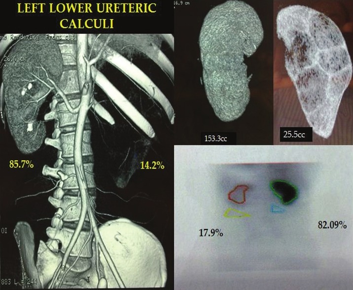
Furthermore, they were subjected to 64 slice helical CT scan (GE medical system, light speed VCT Ct99). The volume estimation software comes preinstalled with the GE 64 slice helical CT scanner (GE medical system,light speed VCT Ct99) workstation (ADW 4.3). The three dimensional tools that was available in this software package have “add object” function that was used to build the three-dimensional virtual representation of the kidneys depending on the arterial phase images. First the area of interest of renal parenchyma was delineated manually then by selecting a piece of enhancing renal parenchyma from the area of interest allows the software to identify the remaining surrounding renal parenchyma that have similar voxel values which is then added into the volume voxel to create the three dimensional renal volume. The extra parenchymal structures like renal pelvis, vessels were not included. The final 3D model of each kidney was compared with the cross sectional images to ensure that only the renal parenchyma was included and sometimes it was necessary to use the cutting tool to free the margins of kidney from adjacent structures. Then the numeric volume of this 3D model was estimated by the software itself. Renal parenchymal volume estimation was done by one radiologist in all the cases who was blinded regarding the nuclear scan report.
The right and left percent renal volume was calculated by dividing right and left volumes respectively by the combined right and left renal volume. This percent renal volume was compared with percent renal function determined by 99mTC - DTPA renal scan [Figure 1]. Correlations between percent renal volume and percent renal function is evaluated using Pearson coefficient in all cases.
Figure 1.
Good correlation between CT % renal volume and DTPA estimated %renal function
RESULTS
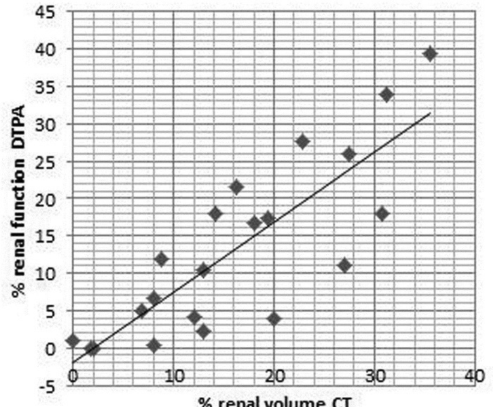
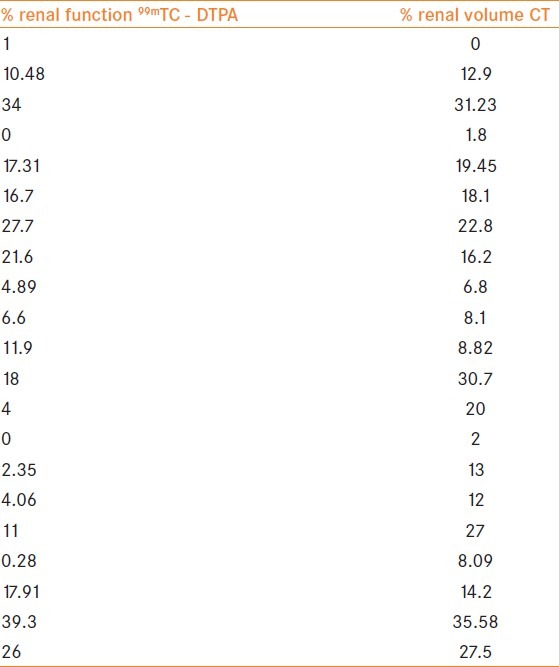
In our study male:female ratio was 1:1.1 with and the age ranged from 20 to 55 years. We evaluated a total 21 (13 left and 8 right) obstructed renal units and 21 nonobstructed renal units i.e 42 renal units. Of 21 obstructed renal units 2 had relief of obstruction before DTPA and CT scan. The etiology of chronic obstruction was pelvi-ureteric junction obstruction in four cases, ureteric calculus in nine cases, renal obstructing calculi in seven cases and lower ureteric stricture in one case. The range of s. creatinine was 0.5-1.2 mg/dl (0.78). The range of GFR estimated by 99mTC - DTPA in obstructed renal units was between 0 and 27 mg/dl (7.35) and in unobstructed renal units ranged between 23.3 and 61.7 mg/dl (46.43). The parenchymal volume as estimated by 64 slice helical CT scan in obstructed renal units are in the range of 0-147.5 cc (31.45) and in unobstructed renal units was 94.2-267cc (142.41). The percent renal fuction as estimated by 99mTC - DTPA in obstructed renal units ranges from 0%-39.33% (13.03) and in nonobstructed units ranges from 60.67 to 100% (86.89). The percent renal volume estimated by 64 slice CT scan in obstructed unit ranges from 0 to 35.58% (16.01) and in nonobstructed units ranges from 64.44 to 100% (83.94). We found a strong correlation between percent renal function estimated by 99mTC - DTPA renal and percent renal volume estimated by 64 slice CT scan in obstructed renal units (r = 0.828, P < 0.001, 95% CI: 0.618-0.928) [Figure 2] [Table 1].
Figure 2.
Positive correlation between % renal volume and % renal function in obstructed renal units
Table 1.
Raw Data for scatter graph (Figure 2)
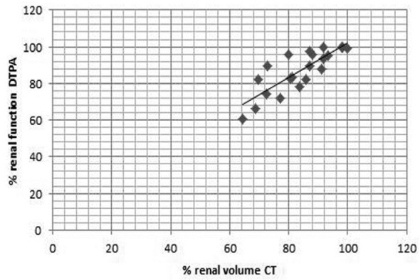
We found a strong correlation between percent renal function estimated by 99mTC - DTPA renal and percent renal volume estimated by 64 slice CT scan in nonobstructed renal units (r = 0.827, P < 0.001, 95% CI: 0.615-0.927) [Figure 3 and Table 2]. Strong correlation is found between percent renal function estimated by 99mTC - DTPA renal and percent renal volume estimated by 64 slice CT scan on combining both obstructed and non obstructed renal units (r = 0.985, P < 0.001, 95% CI: 0.974-0.992) [Figure 4].
Figure 3.
Positive correlation between % renal volume and % renal function in nonobstructed renal units
Table 2.
Data for scatter graph (Figure 3)
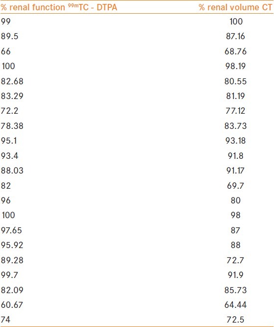
Figure 4.
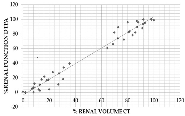
Positive correlation between % renal volume and % renal function in both obstructed and nonobstructed renal units
DISCUSSION
Obstructive uropathy is a common entity that urologists encounters in daily practice and accurate assessment of differential renal function is vital in the management of these chronically obstructed renal units. Twenty-four hours creatinine clearance is one of the methods to evaluate the total renal function but we need to have differential renal function for management of these obstructed units.[1]
At present nuclear renal scan is considered as the imaging modality of choice for differential renal function.[1] But this also have some drawbacks like it is expensive and not freely available particularly in a country like India, also it does not give any information regarding the anatomy of the renal units which is of utmost importance for the management of these obstructed renal units.
Hackstein N et al. in his study found a strong linear relationship between differential renal function by dynamic CT using modified Patlak graphic analysis, nuclear renal scan and 24-hour creatinine clearance. But it has certain disadvantages like it needs assumption of functional renal homogeneity as it involves only a particular section of kidney, complex calculations and nonavailability of the software everywhere.[2]
Ng et al. in his study compared differential renal parenchymal volume with 24-hr creatinine clearance by percutaneous nephrostomy in obstructed units and found correlation between differential renal function with differential creatinine clearance.[3] Herts et al. in their study proposed that estimated GFR by CT-based parenchymal volume can replace GFR measurement by 125I-iothalamate clearance imaging after studying 244 renal donors.[4] El – Ghar et al. observed contrast enhanced spiral CT is more sensitive than IVP for identifying the cause of chronic obstructive uropathy and it is as accurate as radioisotope renal scan for calculating the total and separate kidney function. So they recommend spiral CT with contrast medium as a single radiological diagnostic modality for the assessment of patients with chronic renal obstruction and normal serum creatinine.[5] Feder MT in their study predicting differential renal function using computerized tomography measurements of renal parenchymal area found differential renal parenchymal area measured by CT strongly correlates with differential function on renal scintigraphy and it may obviate the need for nuclear renal scan in some circumstances.[6] Morrisroe S N et al. found strong correlations between percent renal function and percent renal volume in all cases (r = 0.90, P < 0.001). They concluded that differential renal volume measured from computerized tomography strongly correlates with differential renal function on nuclear renal scan for normal and chronically obstructed kidneys and CT may serve as a single radiological diagnostic study for anatomical and functional assessment in patients in whom a poorly functioning renal unit is suspected.[1]
99mTC - DTPA renal scan and CT angiography are the investigations that are mandatory to evaluate the donor for the assessment of split renal function and vascular anatomy of the donor kidney. Summerlin AL et al. in his study found split renal function based on 3D CT models can provide a “one-stop” evaluation of both the anatomical and the functional characteristics of the kidneys of living potential kidney donors.[7]
Differential renal function estimation by CT-based parenchymal volume cannot replace nuclear scan in cases of acute cortical dysfunction and for documentation of improved renal function after surgery[1]. The disadvantages of CT estimated differential functions are its inability to be performed in renal insufficiency and false results in acute obstruction. According to the health physics society fact sheet 2010: the effective dose of IVU is 3 mSv, the effective dose of CT abdomen/pelvis is 10 mSvand the effective dose in DTPA scan is 1.8 mSv. If we combine IVU and DTPA the radiation dosage is half that of CT scan which is a disadvantage but other factors which are advantages in case of CT have to be considered as well.
CONCLUSIONS
Measurement of differential renal function by CT-based parenchymal volume correlates strongly with differential renal function estimated by 99mTC - DTPA renal scan in both obstructed and nonobstructed renal units. It has the added advantage of precise delineation of the anatomical details of the renoureteral unit. So, helical CT scan can be used as a single modality to asses the functional and anatomical status of the obstructed kidneys. It will help urologists in better management of obstructed renal units and lowering the expenses incurred by the patients. Although our study is small it throws light into the efficacy of helical CT in assessment of split renal function. Larger studies are needed to ascertain the usefulness of CECT as a functional study in patients with severely impaired renal function.
Footnotes
Source of Support: Nil
Conflict of Interest: None declared.
REFERENCES
- 1.Morrisroe SN, Su RR, Bae KT, Eisner BH, Hong C, Lahey S, et al. Differential renal function estimation using computerized tomography based renal parenchymal volume measurement. J Urol. 2010;183:2289–93. doi: 10.1016/j.juro.2010.02.024. [DOI] [PubMed] [Google Scholar]
- 2.Hackstein N, Wiegand C, Rau W, Langheinrich AC. Glomerular filtration rate measured by using triphasic helical CT with two point Patlak plot technique. Radiology. 2004;230:221. doi: 10.1148/radiol.2301021266. [DOI] [PubMed] [Google Scholar]
- 3.Ng C, Chan L, Wong KT, Cheng CW, Yu SC, Wong WS. Prediction of differential creatinine clearance in chronically obstructed kidneys by non contrasts helical computerized tomography. Int Braz J Urol. 2004;30:102–7. doi: 10.1590/s1677-55382004000200003. discussion 108. [DOI] [PubMed] [Google Scholar]
- 4.Herts B, Sharma N, Lieber M, Freire M, Goldfarb DA, Poggio ED. Estimating glomerular filtration rate in kidney donors: A model constructed with renal volume measurement from donor CT scans. Radiology. 2009;252:109–16. doi: 10.1148/radiol.2521081873. [DOI] [PubMed] [Google Scholar]
- 5.El-Ghar ME, Shokeir AA, El- Diasty TA, Refaie HF, Gad HM, El-Dein AB. Contrast enhanced spiral computerized tomography in pateints with chronic obstructive uropathy and normal serum creatinine: A single session for anatomical and functional assessment. J Urol. 2004;172:985–8. doi: 10.1097/01.ju.0000135368.77589.7c. [DOI] [PubMed] [Google Scholar]
- 6.Feder MT, Blitstein J, Mason B, et al. Predicting differential renal function using CT measurements of renal parenchymal area. J Urol. 2008;180 doi: 10.1016/j.juro.2008.07.057. [DOI] [PubMed] [Google Scholar]
- 7.Summerlin AL, Lockhart ME, Strang AM, Kolettis PN, Fineberg NS, Smith JK. Determination of split renal function by 3D reconstruction of CT angiograms: A comparison with gamma camera renography. Am J Roentgenol. 2008;191:1552–8. doi: 10.2214/AJR.07.4023. [DOI] [PMC free article] [PubMed] [Google Scholar]


