Abstract
Background: Various formulations of tretinoin have been reported to be unstable after exposure to artificial light or sunlight. The observation that tretinoin is photolabile in the presence of light led to the recommendation that tretinoin be applied in the evening in order to avoid photodegradation, which could potentially reduce efficacy. More recently, the development of innovative vehicle formulations has led, in some cases, to a marked decrease in the photodegradation of tretinoin. Objective: To compare the photostability of a micronized aqueous-based formulation of tretinoin gel 0.05% with tretinoin gel 0.025% following exposure to fluorescent and simulated solar light conditions in vitro. Methods: Micronized tretinoin gel 0.05% and tretinoin gel 0.025% were exposed to fluorescent light over eight hours or simulated solar light up to 600mJ/cm2 (equivalent to 30 minimal erythemal dose). Product samples were prepared and analyzed for tretinoin concentration using high-performance liquid chromatography. Additional duplicate samples were similarly prepared and analyzed after 2, 4, 6, and 8 hours. Results: There was an 11-percent degradation of tretinoin 0.05% formulated as the micronized gel compared to an 86-percent degradation of tretinoin 0.025% formulated as the conventional gel following eight hours of exposure to fluorescent light in vitro. The degradation of tretinoin 0.025% in the conventional gel was greater than 83 percent within two hours. In the second light exposure study, in vitro exposure to simulated solar light provided a gradual dose-response effect with tretinoin 0.05% formulated as the micronized tretinoin gel. The photodegradation of tretinoin 0.025% in the conventional gel was more immediate and of substantial magnitude (>85%) after exposures at all minimal erythemal dose levels. Conclusion: Tretinoin 0.05% formulated as a micronized gel 0.05% showed minimal degradation when exposed to fluorescent light over eight hours. This same formulation exhibited a clear dose-response degradation pattern when exposed to simulated solar light. In contrast, tretinoin 0.025% formulated in a conventional gel exhibited marked photodegradation within the first two hours when exposed to both light conditions in vitro. This information adds to the body of evidence that supports the observation that certain vehicle formulations may reduce the potential for photodegradation of tretinoin.
Tropical tretinoin is widely used in the treatment of acne vulgaris (AV). It may be prescribed as monotherapy in mild comedonal acne and is often combined with benzoyl peroxide (BP) or BP-antibiotic fixed combination products to treat inflammatory AV.1 A common approach is to recommend use of tretinoin in the evening, typically at bedtime, because it is known to be photolabile with substantial degradation shown to occur after exposure to light.2–5 This photodegradation appears to correlate with specific light conditions, may vary depending on the intensity and type of light exposure, and can be mitigated by incorporating some advanced formulation technologies, such as incorporation of tretinoin into a microsphere.5–8
In order to avoid confusion between terms that may sound similar but are very different, the following describes both microspheres and micronization.
The microsphere (microsponge) is essentially a small particulate sponge which incorporates the tretinoin into its interstitial spaces and has been used in the tretinoin microsphere gel 0.04% and 0.1% formulations (Retin A Micro® Gel; Medicis, a division of Valeant Pharmaceuticals LLC). The use of properly manufactured microspheres allows for a protective effect from exogenous exposures (i.e., UV light, benzoyl peroxide) prior to release of tretinoin from the microsphere, which occurs over time after topical application. To contrast, micronization involves a process that is distinct from microsphere use, and refers to the consistent manufacturing of a designated size range of smaller particles of active ingredient. With topical products used to treat AV, micronization is designed to create a uniformly small enough particle size that allows for follicular access of an ingredient. In addition, a consistent and small particle size of a potentially irritating active ingredient (i.e., retinoid, benzoyl peroxide) allows for more even distribution upon topical application. Formulations that do not contain a micronized active or do not stringently control particle size often contain clumps of larger particles that are more likely to produce “hot spots” due to uneven distribution of the larger particles and clusters on the skin surface.
As tretinoin has been shown to degrade rapidly when exposed to light, its use in the clinical setting has essentially been limited to evening application, thus reducing the flexibility of the treatment regimen. A formulation of tretinoin that is not associated with a clinically relevant magnitude of photodegradation would allow for greater flexibility with regard to the timing of use.
An in vitro light stability study compared the photodegradation of tretinoin 0.025% formulated in the conventional gel vehicle compared to tretinoin 0.1% incorporated in a specialized microsphere gel formulation after exposures to ambient (fluorescent) light, incandescent light, or in darkness. The spectrum of indoor lights covered the visible light spectrum and included only minimal ultraviolet-A (UVA) radiation. The microsphere formulation was found to be more stable.3 After 24 hours of exposure to fluorescent light, 98 percent of the initial tretinoin concentration remained unchanged as compared to only 31 percent remaining in the tretinoin gel 0.025%.3 This level of degradation with tretinoin gel 0.025% is consistent with what has been reported in earlier studies and may be clinically significant.4 A more recent study reported a 16-percent degradation of tretinoin 0.1% in the microsphere gel following six hours of simulated solar ultraviolet (UV) irradiation, a period intended to mimic reasonable daily outdoor use, compared to 90-percent degradation with the tretinoin 0.025% gel. This data suggest that photostability might be related to the actual light exposure conditions as well as formulation.5
The UVA component of light has been suggested to be a major contributor to the photodegradation of tretinoin.8,9 UVA is an important component of UV solar radiation. It penetrates deeper than ultraviolet-B (UVB) light into the skin due to its longer wavelength and can interact with both epidermal and dermal cells.10 UVA is capable of penetrating window glass, with indoor solar UVA irradiances representing about 25 percent of outdoor irradiance levels and about 60fold greater irradiance levels than with fluorescent light exposure.11
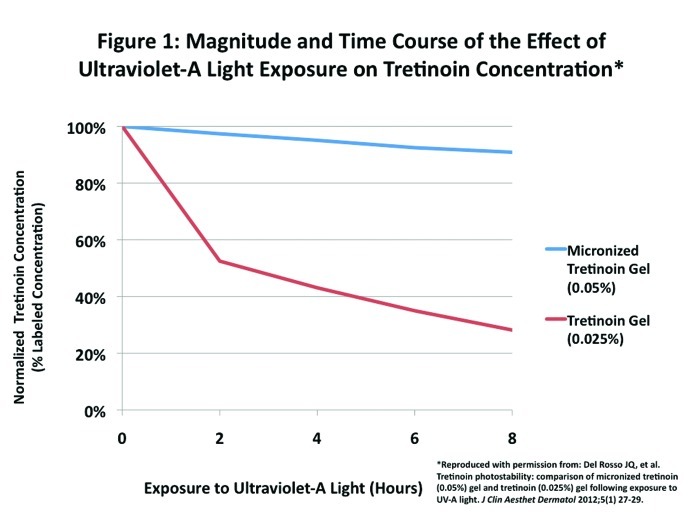
Minimal photodegradation of a micronized formulation of tretinoin gel 0.05% under UVA conditions was recently reported.12 The nine-percent degradation (Figure 1) seen after eight hours was considerably lower than that observed with tretinoin gel 0.025% (72% degradation)12 and with what has been noted by other investigators with tretinoin gel microsphere 0.1% (16% degradation) when exposed to a simulated solar UV light source for a shorter period of time.5 These data were significant based on peak absorption characteristics of tretinoin, UVA skin penetration characteristics, and overall knowledge about day-to-day levels of indoor and outdoor UVA exposure.12
Figure 1.
Magnitude and time course of the effect of ultraviolet-A light exposure on tretinoin concentration.
Reproduced with permission from: Del Rosso JQ, et al. Tretinoin photostability: comparison of micronized tretinoin (0.05%) gel and tretinoin (0.025%) gel following exposure to UV-A light. J Clin Aesthet Dermatol. 2012;5(1):27-29.
The authors of this manuscript deemed it a priority to more fully characterize the impact of other wavelengths of light on the potential for photodegradation of tretinoin 0.05% when micronized and incorporated in the specialized aqueous-based gel. The significance of this information would be a more complete understanding of if, how, and when tretinoin in this formulation may be photodegraded and to what magnitude any substantial degradation does occur. This study compares the photodegradation of tretinoin formulated as both the micronized tretinoin gel 0.05% (aqueous-based formulation) and the tretinoin gel 0.025% (conventional gel formulation) under exposure conditions with fluorescent light and simulated solar light conditions.
METHODS
Micronized tretinoin aqueous-based gel 0.05% (Atralin® Gel; Medicis, a division of Valeant Pharmaceuticals LLC) and tretinoin gel 0.025% (conventional gel vehicle) were exposed to cool white fluorescent (CWF) light and simulated solar light over time at two different testing locations (Dow Pharmaceutical Sciences, Petaluma, California and Florida Suncare Testing Inc., Ormond Beach, Florida).
The CWF light source included six 20W tubular light bulbs and covered a wavelength range of 350 to 700nm with an intensity of 16,000lux. Measurements were undertaken at 2-, 4-, 6-, and 8-hour time points. The solar simulator system was an Atlas SunTest CPS machine (Atlas Material Testing Technology LLC, Chicago, Illinois), which meets ID65 spectral criterion with an optical filter system consisting of a coated quartz glass dish, a window glass filter, and an ID65 filter used in combination. The filtered xenon light source (SunTest) is a full-spectrum light source containing both visible and UV outputs. The intensity of simulated solar light provided by the Atlas SunTest CPS machine is 240mJ/cm2 per hour (equivalent to an minimal erythemal dose [MED] of 12 MED/hour). Test substances were exposed to solar light at this intensity for 0 to 150 minutes. The highest intensity (600mJ/cm2 or 30 MED) is equivalent to eight hours of exposure to sunlight in mid-June on a clear day in Florida (29.5°N) by a typical person with Fitzpatrick type II skin.13,14 These are considered extreme conditions and it is well known that MED is influenced by skin type, time of day, season, latitude, cloud cover, surface reflection, and altitude.13,14
Equal amounts of each gel were placed in 5mL clear polypropylene syringes and exposed separately to the two light conditions. After irradiation for designated times or intensities, test samples were analyzed for tretinoin using high-performance liquid chromatography (HPLC). Duplicate 2g samples were delivered into individual 50mL volumetric flasks and dissolved in 25mL of phosphate buffer (pH 3.0). Each flask was filled to volume with tetrahydrofuran. A portion of each sample solution was filtered through a 0.45pm polyvinylidene fluoride filter and assayed by HPLC immediately after preparation. Additional duplicate samples were similarly prepared and analyzed after 2, 4, 6, and 8 hours of light exposure. Each sample was analyzed for tretinoin and expressed as percent of labeled concentration (%LC).
RESULTS
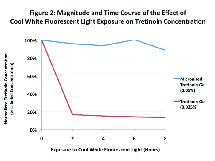
When exposed to CWF light for eight hours, there was an 11-percent degradation (%LC) of micronized tretinoin gel 0.05% compared to an 84-percent degradation of tretinoin gel 0.025% (Figure 2). The majority of degradation of tretinoin gel 0.025% occurred early during the exposure period, with an 83-percent degradation seen at the first measurement point (2 hours).
Figure 2.
Magnitude and time course of the effect of cool white fluorescent light exposure on tretinoin concentration
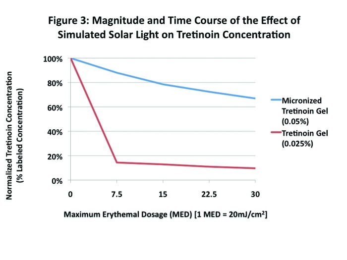
Following exposure to simulated solar light, a dramatic degradation of tretinoin gel 0.025% was seen across all MED assessment points (MED range from 7.5–30) with a degradation (%LC) ranging from 85 to 90 percent. With micronized tretinoin gel 0.05%, a dose-dependent response to solar light was seen. Degradation was 12 percent with an MED of 7.5 and increased progressively in a MED-dependent (simulated solar light dose-dependent) pattern under the most extreme MED conditions that were used in the study (MED 30). However, these levels of tretinoin degradation were modest as compared to the magnitude of degradation observed with tretinoin gel 0.025% formulated in the conventional gel vehicle (Figure 3).
Figure 3.
Magnitude and time course of the effect of simulated solar light on tretinoin concentration
DISCUSSION
Tretinoin gel 0.025% has been shown to degrade under a variety of light conditions. In this study, tretinoin degradations of 85 to 90 percent were seen in fluorescent light and 84 to 89 percent in simulated solar light with tretinoin 0.025% formulated in a conventional gel vehicle. The impact of fluorescent light on this same formulation of tretinoin was seen from the first time point of evaluation (2 hours), and all the simulated solar light conditions tested resulted in similar levels of degradation. Other investigators have shown comparable levels of degradation with tretinoin gel 0.025% (conventional gel) in fluorescent light, incandescent light, and simulated solar UV irradiation.3,5
Photodegradation of tretinoin appears to correlate with specific light conditions and can be influenced by the vehicle technology used to formulate the product.6–8 New formulations of tretinoin have been studied under a variety of light conditions. For example, tretinoin microsphere gel 0.1% showed minimal (<3%) degradation following exposure to fluorescent or incandescent light over 24 hours, but more degradation (16%) was observed following exposure to simulated solar UV irradiation over six hours.3,5
The micronization of tretinoin and its formulation in an aqueous-based gel 0.05% appears to contribute to the protection of tretinoin against photodegradation. Following exposure to UVA light, only nine-percent tretinoin degradation was seen after eight hours. A similar level of protection was afforded in this current study following fluorescent light exposure for eight hours. There was a time-dependent degradation of micronized tretinoin gel 0.05% following simulated solar light; however, the level of degradation seen was comparable to other light conditions we have tested with the exception of extreme conditions unlikely to be encountered in clinical practice (8 hours continuous exposure to sunlight in mid-June on a clear day in Florida). Importantly, even under these extreme light exposure conditions, almost 80 percent of the tretinoin was still present.
These data add to our knowledge on tretinoin photostability under various light conditions and confirm other data that support a protective effect afforded by the micronized tretinoin gel 0.05% formulation against photodegradation of tretinoin. An important outcome of these findings is the potential for flexibility with the application of micronized tretinoin gel 0.05% during the day or evening. A limitation of the testing methodology is that the study is completed in an artificial environment and does not take into account any effects that may potentially influence the magnitude and/or time course of tretinoin photodegradation after application of the tretinoin formulation to human skin.
ACKNOWLEDGMENT
The authors acknowledge Inergy Limited, specifically Brian Bulley, MSc, for support in preparation of this publication. Valeant Dermatology (now Medicis), a wholly owned subsidiary of Valeant Pharmaceuticals North America LLC, funded the activities of Inergy Limited pertaining to this manuscript. This manuscript including graphics (Figures) was finalized and submitted to the journal by the lead author after the receipt of input from the other authors and Inergy Limited. Galley review and final necessary adjustments were completed by the lead author and returned directly to the journal.
Footnotes
DISCLOSURE: Dr. Del Rosso has served as a consultant, speaker, and/or researcher for Allergan, Bayer, Eisai, Galderma, Medicis, Onset Dermatologics , Obagi Medical Products, Pharmaderm, Primus, Promius, Ranbaxy, TriaBeauty, Unilever, Valeant, and Warner-Chilcott. With regard to tretinoin, Dr. Del Rosso has served as a consultant, advisory board member, and speaker related to micronized tretinoin 0.05% gel with Coria prior to their purchase by Valeant and, still currently with Valeant, and has been a consultant, speaker, and researcher related to tretinoin 0.04% and 0.1% microsphere gels in the past with Ortho Dermatology. He currently serves as a consultant with Valeant on products related to acne and other areas of medical dermatology. Dr. Harper has served as a consultant, speaker, and/or researcher for Allergan, Bayer, Galderma, Medicis, Ranbaxy, and Valeant. With regard to tretinoin, Dr. Harper has served as a consultant, advisory board member, and speaker related to micronized tretinoin 0.05% gel with Coria prior to their purchase by Valeant and currently with Valeant. Drs. Pillai and Moore are employees of Dow Pharmaceutical Sciences, developers of micronized tretinoin 0.05% gel.
REFERENCES
- 1.Shroot B. Further light is shed on topical therapy. J Invest Dermatol. 2003;121(3):xiii–xiv. doi: 10.1046/j.1523-1747.2003.12455.x. [DOI] [PubMed] [Google Scholar]
- 2.Nyirady J, Lucas C, Yusuf M, et al. The stability of tretinoin in tretinoin gel microsphere 0.1% Cutis. 2002;70(5):295–298. [PubMed] [Google Scholar]
- 3.Martin B, Meunier C, Montels D, et al. Chemical stability of adapalene and tretinoin when combined with benzoyl peroxide in presence and in absence of visible light and ultra-violet radiation. Br J Dermatol. 1998;139(Suppl 52):8–11. doi: 10.1046/j.1365-2133.1998.1390s2008.x. [DOI] [PubMed] [Google Scholar]
- 4.Nighland M, Yusuf M, Wisniewski S, et al. The effect of simulated solar UV irradiation on tretinoin in tretinoin gel microsphere 0.1% and tretinoin gel 0.025% Cutis. 2006;77(5):313–316. [PubMed] [Google Scholar]
- 5.Brisaert MG, Everaerts I, Plaizier-Vercammen JA. Chemical stability of tretinoin in dermatological preparation. Pharm Acta Helv. 1995;70:161–166. [Google Scholar]
- 6.Brisaert M, Plaizier-Vercammen J. Investigation on the photostability of a tretinoin lotion and stabilization with additives. Int J Pharm. 2000;199:49–57. doi: 10.1016/s0378-5173(00)00366-5. [DOI] [PubMed] [Google Scholar]
- 7.Tashtoush BM, Jacobson EL, Jacobson MK. UVA is the major contributor to the photodegradation of tretinoin and isotretinoin: implications for development of improved pharmaceutical formulations. Int J Pharm. 2008;352:123–128. doi: 10.1016/j.ijpharm.2007.10.045. [DOI] [PMC free article] [PubMed] [Google Scholar]
- 8.Teraoka R, Konishi Y, Matsuda Y. Photochemical and oxidative degradation of the solid-state tretinoin tocoferil. Chem Pharm Bull. 2001;49(4):368–372. doi: 10.1248/cpb.49.368. [DOI] [PubMed] [Google Scholar]
- 9.Huang XX, Bernerd F, Halliday GM. Ultraviolet A within sunlight induces mutations in the epidermal basal layer of engineered human skin. Am J Pathol. 2009;174:1534–1543. doi: 10.2353/ajpath.2009.080318. [DOI] [PMC free article] [PubMed] [Google Scholar]
- 10.Godar DE, Landry RJ, Lucas AD. Increased UVA exposures and decreased cutaneous Vitamin D(3) levels may be responsible for the increasing incidence of melanoma. Med Hypotheses. 2009;72(4):434443. doi: 10.1016/j.mehy.2008.09.056. [DOI] [PubMed] [Google Scholar]
- 11.Del Rosso JQ, Harper J, Pillai R, Moore R. Tretinoin photostability: comparison of micronized tretinoin (0.05%) gel and tretinoin (0.025%) gel following exposure to ultraviolet A light. J Clin Aesthet Dermatol. 2012;5(1):27–29. [PMC free article] [PubMed] [Google Scholar]
- 12.Diffey BL. Solar ultraviolet radiation effects on biological systems. Phys Med Biol. 1991;36(3):299–328. doi: 10.1088/0031-9155/36/3/001. [DOI] [PubMed] [Google Scholar]
- 13.Diffey BL, Elwood JM. Tables of ambient solar ultraviolet radiation for use in epidemiological studies of malignant melanoma and other diseases. Epidemiological Aspects of Cutaneous Malignant Melanoma. In: Gallagher RP, Elwood JM, editors. Boston: Kluwer Academic Press; 1994. pp. 81–105. [Google Scholar]


