

Full text
PDF
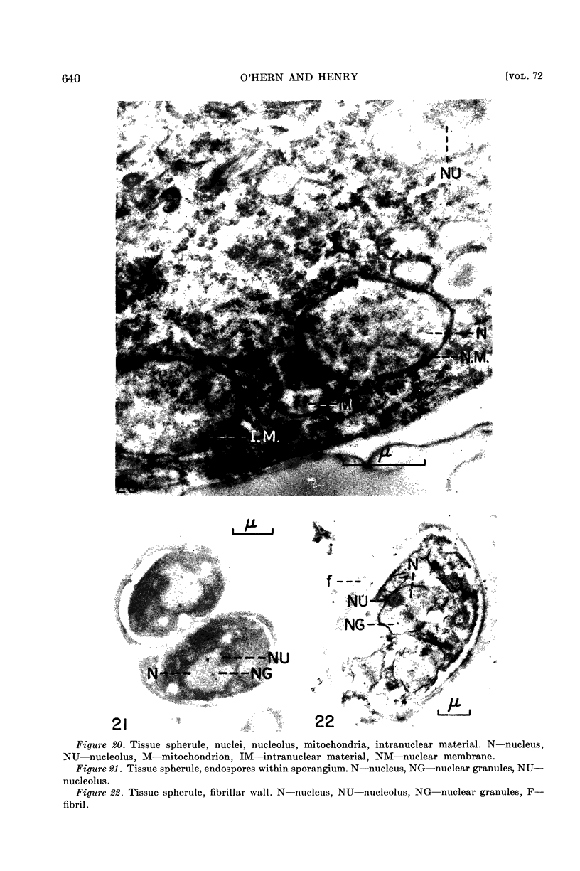
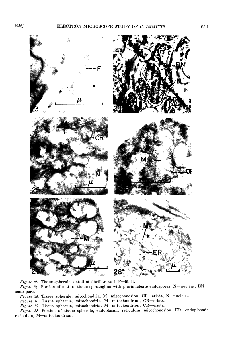
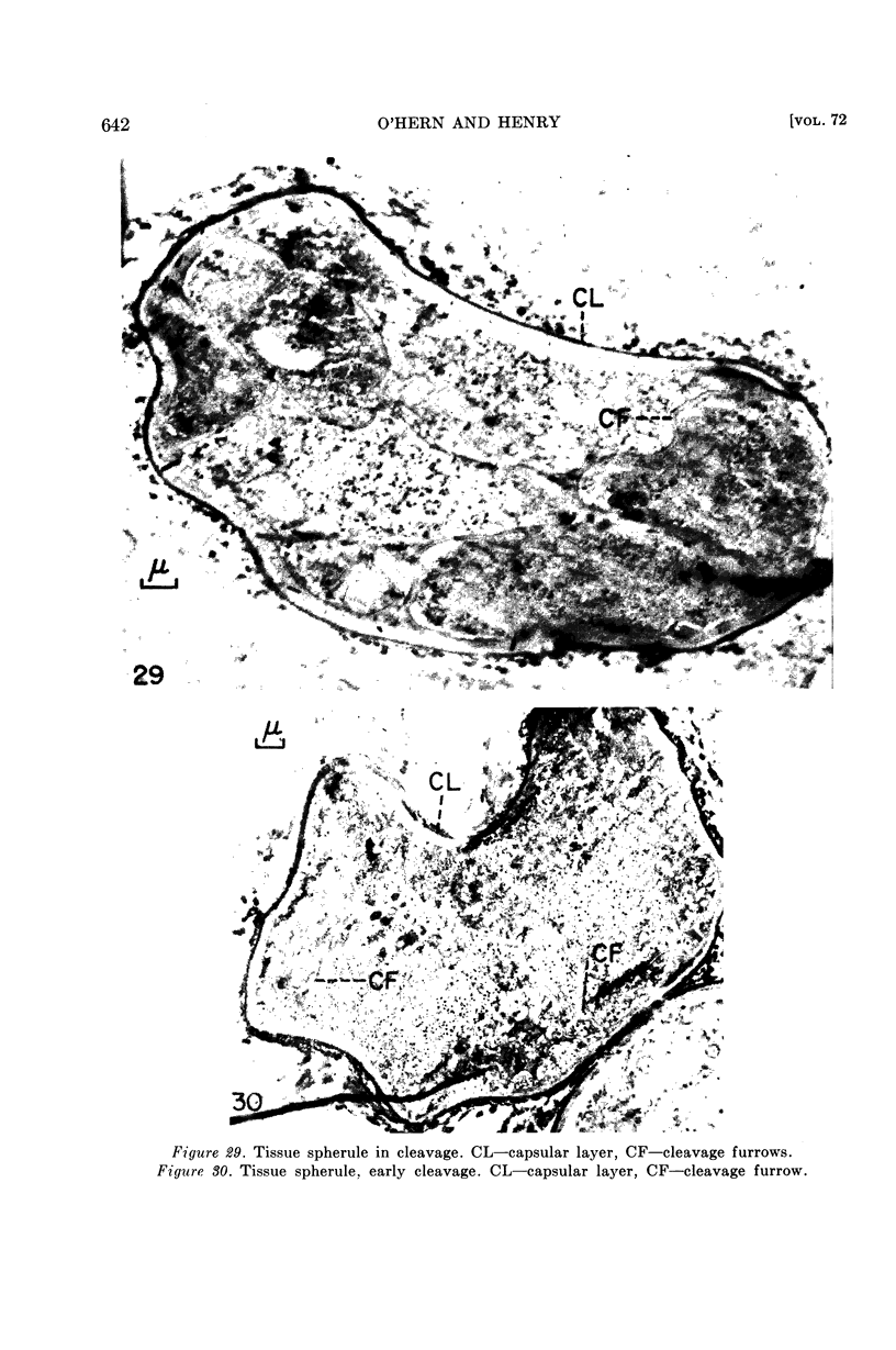
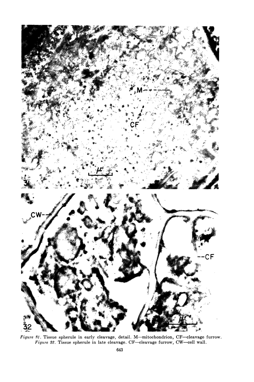
Images in this article
Selected References
These references are in PubMed. This may not be the complete list of references from this article.
- BLANK F., BURKE R. C. Chemical composition of the cell wall of Coccidioides immitis. Nature. 1954 May 1;173(4409):829–829. doi: 10.1038/173829a0. [DOI] [PubMed] [Google Scholar]
- CHAPMAN G. B., HILLIER J. Electron microscopy of ultra-thin sections of bacteria I. Cellular division in Bacillus cereus. J Bacteriol. 1953 Sep;66(3):362–373. doi: 10.1128/jb.66.3.362-373.1953. [DOI] [PMC free article] [PubMed] [Google Scholar]
- CHIFFELLE T. L., PUTT F. A. Propylene and ethylene glycol as solvents for Sudan IV and Sudan black B. Stain Technol. 1951 Jan;26(1):51–56. doi: 10.3109/10520295109113178. [DOI] [PubMed] [Google Scholar]
- LATTA H., HARTMANN J. F. Use of a glass edge in thin sectioning for electron microscopy. Proc Soc Exp Biol Med. 1950 Jun;74(2):436–439. doi: 10.3181/00379727-74-17931. [DOI] [PubMed] [Google Scholar]
- PALADE G. E. A study of fixation for electron microscopy. J Exp Med. 1952 Mar;95(3):285–298. doi: 10.1084/jem.95.3.285. [DOI] [PMC free article] [PubMed] [Google Scholar]
- PALADE G. E. An electron microscope study of the mitochondrial structure. J Histochem Cytochem. 1953 Jul;1(4):188–211. doi: 10.1177/1.4.188. [DOI] [PubMed] [Google Scholar]
- PORTER K. R. Electron microscopy of basophilic components of cytoplasm. J Histochem Cytochem. 1954 Sep;2(5):346–375. doi: 10.1177/2.5.346. [DOI] [PubMed] [Google Scholar]
- TARBET J. E., BRESLAU A. M. Histochemical investigation of the spherule of Coccidioides immitis in relation to host reaction. J Infect Dis. 1953 Mar-Apr;92(2):183–190. doi: 10.1093/infdis/92.2.183. [DOI] [PubMed] [Google Scholar]