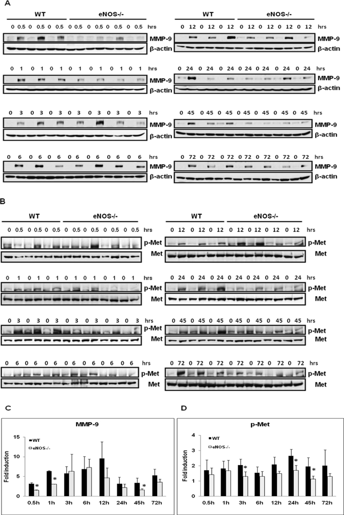
Fig. 3. Dysregulated MMP-9 Induction and Met phosphorylation in eNOS−/−.
Western blotting of (A) MMP-9 and (B) p-Met (Tyr1349) expression of total protein extracts of resected lobes (0 min) and remnant livers at indicated timepoints post-PH of WT (n=3), eNOS−/− (n=4); (C, D) Densitometric analysis of fold induction as compared to the respective remnant livers. Data represents mean+/− SD. *P < 0.05 vs WT.

