Abstract
Purpose
To screen deletions/duplications of the RB1 gene in a large cohort of Iranian patients using the multiplex ligation-dependent probe amplification (MLPA) technique.
Methods
A total of 121 patients with retinoblastoma, involving 55 unilateral and 66 bilateral or familial retinoblastomas, were included in this study. Among these patients, 121 blood and 43 tissue samples were available. DNA was extracted from the blood and tissue samples and analyzed with an RB1-specific MLPA probe set. The mutation findings were validated with SYBR Green Real-Time PCR.
Results
Twenty-two mutations were found in 21 patients; of these, ten mutations were detected in patients with isolated unilateral retinoblastoma.
Conclusions
Our results suggested that MLPA is a fast, reliable, and powerful method for detecting deletions/duplications in patients with retinoblastoma.
Introduction
Retinoblastoma, with an estimated frequency of 1:15,000, is the most common intraocular solid tumor in children under 6 years of age [1]. Inactivation of both alleles of the RB1 gene in a single immature retinal cell can trigger retinoblastoma tumorigenesis. In about 50% of patients, both inactivating mutations occur somatically, whereas in the hereditary form, a mutated allele is inherited, and the second mutational event occurs somatically. The latter group often presents with the disease at an earlier age in both eyes [2,3].
The large RB1 gene spans more than 180 kb on chromosome 13q14, which consists of 27 exons and transcribes into 4.8 kb messenger RNA [4]. Thus far, wide spectrums of the mutations including large cytogenetic rearrangements, subcytogenetic deletions/duplications, and point mutations have been reported in the RB1 gene. With the exception of recurrent mutations in 11 arginine codons, the RB1 gene has no remarkable mutation hot spots and point mutations, which are usually unique to each family, distributed all over it [4-12].
Detecting RB1 mutations could enhance the quality of clinical management of retinoblastoma in patients, and risk prediction for all members of affected families could be estimated [13]. Despite these advantages, there are many challenges in molecular genetic diagnosis of retinoblastoma because of the large size of the RB1 gene and widely dispersed mutations [13,14].
Currently, the routine procedure for RB1 gene testing is joint screening of the coding regions with direct sequencing and deletions/duplications analysis. Fluorescent in situ hybridization (FISH) and karyotyping were previously used to evaluate cytogenetic abnormalities but have been replaced by more enhanced and specialized techniques such as multiplex ligation-dependent probe amplification (MLPA) or quantitative multiplex fluorescence PCR (QMFPCR). Together, these approaches could detect more than 90% of all known mutations [11,15].
In the past decade, MLPA has been accepted as a sensitive method for detecting cytogenetic and subcytogenetic abnormalities and has been merged in the gene testing procedure in many laboratories [16]; however, there is little information about the ability of this technique in the RB1 gene literature [11,17,18]. Accordingly, in the present study, the detection rate of RB1 gene gross rearrangements in a large cohort of Iranian patients with retinoblastoma was investigated with MLPA.
Methods
Subjects
A total of 121 patients with retinoblastoma (55 patients with isolated unilateral retinoblastoma, one patient with hereditary unilateral retinoblastoma, 53 patients with isolated bilateral retinoblastoma, and 12 patients with hereditary bilateral retinoblastoma) who were referred to Mahak, Farabi, and Rasoul Akram hospitals were included in this study. The mean ages of the patients with unilateral and bilateral retinoblastoma were 21.7 and 14.4 months, respectively. Among these, enucleated eye samples from 43 patients were also recruited for molecular analysis. In the patients with unilateral retinoblastoma, 23 out of 56 were enucleated, and tumor samples were available; only one patient had a family history of retinoblastoma. In the bilateral group, 20 patients were enucleated, and 12 patients had a family history of retinoblastoma.
From each patient, 5 ml of peripheral blood in tubes containing EDTA and 2 ml in heparin tubes were collected. Genomic DNA from peripheral blood samples was extracted using standard salting out method. DNA from the tumor samples were extracted by heat-induced retrieval protocol (boiling of tissue sections in 0.1 M alkaline solution) [19]. All patients’ families were subjected to genetic counseling, and informed consent was obtained from each parent/guardian. The study protocol was approved by the Avicenna Research Institute's Ethics and Human Rights Committee. The study was in accordance with the provisions of the Declaration of Helsinki. Clinically, retinoblastoma was diagnosed by the presence of tumors in one or both eyes, and diagnosis for enucleated patients was confirmed with pathological analysis.
Multiplex ligation-dependent probe amplification
To investigate large deletions/duplications in the RB1 gene, MLPA analysis was performed using the SALSA MLPA kit P047-B1 RB1 (MRC-Holland, Amsterdam, the Netherlands) according to the manufacturer’s protocol. The kit contained 24 probes for the RB1 gene (the promoter and each exon had a specific probe, except exons 5, 10, 15, and 16), three probes for the flanking genes of the RB1 gene, ITM2B, CHC1L, and DLEU1, and 13 control probes on locations other than chromosome 13. CHC1L and DLEU1 are centromeric whereas ITM2B is telomeric to RB1. The average distances between RB1 and ITM2B, CHC1L, and DLEU1 are 50 kb, 50 kb, and 1.5 Mb, respectively.
Briefly, 100 ng of genomic DNA in a final volume of 5 µl was denatured and hybridized with the SALSA probe mix, followed by incubation at 60 °C for 18 h. Subsequently, the annealed probes were ligated using the Ligase-65 mix provided at 54 °C for 15 min.
In the next step, 10 µl of ligated products, as the template, were used for DNA amplification. The PCR amplicons were run on a Genetic Analyzer 3130 (Applied Biosystems, Foster City, CA), and the results were analyzed with GeneMarker software version 1.91 (SoftGenetics LLC, State College, PA). The normal pattern was expected to produce a normalized signal value ratio of 1:1; any value out of the ranges <0.75 or >1.30 was considered abnormal and corresponded to a deletion and a duplication, respectively.
Inclusion criteria for control samples
In each MLPA reaction, regarding the number of samples, three to six control samples were simultaneously used. All controls were adults, with no ocular tumor or other malignancy. In addition, the locations of all internal probes of the RB1 gene in the control group were verified with direct sequencing. To confirm the presence of two normal copies of the RB1 gene and the absence of any chromosomal deletions, all control samples were checked with the two STS markers, D13S153 (Rbi2, inside RB1 intron 2) and D13S128 (in the flanking sequence of the RB1 gene). Only samples heterozygous for both markers were included.
Validation of multiplex ligation-dependent probe amplification results
Samples with abnormal MLPA results were checked with direct sequencing to be assured of an intact probe binding site and appropriate binding of MLPA probes to their related regions; the primers and PCR conditions were described previously [11]. Gene dosage for different samples was performed with relative quantification, and 2-ΔΔCt was calculated by normalizing the RB1 exons to the RPPH1 gene, a single copy reference gene. In addition, fragments with similar size and GC content to RPPH1 were designed for the RB1 exons in which deletions and duplications were observed with MLPA. Only those with similar efficiency to RPPH1 were used to evaluate the gene dosage. The primer sequences and sizes are shown in Table 1. Real-time PCR (SYBR Green) was performed using a serial dilution of DNA samples including 200, 100, 50, 25, 12.5, 6.25, and 3.125 ng in quadruple repeats. Then, according to the standard curves created and by comparing the slope and efficiency of each reaction, 25 ng of DNA was chosen as the best concentration that gave dose-dependent results. In this DNA concentration, real-time PCR detected samples with deletion or duplication. The copy numbers of the exons compared to the reference gene were determined as follows: ΔΔCt=(Ct RPPH1 (calibrator sample) − Ct RB1 exon (calibrator sample)) – (Ct RPPH1 (unknown sample) − Ct RB1 exon (unknown sample)). Absolute quantification was made by converting the measured values to absolute ones. Then using the ratio equation (2−ΔΔCt), the relative gene copy numbers were calculated. The expected values were about 1 for normal samples, 0.5 for heterozygous deletions, and 1.5 for heterozygous duplications [20].
Table 1. The primers used for quantitative analysis of RB1 gene.
| Genomic region | Primers (5′ → 3′) | Size (bp) | Ta | |
|---|---|---|---|---|
|
RPPH1 |
Forward |
GAGGTGAGTTCCCAGAGAACG |
134 |
60 |
| Reverse |
TTCGCTGGCCGTGAGTCTGTTC |
|||
|
RB1 exon 7 |
Forward |
TCAGGGGAAGTATTACAAATGGAAG |
117 |
60 |
| Reverse |
ACTATATGGTTCTTTGAGCAACATG |
|||
|
RB1 exon 13 |
Forward |
CTAAAGCTGTGGGACAGGGTTG |
116 |
60 |
| Reverse |
TTATACGAACTGGAAAGATGCTGC |
|||
|
RB1 exon 17 |
Forward |
GCCTTTGATTTTTACAAAGTGATCGAAAG |
128 |
60 |
| Reverse |
CTTACTGAGAGCCATGCAAGGGA |
|||
|
RB1 exon 19 |
Forward |
TATATCTAGGTATCTTTCTCCTGTAAG |
130 |
60 |
| Reverse |
GGTAGATTTCAATGGCTTCTGGG |
|||
| RB1 exon 22 | Forward |
TTGCAGTATGCTTCCACCAGG |
123 | 60 |
| Reverse | GGTAGGGGGCTAGAGCAAAAAC | |||
Karyotype analysis
To investigate cytogenetic abnormalities in the patients, fresh blood samples were taken in heparin tubes, cultured on RPMI-1640 medium, and finally GTG banded according to standard protocols. The prepared slides were directly analyzed under the microscope to evaluate any potential chromosomal changes. Each slide had approximately 440 to 500 bands.
Results
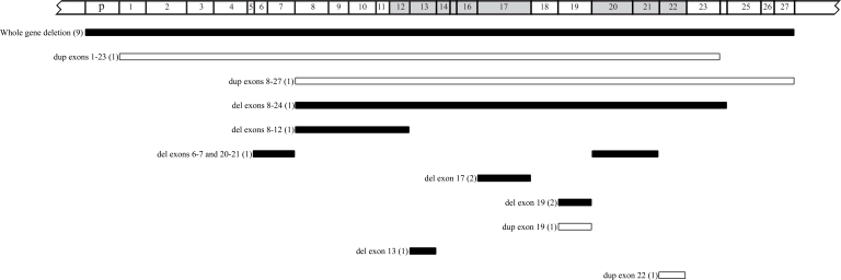
Cytogenetic analysis for all patients showed a normal 46, XY or 46, XX karyotype. MLPA reactions were performed for all 121 samples, resulting in 22 mutations identified in 21 patients; among these mutations, nine whole gene deletions, nine intragenic deletions, and four intragenic duplications were observed (Table 2 and Figure 1). In eight of the nine patients with whole gene deletion, all three flanking probes signals showed a deletion, which indicated a deletion of at least 1.5 Mb; however, in one bilateral patient, only the two probes for ITM2B and CHC1L were deleted. All mutations identified with MLPA were validated using real-time PCR analysis; the sequencing results also showed that there is no point mutation in the probe-binding site of the samples.
Table 2. The rearrangements found in the 21 retinoblastoma patients detected by MLPA in tumor as well as blood samples.
| Patient’s ID | Mutation in tumor sample | Mutation in blood sample | Type of disease | Familial History |
|---|---|---|---|---|
| IRB1 |
Del Ex 8 −12 |
Normal |
Unilateral |
No |
| IRB12 |
Whole gene deletion |
Whole gene deletion |
Unilateral |
No |
| IRB13 |
Del Ex 17 |
Normal |
Unilateral |
No |
| IRB14 |
Del Ex 8–24 |
Del Ex 8–24 |
Bilateral |
No |
| IRB15 |
Del Ex 6–7, Del Ex 20–21 |
Del Ex 6–7 |
Bilateral |
No |
| IRB19 |
Dup Ex 19 |
Dup Ex 19 |
Unilateral |
No |
| IRB22 |
Dup Ex 8–27 |
Normal |
Unilateral |
No |
| IRB28 |
Whole gene deletion |
Normal |
Unilateral |
No |
| IRB35 |
Dup Ex 1–23 |
Normal |
Unilateral |
No |
| IRB38 |
Whole gene deletion |
Whole gene deletion |
Bilateral |
No |
| IRB40 |
Dup Ex 22 |
Dup Ex 22 |
Bilateral |
No |
| IRB41 |
Whole gene deletion |
Whole gene deletion |
Bilateral |
No |
| IRB113 |
No tumor sample available |
Del Ex 13 |
Unilateral |
No |
| IRB120 |
No tumor sample available |
Whole gene deletion |
Bilateral |
No |
| IRB133 |
No tumor sample available |
Whole gene deletion |
Bilateral |
No |
| IRB139 |
No tumor sample available |
Whole gene deletion |
Bilateral |
No |
| IRB143 |
No tumor sample available |
Whole gene deletion |
Unilateral |
Yes |
| IRB158 |
No tumor sample available |
Whole gene deletion |
Unilateral |
No |
| IRB159 |
No tumor sample available |
Del Ex 19 |
Bilateral |
No |
| IRB179 |
No tumor sample available |
Del Ex 19 |
Unilateral |
No |
| IRB194 | No tumor sample available | Del Ex 17 | Bilateral | No |
Del: Deletion; Ex: Exon; Dup: Duplication.
Figure 1.
Schematic representation of the deletions and duplications found in this study. Arabic numbers in the parentheses show the occurrence times for each rearrangement. The white bars represent duplications, and the black ones indicate deletions. The gray regions on the RB1 gene show the pocket domains.
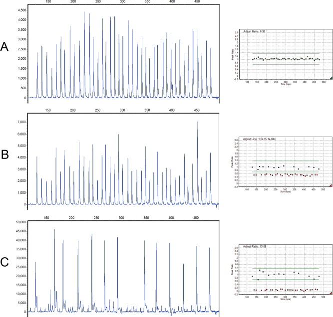
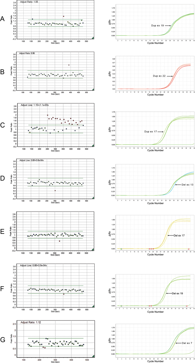
Regarding disease type, ten large mutations were found in patients with unilateral retinoblastoma (ten of 55); five of these patients showed abnormalities in either blood and tumor samples. According to these results, the MLPA detection rate for constitutional mutations in patients with unilateral retinoblastoma (five of 55) was 9.1%. The total detection rate for unilateral cases (10 of 55) was 18.2% and for bilateral and familial cases was 11 of 66 (16.6%). Sixteen mutations in 121 (13.2%) blood samples were detected with MLPA. The MLPA results are shown in Figure 2 and Figure 3. Examples of standard curves and amplification plots of real-time PCR demonstrating deletions in two patients are elucidated in Figure 3.
Figure 2.
Chromatograms illustrating whole gene deletion in patient IRB12. A: Normal control. B: Multiplex ligation-dependent probe amplification (MLPA) results of a blood DNA sample shows heterozygous deletion of the RB1 gene. C: The later MLPA results for the tumor DNA sample of the same patient show homozygous deletion of RB1.
Figure 3.
GeneMarker plots of multiplex ligation-dependent probe amplification reactions (left) and corresponding real-time polymerase chain reactions (right) to validate the results of (A) exon 19 duplication, (B) exon 22 duplication, (C) exon 8 through 22 duplication, (D) exon 13 deletion, (E) exon 17 deletion, (F) exon 19 deletion, and (G) exon 6 and 7 deletion.
Discussion
MLPA as a sensitive, reproducible, and sequence-specific technique was described by Schouten et al. for detecting gains or losses of single exons in small amounts of human DNA samples [16]. Now MLPA is a reliable method for detecting large deletions/duplications. Despite the growing number of studies that have used MLPA to analyze genes in various human diseases [21-25], only a few have used it in evaluating RB1 mutations in retinoblastoma and other cancers [17,18,26-28]. Although many laboratories use MLPA for detecting and screening large RB1 mutations, there is no comprehensive data in the literature. In this survey, 121 patients with retinoblastoma were evaluated with MLPA.
Cytogenetic abnormalities and subcytogenetic mutations cause many retinoblastomas [29-32]. Approximately 15%–25% of retinoblastoma cases are due to large deletions and insertions [11,13,14]. These rearrangements were previously investigated with various techniques such as karyotyping, G-banding, FISH, QMFPCR, MLPA, and real-time PCR [5,10,14,33-35]. However, each technique has its own advantages and limitations. Karyotyping and FISH can detect only large rearrangements including entire gene deletion. These techniques are especially useful in determining the borders of large deletions that have been identified by other methods. Other methods such as MLPA, real-time PCR, and QMFPCR, which are based on quantification of amplifying PCR products, are more sensitive and can detect more mutations. Real-time PCR assays are characterized by high precision; however, they are difficult to implement as a multiplex. MLPA and QMFPCR can be easily multiplexed. Hence, in a single reaction, all regions of a gene can be analyzed, and an abnormality is found, it can reanalyzed with real-time PCR. QMFPCR should be set up manually, which makes the technique error-prone and reduces its reproducibility, whereas MLPA probes and reagents are commercially available and easy to use. Despite the ability of MLPA and QMFPCR to identifying the numerical changes in DNA, the techniques cannot detect mutations such as translocations and inversions, while karyotyping can detect this type of aberration.
In the present study, no RB1 mutations including large deletion, translocation, and inversion were detected with karyotyping. By analyzing the MLPA results, 22 deletions/duplications in 21 patients were found. Among these mutations, 16 were detected in the blood and tumor samples. The frequency of deletions/duplications in previous studies varies from 10% to 20% [5,7,13,14]. In our study, the frequency of constitutional deletions/duplications in isolated unilateral and bilateral/familial tumors were 9.1% (five of 55) and 16.6% (11 of 66), respectively. According to our results, 17.3% (21 of 121) of the total mutations were detected with MLPA, a rate similar to previous studies. Germline mutations in 10%–13% of patients with unilateral retinoblastoma have been reported [4,13,14,36-38]. Our estimation of the deletions/duplications rate using MLPA was 9.1% for patients with isolated unilateral retinoblastoma. Comparison of data shows that the MLPA detection rate in these patients was nearly equal to the total mutations (including deletion/duplication and point mutations) found in the other studies. However, our frequency of deletions/duplications in patients with unilateral retinoblastoma is higher than previous reports. This could be explained by two hypotheses: First, the small size of the population (55 samples) could result in chance findings. Second, the higher detection rate could be due to the higher sensitivity of MLPA compared to the methods used in other studies.
Analysis of larger sample sizes may show that this high rate of alterations is a chance finding. However, according to the results, MLPA should be considered for unilateral retinoblastoma. Therefore, to find causal mutations, if tumor samples are not available, MLPA could be recommended as the first step of mutation detection. However, if no mutation is detected with MLPA, other popular methods including sequencing of the entire RB1 coding region is suggested.
Recently, Rushlow et al. [15] showed that a remarkable number of patients with retinoblastoma carry mosaic mutations; the researchers found mutations in 92.6% of cases via a combination of full sequencing and deletions/duplication analysis of RB1. Moreover, they found additional mutations in cases with clearly normal results using PCR-based methods, so the detection rate increased to 94.8% [15]. Results of MLPA or real-time PCR in low-level mosaic cases, depending on the percentage of mutant cells, may be mistaken as normal. Actually, MLPA is a relative quantification method, and deletions/duplications in unknown samples are identified by comparison to the normal controls. MLPA is not expected to detect all imbalances in mosaic cases [39].
Germline mutations in unilateral cases such as splice site affecting and missense mutations out-of-pocket domains usually have mild to moderate deleterious effects [40,41]. In this regard, based on our findings, deletions/duplications have a relatively weak to moderate effect as well. There is considerable evidence of the genetic mechanisms that explain the incomplete penetrance of single exonic deletions (for example, exon 4) or multiexonic large deletions such as exons 24–25 [42-44]. In such mutations, the in-frame exonic deletions cause the pRB to lose some of its function as well as penetrance of the mutation, which depends on the remaining activity of the shortened protein. In addition, as previously described, the whole gene deletions show incomplete penetrance as well [5,45-47]. However, when age is addressed, the unilateral tumors may be due to the patients’ young age. Patients with unilateral retinoblastoma, as a progressive consequence of the disease, may probably show bilateral tumors in the future. Both unilaterally affected children (3 and 8 months) in our study with whole gene deletion were treated with systemic chemotherapy, which may prevent formation of new tumors in the other eye. Thus, these patients may be incorrectly categorized in the unilateral group. To clarify this, further long-standing investigation and follow-up are necessary.
In conclusion, MLPA is a strong method for primary evaluation of RB1 gene deletions/duplications in patients with retinoblastoma. Therefore, MLPA is recommended as a fast method in primary screening of retinoblastoma.
Acknowledgments
The authors thank all participants for providing blood and tissue samples. The current work was funded by Avicenna Research Institute (Grant No. 880104-025), Tarbiat Modares University and Mahak Hospital; also all analysis of the fragments was performed in the Tehran Medical Genetics Laboratory. We thank Dr. Saeed Talebi and Mrs. Halleh Edalatkhah for their helpful comments on real time PCR analysis.
References
- 1.Vogel F. Genetics of retinoblastoma. Hum Genet. 1979;52:1–54. doi: 10.1007/BF00284597. [DOI] [PubMed] [Google Scholar]
- 2.Knudson AG., Jr Mutation and cancer: statistical study of retinoblastoma. Proc Natl Acad Sci USA. 1971;68:820–3. doi: 10.1073/pnas.68.4.820. [DOI] [PMC free article] [PubMed] [Google Scholar]
- 3.Comings DE. A general theory of carcinogenesis. Proc Natl Acad Sci USA. 1973;70:3324–8. doi: 10.1073/pnas.70.12.3324. [DOI] [PMC free article] [PubMed] [Google Scholar]
- 4.Lohmann DR, Brandt B, Hopping W, Passarge E, Horsthemke B. The spectrum of RB1 germ-line mutations in hereditary retinoblastoma. Am J Hum Genet. 1996;58:940–9. [PMC free article] [PubMed] [Google Scholar]
- 5.Albrecht P, Ansperger-Rescher B, Schuler A, Zeschnigk M, Gallie B, Lohmann DR. Spectrum of gross deletions and insertions in the RB1 gene in patients with retinoblastoma and association with phenotypic expression. Hum Mutat. 2005;26:437–45. doi: 10.1002/humu.20234. [DOI] [PubMed] [Google Scholar]
- 6.Leone PE, Vega ME, Jervis P, Pestana A, Alonso J, Paz-y-Mino C. Two new mutations and three novel polymorphisms in the RB1 gene in Ecuadorian patients. J Hum Genet. 2003;48:639–41. doi: 10.1007/s10038-003-0092-5. [DOI] [PubMed] [Google Scholar]
- 7.Lohmann DR. RB1 gene mutations in retinoblastoma. Hum Mutat. 1999;14:283–8. doi: 10.1002/(SICI)1098-1004(199910)14:4<283::AID-HUMU2>3.0.CO;2-J. [DOI] [PubMed] [Google Scholar]
- 8.Lohmann DR, Brandt B, Oehlschlager U, Gottmann E, Hopping W, Passarge E, Horsthemke B. Molecular analysis and predictive testing in retinoblastoma. Ophthalmic Genet. 1995;16:135–42. doi: 10.3109/13816819509057854. [DOI] [PubMed] [Google Scholar]
- 9.Szijan I, Lohmann DR, Parma DL, Brandt B, Horsthemke B. Identification of RB1 germline mutations in Argentinian families with sporadic bilateral retinoblastoma. J Med Genet. 1995;32:475–9. doi: 10.1136/jmg.32.6.475. [DOI] [PMC free article] [PubMed] [Google Scholar]
- 10.Lohmann D, Horsthemke B, Gillessen-Kaesbach G, Stefani FH, Hofler H. Detection of small RB1 gene deletions in retinoblastoma by multiplex PCR and high-resolution gel electrophoresis. Hum Genet. 1992;89:49–53. doi: 10.1007/BF00207041. [DOI] [PubMed] [Google Scholar]
- 11.Ahani A, Behnam B, Khorshid HR, Akbari MT. RB1 gene mutations in Iranian patients with retinoblastoma: report of four novel mutations. Cancer Genet. 2011;204:316–22. doi: 10.1016/j.cancergen.2011.04.007. [DOI] [PubMed] [Google Scholar]
- 12.Abidi O, Knari S, Sefri H, Charif M, Senechal A, Hamel C, Rouba H, Zaghloul K, El Kettani A, Lenaers G, Barakat A. Mutational analysis of the RB1 gene in Moroccan patients with retinoblastoma. Mol Vis. 2011;17:3541–7. [PMC free article] [PubMed] [Google Scholar]
- 13.Richter S, Vandezande K, Chen N, Zhang K, Sutherland J, Anderson J, Han L, Panton R, Branco P, Gallie B. Sensitive and efficient detection of RB1 gene mutations enhances care for families with retinoblastoma. Am J Hum Genet. 2003;72:253–69. doi: 10.1086/345651. [DOI] [PMC free article] [PubMed] [Google Scholar]
- 14.Houdayer C, Gauthier-Villars M, Lauge A, Pages-Berhouet S, Dehainault C, Caux-Moncoutier V, Karczynski P, Tosi M, Doz F, Desjardins L, Couturier J, Stoppa-Lyonnet D. Comprehensive screening for constitutional RB1 mutations by DHPLC and QMPSF. Hum Mutat. 2004;23:193–202. doi: 10.1002/humu.10303. [DOI] [PubMed] [Google Scholar]
- 15.Rushlow D, Piovesan B, Zhang K, Prigoda-Lee NL, Marchong MN, Clark RD, Gallie BL. Detection of mosaic RB1 mutations in families with retinoblastoma. Hum Mutat. 2009;30:842–51. doi: 10.1002/humu.20940. [DOI] [PubMed] [Google Scholar]
- 16.Schouten JP, McElgunn CJ, Waaijer R, Zwijnenburg D, Diepvens F, Pals G. Relative quantification of 40 nucleic acid sequences by multiplex ligation-dependent probe amplification. Nucleic Acids Res. 2002;30:e57. doi: 10.1093/nar/gnf056. [DOI] [PMC free article] [PubMed] [Google Scholar]
- 17.Livide G, Epistolato MC, Amenduni M, Disciglio V, Marozza A, Mencarelli MA, Toti P, Lazzi S, Hadjistilianou T, De Francesco S, D'Ambrosio A, Renieri A, Ariani F. Epigenetic and copy number variation analysis in retinoblastoma by MS-MLPA. Pathol Oncol Res. 2012;18:703–12. doi: 10.1007/s12253-012-9498-8. [DOI] [PubMed] [Google Scholar]
- 18.Sellner LN, Edkins E, Smith N. Screening for RB1 mutations in tumor tissue using denaturing high performance liquid chromatography, multiplex ligation-dependent probe amplification, and loss of heterozygosity analysis. Pediatr Dev Pathol. 2006;9:31–7. doi: 10.2350/07-05-0078.1. [DOI] [PubMed] [Google Scholar]
- 19.Shi SR, Datar R, Liu C, Wu L, Zhang Z, Cote RJ, Taylor CR. DNA extraction from archival formalin-fixed, paraffin-embedded tissues: heat-induced retrieval in alkaline solution. Histochem Cell Biol. 2004;122:211–8. doi: 10.1007/s00418-004-0693-x. [DOI] [PubMed] [Google Scholar]
- 20.Schneider M, Joncourt F, Sanz J, von Kanel T, Gallati S. Detection of exon deletions within an entire gene (CFTR) by relative quantification on the LightCycler. Clin Chem. 2006;52:2005–12. doi: 10.1373/clinchem.2005.065136. [DOI] [PubMed] [Google Scholar]
- 21.Sørensen KM, Agergaard P, Olesen C, Andersen PS, Larsen LA, Ostergaard JR, Schouten JP, Christiansen M. Detecting 22q11.2 deletions by use of multiplex ligation-dependent probe amplification on DNA from neonatal dried blood spot samples. J Mol Diagn. 2010;12:147–51. doi: 10.2353/jmoldx.2010.090099. [DOI] [PMC free article] [PubMed] [Google Scholar]
- 22.Svensson AM, Chou LS, Miller CE, Robles JA, Swensen JJ, Voelkerding KV, Mao R, Lyon E. Detection of large rearrangements in the cystic fibrosis transmembrane conductance regulator gene by multiplex ligation-dependent probe amplification assay when sequencing fails to detect two disease-causing mutations. Genet Test Mol Biomarkers. 2010;14:171–4. doi: 10.1089/gtmb.2009.0099. [DOI] [PubMed] [Google Scholar]
- 23.Coutton C, Monnier N, Rendu J, Lunardi J. Development of a multiplex ligation-dependent probe amplification (MLPA) assay for quantification of the OCRL1 gene. Clin Biochem. 2010;43:609–14. doi: 10.1016/j.clinbiochem.2009.12.012. [DOI] [PubMed] [Google Scholar]
- 24.Nissen PH, Christensen SE, Wallace A, Heickendorff L, Brixen K, Mosekilde L. Multiplex ligation-dependent probe amplification (MLPA) screening for exon copy number variation in the calcium sensing receptor gene: no large rearrangements identified in patients with calcium metabolic disorders. Horumon To Rinsho. 2010;72:758–62. doi: 10.1111/j.1365-2265.2009.03750.x. [DOI] [PubMed] [Google Scholar]
- 25.Trarbach EB, Teles MG, Costa EM, Abreu AP, Garmes HM, Guerra G, Jr, Baptista MT, de Castro M, Mendonca BB, Latronico AC. Screening of autosomal gene deletions in patients with hypogonadotropic hypogonadism using multiplex ligation-dependent probe amplification: detection of a hemizygosis for the fibroblast growth factor receptor 1. Horumon To Rinsho. 2010;72:371–6. doi: 10.1111/j.1365-2265.2009.03642.x. [DOI] [PubMed] [Google Scholar]
- 26.Pavicic W, Perkiö E, Kaur S, Peltomäki P. Altered methylation at microRNA-associated CpG islands in hereditary and sporadic carcinomas: a methylation-specific multiplex ligation-dependent probe amplification (MS-MLPA)-based approach. Mol Med. 2011;17:726–35. doi: 10.2119/molmed.2010.00239. [DOI] [PMC free article] [PubMed] [Google Scholar]
- 27.Berge EO, Knappskog S, Lillehaug JR, Lonning PE. Alterations of the retinoblastoma gene in metastatic breast cancer. Clin Exp Metastasis. 2011;28:319–26. doi: 10.1007/s10585-011-9375-y. [DOI] [PMC free article] [PubMed] [Google Scholar]
- 28.Berge EO, Knappskog S, Geisler S, Staalesen V, Pacal M, Borresen-Dale AL, Puntervoll P, Lillehaug JR, Lonning PE. Identification and characterization of retinoblastoma gene mutations disturbing apoptosis in human breast cancers. Mol Cancer. 2010;9:173. doi: 10.1186/1476-4598-9-173. [DOI] [PMC free article] [PubMed] [Google Scholar]
- 29.Blanquet V, Creau-Goldberg N, de Grouchy J, Turleau C. Molecular detection of constitutional deletions in patients with retinoblastoma. Am J Med Genet. 1991;39:355–61. doi: 10.1002/ajmg.1320390321. [DOI] [PubMed] [Google Scholar]
- 30.Abouzeid H, Munier FL, Thonney F, Schorderet DF. Ten novel RB1 gene mutations in patients with retinoblastoma. Mol Vis. 2007;13:1740–5. [PubMed] [Google Scholar]
- 31.Brichard B, Chantrain C, Gala JL, Sibille C, Vermylen C, De Potter P. Retinoblastoma and deletion of the long arm of chromosome 13: an underestimated diagnosis? Pediatr Blood Cancer. 2008;50:694–6. doi: 10.1002/pbc.20963. [DOI] [PubMed] [Google Scholar]
- 32.Caselli R, Speciale C, Pescucci C, Uliana V, Sampieri K, Bruttini M, Longo I, De Francesco S, Pramparo T, Zuffardi O, Frezzotti R, Acquaviva A, Hadjistilianou T, Renieri A, Mari F. Retinoblastoma and mental retardation microdeletion syndrome: clinical characterization and molecular dissection using array CGH. J Hum Genet. 2007;52:535–42. doi: 10.1007/s10038-007-0151-4. [DOI] [PubMed] [Google Scholar]
- 33.Dehainault C, Lauge A, Caux-Moncoutier V, Pages-Berhouet S, Doz F, Desjardins L, Couturier J, Gauthier-Villars M, Stoppa-Lyonnet D, Houdayer C. Multiplex PCR/liquid chromatography assay for detection of gene rearrangements: application to RB1 gene. Nucleic Acids Res. 2004;32:e139. doi: 10.1093/nar/gnh137. [DOI] [PMC free article] [PubMed] [Google Scholar]
- 34.Zielinski B, Gratias S, Toedt G, Mendrzyk F, Stange DE, Radlwimmer B, Lohmann DR, Lichter P. Detection of chromosomal imbalances in retinoblastoma by matrix-based comparative genomic hybridization. Genes Chromosomes Cancer. 2005;43:294–301. doi: 10.1002/gcc.20186. [DOI] [PubMed] [Google Scholar]
- 35.Lohmann DR, Brandt B, Hopping W, Passarge E, Horsthemke B. Spectrum of small length germline mutations in the RB1 gene. Hum Mol Genet. 1994;3:2187–93. doi: 10.1093/hmg/3.12.2187. [DOI] [PubMed] [Google Scholar]
- 36.Lohmann DR, Horsthemke B. No association between the presence of a constitutional RB1 gene mutation and age in 68 patients with isolated unilateral retinoblastoma. Eur J Cancer. 1999;35:1035–6. doi: 10.1016/s0959-8049(99)00063-5. [DOI] [PubMed] [Google Scholar]
- 37.Klutz M, Horsthemke B, Lohmann DR. RB1 gene mutations in peripheral blood DNA of patients with isolated unilateral retinoblastoma. Am J Hum Genet. 1999;64:667–8. doi: 10.1086/302254. [DOI] [PMC free article] [PubMed] [Google Scholar]
- 38.Lohmann DR, Gerick M, Brandt B, Oelschlager U, Lorenz B, Passarge E, Horsthemke B. Constitutional RB1-gene mutations in patients with isolated unilateral retinoblastoma. Am J Hum Genet. 1997;61:282–94. doi: 10.1086/514845. [DOI] [PMC free article] [PubMed] [Google Scholar]
- 39.Gerdes T, Kirchhoff M, Lind AM, Vestergaard Larsen G, Kjaergaard S. Multiplex ligation-dependent probe amplification (MLPA) in prenatal diagnosis-experience of a large series of rapid testing for aneuploidy of chromosomes 13, 18, 21, X, and Y. Prenat Diagn. 2008;28:1119–25. doi: 10.1002/pd.2137. [DOI] [PubMed] [Google Scholar]
- 40.Schüler A, Weber S, Neuhauser M, Jurklies C, Lehnert T, Heimann H, Rudolph G, Jockel KH, Bornfeld N, Lohmann DR. Age at diagnosis of isolated unilateral retinoblastoma does not distinguish patients with and without a constitutional RB1 gene mutation but is influenced by a parent-of-origin effect. Eur J Cancer. 2005;41:735–40. doi: 10.1016/j.ejca.2004.12.022. [DOI] [PubMed] [Google Scholar]
- 41.Zhang K, Nowak I, Rushlow D, Gallie BL, Lohmann DR. Patterns of missplicing caused by RB1 gene mutations in patients with retinoblastoma and association with phenotypic expression. Hum Mutat. 2008;29:475–84. doi: 10.1002/humu.20664. [DOI] [PubMed] [Google Scholar]
- 42.Otterson GA, Chen W, Coxon AB, Khleif SN, Kaye FJ. Incomplete penetrance of familial retinoblastoma linked to germ-line mutations that result in partial loss of RB function. Proc Natl Acad Sci USA. 1997;94:12036–40. doi: 10.1073/pnas.94.22.12036. [DOI] [PMC free article] [PubMed] [Google Scholar]
- 43.Bremner R, Du DC, Connolly-Wilson MJ, Bridge P, Ahmad KF, Mostachfi H, Rushlow D, Dunn JM, Gallie BL. Deletion of RB exons 24 and 25 causes low-penetrance retinoblastoma. Am J Hum Genet. 1997;61:556–70. doi: 10.1086/515499. [DOI] [PMC free article] [PubMed] [Google Scholar]
- 44.Fernández C, Repetto K, Dalamon V, Bergonzi F, Ferreiro V, Szijan I. RB1 germ-line deletions in Argentine retinoblastoma patients. Mol Diagn Ther. 2007;11:55–61. doi: 10.1007/BF03256222. [DOI] [PubMed] [Google Scholar]
- 45.Harbour JW. Molecular basis of low-penetrance retinoblastoma. Arch Ophthalmol. 2001;119:1699–704. doi: 10.1001/archopht.119.11.1699. [DOI] [PubMed] [Google Scholar]
- 46.Taylor M, Dehainault C, Desjardins L, Doz F, Levy C, Sastre X, Couturier J, Stoppa-Lyonnet D, Houdayer C, Gauthier-Villars M. Genotype-phenotype correlations in hereditary familial retinoblastoma. Hum Mutat. 2007;28:284–93. doi: 10.1002/humu.20443. [DOI] [PubMed] [Google Scholar]
- 47.Dommering CJ, Marees T, van der Hout AH, Imhof SM, Meijers-Heijboer H, Ringens PJ, van Leeuwen FE, Moll AC. RB1 mutations and second primary malignancies after hereditary retinoblastoma. Fam Cancer. 2012;11:225–33. doi: 10.1007/s10689-011-9505-3. [DOI] [PMC free article] [PubMed] [Google Scholar]


