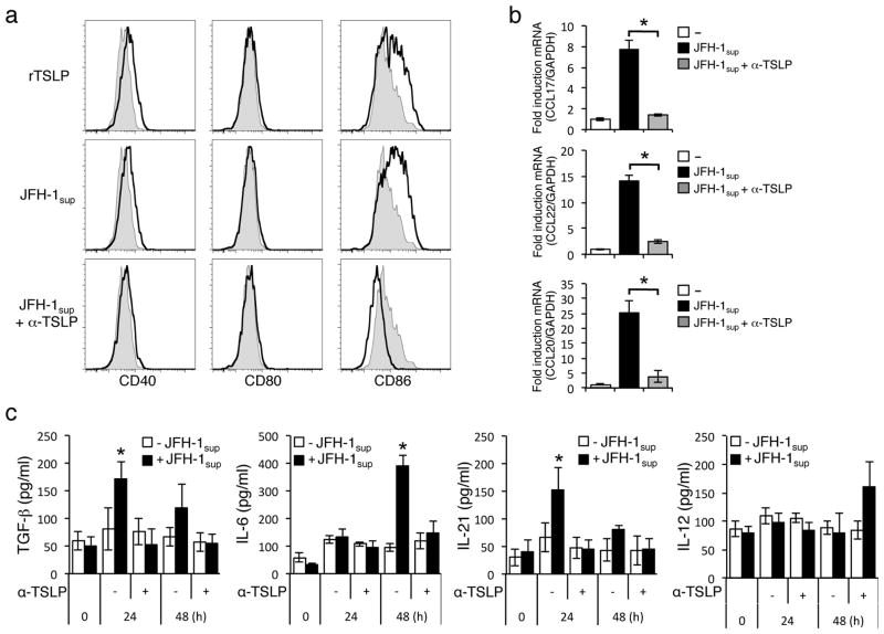
Figure 4. TSLP released by JFH-1-infected cells activates human monocyte-derived DCs by inducing the production of TGF-β, IL-6, and IL-21 cytokines.
a) FACS analysis of DCs after 24 h incubation with the indicated stimuli: rTSLP, JFH-1sup, JFH-1sup plus anti-TSLP mAb. Filled histogram represents staining for DCs cultured with media alone and open histogram represents staining for DCs cultured with indicated stimuli. The expression levels of DC activation markers CD40, CD80, and CD86 were determined by flow cytometry. b) The expression level of CCL17, CCL22, and CCL20 chemokines relative to GAPDH was determined by real time PCR. Data shown are the mean ± SD of three independent experiments (b) or one experiment representative of three independent experiments with similar results (a). Both the JFH-1-infected and non-infected HepG2 cells were co-cultured with THP-1 cells in the transwell system at a 1:1 ratio for the indicated time course in the presence or absence of anti-TSLP antibody (15 ng/ml). The levels of TGF-β, IL-6, IL-21, and IL-12 production were measured in the culture supernatant by ELISA (c). Data represent mean ± SD of three independent experiments. These results were reproducible from five independent experiments.

