Abstract
Background
Cutaneous lesions are commonly seen in the newborn period and exhibit inconsistency from the skin lesions of an adult.
Objective
The present study was carried out with an aim to determine the frequency of physiologic and pathologic cutaneous findings in newborns.
Methods
Typically, 1234 newborns were included in this study. A questionnaire about maternal gestational history, maternal and family history was issued to the parents of each newborn. The presence of cutaneous lesions was recorded.
Results
Overall, 642 (52%) of the newborns were male and 592 (48%) were female. Typically, 831 newborns (67.3%) had at least one cutaneous lesion. The prevalence of genital hyperpigmentation and milia was significantly higher in males. In premature newborns, the pervasiveness of cutis marmorata and genital hyperpigmentation was found to be significantly higher. Caput succedaneum, transient neonatal pustular melanosis and cyanosis appeared predominantly in vaginally born infants. Erythema toxicum neonatorum was seen in infants, who were born by cesarean section. The predominance of Mongolian spots and erythema toxicum neonatorum were significantly higher in the newborns of the multiparous mothers; however, caput succedaneum was significantly higher in newborns of the primiparous mothers.
Conclusion
A number of studies about neonatal dermatoses have been carried out involving different methods in various countries. We consider that our study may be useful in literature, as it has been carried out involving large number of maternal parameters.
Keywords: Maternal relation, Newborn, Skin
INTRODUCTION
Cutaneous lesions are of common occurrence in the newborn period. Newborn period is described as the first 4 weeks of extra-uterine life1. The skin and appendage of newborn skin have different features when compared to the skin of adults2. Many physiologic or pathologic cutaneous changes may be seen during this period. Several studies have reported on cutaneous lesions in newborns. A few of them have investigated all the abnormalities; whereas others have recorded some specific lesions, such as erythema toxicum neonatorum (ETN), birthmarks and vascular lesions in newborns3-7. Previously, in our country, there were two studies, which reported on skin findings in newborns. To the best of our knowledge, our study is the most far-reaching research, which has been carried out in Turkey, using the data of only one hospital. In this study, evaluations of physiologic and pathologic cutaneous lesions were conducted within the first 48 hours of extra-uterine life.
MATERIALS AND METHODS
A detailed study was conducted between April 2006 and September 2006 on 1234 newborns in their first 2 days of life. This study was initiated at the perinatal clinic of Dr. Zekai Burak Womens Health, Education and Research Hospital. Prevalence of neonatal skin lesions was determined by means of clinical diagnosis. A data collection protocol was followed in each case to identify: (1) maternal factors (diseases, toxic habits, medications, and dietary supplements), and (2) neonatal parameters (gestational age and birth weight).
All the newborns were examined by the same dermatologist. The entire skin surface, including the mucous membranes and the nails, were carefully examined. The diagnosis of the cutaneous lesions was recorded. Histopathological examination was not performed.
Based on the gestational age, three groups were established as; pre-term ≤36 weeks, term 37~42 weeks, and post-term ≥42 weeks. Data for the quantitative variables were categorized into groups. The qualitative variables were presented as a percentage and were analyzed using the χ2-test. SPSS version 11.5 (SPSS Inc., Chicago, IL, USA) was used for the statistical analysis. Significance was established as p<0.05.
RESULTS
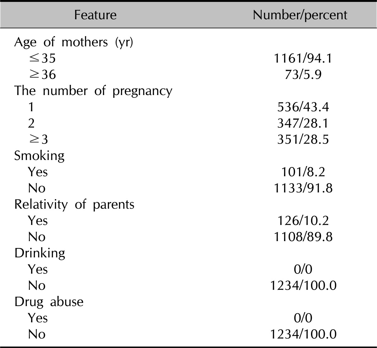
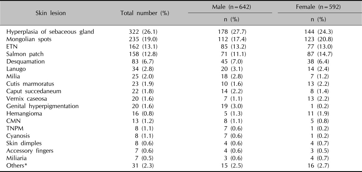
Typically, 642 (52%) of newborns were male, 592 (48%) were female, 752 (60.9%) of newborns were vaginally born, and 482 (39.1%) of newborns were born by cesarean section. Change (varied) in gestational age was observed from 26 to 44 weeks (mean: 38.54±1.99). In general, 129 newborns were premature, 1102 were mature and 3 were post-mature. The features of the mothers are listed in Table 1. The details of cutaneous lesions types are shown in Table 2.
Table 1.
Demographic features of babies mothers

Table 2.
Skin lesions and distribution of them according to gender

ETN: erythema toxicum neonatorum, CMN: congenital melanocytic nevus, TNPM: transient neonatal pustular melanosis. *Echimosis, suction bullae, accessory tragus, accessory nipple, aplasia cutis, microcephalia, breast hypertrophy, spina bifida, myelocel.
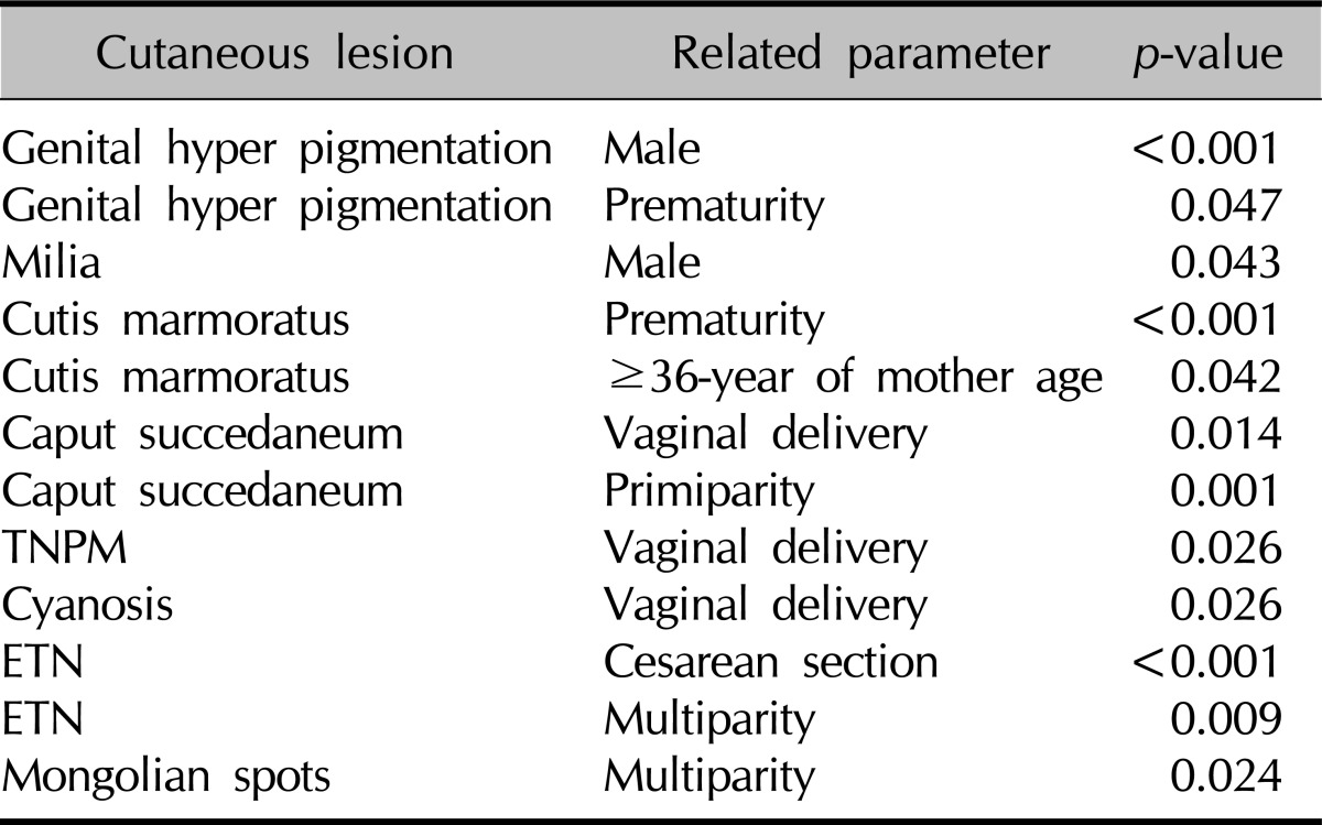
Genital hyperpigmentation and milia were significantly higher in males (p<0.001, p=0.043 respectively). Other cutaneous lesions did not show a statistical difference according to gender. Cutis marmorata and genital hyperpigmentation were significantly higher in premature newborns (p<0.001, p=0.047 respectively). While caput succedaneum, transient neonatal pustular melanosis and cyanosis appeared in vaginally born infants (p=0.014, p=0.026, p=0.026 respectively); ETN was seen in infants, who were born by cesarean section (p<0.001). The prevalence of cutis marmorata was significantly higher in infants whose mother's age was ≥36 (p=0.042). Mongolian spots and ETN were significantly higher in newborns of the multiparous mothers (p=0.024, p=0.009 respectively), but caput succedaneum was significantly higher in newborns of primiparous mothers (p=0.001) (Table 3). There were no significant differences noted for each of the skin lesions if the parent was a smoker or the parents were smoker or consanguineous.
Table 3.
Cutaneous lesions, significantly related parameters and p-value

TNPM: transient neonatal pustular melanosis, ETN: erythema toxicum neonatorum.
DISCUSSION
Many studies about the prevalence of neonatal skin lesions have been reported in different countries involving various racial groups. To the best of our knowledge, our study presents the third observational study conducted in Turkey with the aim of examining the presence of the cutaneous findings and their relationship to maternal factors. The prevalence of neonatal cutaneous findings in the literature has been reported to be between 57 and 99.3%8,9. In our study, most of the neonates (67.3%) had one or more cutaneous findings. However, unlike previous studies, this study has been performed with large number of maternal parameters and our study is the most far-reaching study of a single centre experience in Turkey.
Cutaneous lesions of newborns were compared with different parameters such as gender and gestational age of newborns, the way of delivery, maternal age and maturity1,3,4. In our study, in addition to the aforementioned parameters, smoking and drug abuse during the pregnancy and consanguinity of parents were also investigated. All of the mothers confirmed that they had abstained from alcohol and any drug use during the term of their pregnancy. In the case of 10.2% of the newborns, the parents were closely related (first cousins). No correlation was found between cutaneous lesions and smoking or close relative parents.
Previous studies investigated newborn skin lesions in newborns of different age ranging from 48 hours-2 weeks, or different disease classifications3-5,7. Because of this, comparison between the outcomes of the current study with previous reports is difficult. There are only 3 reports that investigated cutaneous lesions of newborns within the first 48 hours, the results of which are (note: please check if this is right here) similar to our study1,3,10. Moosavi et al.1 reported that 96% of Iranian newborns had at least one cutaneous lesion. Rivers et al.3 found that the prevalence of cutaneous lesions in Australian newborns was 99.3%. Our study shows that the prevalence of cutaneous lesions was lower in Turkish newborns (67.3%). This discrepancy may be due to racial and geographic diversity.
Furthermore, we have done a comparative analysis of the results of our study with other studies in literature. The prevalence of sebaceous hyperplasia in a newborn in various reports has been accounted as 21.4~48%3,11. The rate in our study was found to be within these limits. In literature, statistical difference was not detected between sebaceous hyperplasia and the gender of infants. Our results were consistent with these outcomes. Moosavi et al.1 found that the presence of sebaceous hyperplasia increased with increasing maturity. Nanda et al.12 detected inclination towards a higher side in newborns, who were born vaginally and in infants of multiparous mothers. Our study did not observe any statistical relationship between sebaceous hyperplasia and these parameters.
Caput succedaneum was seen in 1.8% of infants, where 86.4% of them were vaginal births (p=0.014). In addition, caput succedaneum was found to be significantly higher in infants of primiparous mothers. This data supported the role of trauma in etiology of caput succedaneum. In contrary, it was found to be lower in our study (7.9% and 9.8%), which can be related to an increase in the number of cesarean section births7,10. Prevalence of cutis marmorata was found to be 1.9% in our study, while it was 6.5% in the study of Boccardi et al.7. This difference may be due to a seasonal discrepancy, as our study was carried out during the spring and summer seasons. Cutis marmorata appeared to be related to prematurity and mature mothers (36-year and above) (p<0.001, p=0.042). Prevalence of ETN in our study was 13.1%, which is consistent with the published literature (8~43.7%)4,5. Different reports have stated about the relationship between ETN and maturity4,7. We did not detect a significant relation between ETN and maturity, which was similar to the results of Rivers et al.3. In our study, significant relation was found between cesarean section and ETN, differing from literature4. This relationship may be due to an increased rate of cesarean section in our country. ETN was found to be higher in infants of multipara mothers. This data was similar to the literature11,13.
Consequently, many studies have been done with different methods about neonatal dermatoses in various countries. The frequency of these lesions changes with methodology of studies, racial, climatic, and geographic factors. We consider that our study may be useful in literature, as it has been carried out involving large number of maternal parameters.
References
- 1.Moosavi Z, Hosseini T. One-year survey of cutaneous lesions in 1000 consecutive Iranian newborns. Pediatr Dermatol. 2006;23:61–63. doi: 10.1111/j.1525-1470.2006.00172.x. [DOI] [PubMed] [Google Scholar]
- 2.Chang MW, Orlow SJ. Neonatal, Pediatric and adolescent dermatology. In: Wollf K, Fitzpatrick TB, Goldsmith LA, Katz SI, Gilchrest BA, Paller AS, et al., editors. Fitzpatrick's dermatology in general medicine. 7th ed. New York: McGraw Hill; 2008. pp. 935–955. [Google Scholar]
- 3.Rivers JK, Frederiksen PC, Dibdin C. A prevalence survey of dermatoses in the Australian neonate. J Am Acad Dermatol. 1990;23:77–81. doi: 10.1016/0190-9622(90)70190-s. [DOI] [PubMed] [Google Scholar]
- 4.Osburn K, Schosser RH, Everett MA. Congenital pigmented and vascular lesions in newborn infants. J Am Acad Dermatol. 1987;16:788–792. doi: 10.1016/s0190-9622(87)70102-9. [DOI] [PubMed] [Google Scholar]
- 5.Liu C, Feng J, Qu R, Zhou H, Ma H, Niu X, et al. Epidemiologic study of the predisposing factors in erythema toxicum neonatorum. Dermatology. 2005;210:269–272. doi: 10.1159/000084749. [DOI] [PubMed] [Google Scholar]
- 6.Karvonen SL, Vaajalahti P, Marenk M, Janas M, Kuokkanen K. Birthmarks in 4346 Finnish newborns. Acta Derm Venereol. 1992;72:55–57. [PubMed] [Google Scholar]
- 7.Boccardi D, Menni S, Ferraroni M, Stival G, Bernardo L, La Vecchia C, et al. Birthmarks and transient skin lesions in newborns and their relationship to maternal factors: a preliminary report from northern Italy. Dermatology. 2007;215:53–58. doi: 10.1159/000102034. [DOI] [PubMed] [Google Scholar]
- 8.Ferahbas A, Utas S, Akcakus M, Gunes T, Mistik S. Prevalence of cutaneous findings in hospitalized neonates: a prospective observational study. Pediatr Dermatol. 2009;26:139–142. doi: 10.1111/j.1525-1470.2009.00903.x. [DOI] [PubMed] [Google Scholar]
- 9.Gokdemir G, Erdogan HK, Köşlü A, Baksu B. Cutaneous lesions in Turkish neonates born in a teaching hospital. Indian J Dermatol Venereol Leprol. 2009;75:638. doi: 10.4103/0378-6323.57742. [DOI] [PubMed] [Google Scholar]
- 10.Sachdeva M, Kaur S, Nagpal M, Dewan SP. Cutaneous lesions in new born. Indian J Dermatol Venereol Leprol. 2002;68:334–337. [PubMed] [Google Scholar]
- 11.Hidano A, Purwoko R, Jitsukawa K. Statistical survey of skin changes in Japanese neonates. Pediatr Dermatol. 1986;3:140–144. doi: 10.1111/j.1525-1470.1986.tb00505.x. [DOI] [PubMed] [Google Scholar]
- 12.Nanda A, Kaur S, Bhakoo ON, Dhall K. Survey of cutaneous lesions in Indian newborns. Pediatr Dermatol. 1989;6:39–42. doi: 10.1111/j.1525-1470.1989.tb00265.x. [DOI] [PubMed] [Google Scholar]
- 13.Jacobs AH, Walton RG. The incidence of birthmarks in the neonate. Pediatrics. 1976;58:218–222. [PubMed] [Google Scholar]


