

Abstract
Radiological images in a case of excluded calyx are shown that resulted from percutaneous nephrolithotomy for a stag horn stone that was approached through the superior calyx.
Keywords: Infundibular stenosis, Excluded calyx, Pyelolithotomy
A 27-year man with normal renal functions presented with staghorn right renal stone (Fig. 1) and was managed by percutaneous nephrolithotomy using superior calyceal puncture approach. Complete stone clearance was achieved and the patient was discharged. He presented after 2 weeks with high-grade fever, and ultrasonography revealed right upper pole pyocalycosis. Intravenous urogram revealed nonvisualization of the superior calyx (Fig. 2). A nephrostomy tube was placed in the superior calyx percutaneously to drain the superior calyx. Retrograde pyelography (Fig. 3) showed blocked superior infundibulum, while nephrostogram revealed contrast-filled superior calyx that was massively dilated (Fig. 4). CT scan also showed the dilated superior calyx (Fig. 5). He was managed by percutaneous antegrade creation of neo-infundibulum between the superior calyx and the renal pelvis across a DJ stent using holmium:YAG laser. At 6 months of follow-up, the patient is now asymptomatic.
Fig. 1.

Intravenous urogram showing the staghorn right renal stone
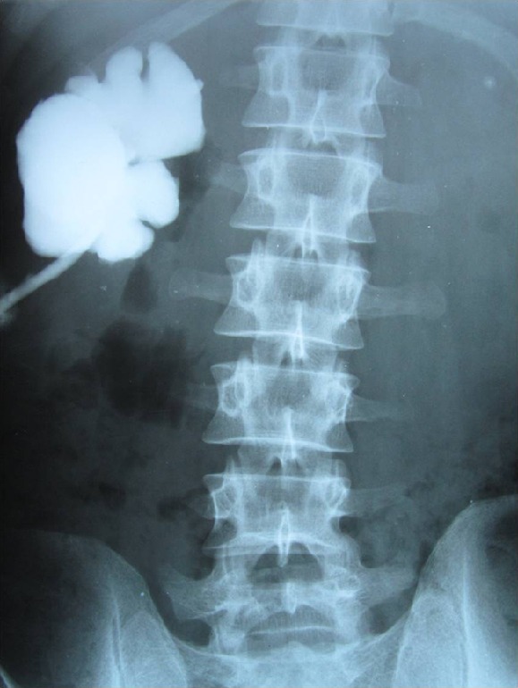
Fig. 2.

Intravenous urogram showing non-visualization of the superior calyx
Fig. 3.
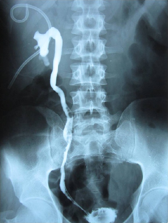
Retrograde pyelogram did not show filling of the superior calyx due to infundibular block (nephrostomy tube in the superior calyx to drain the blocked calyx)
Fig. 4.
Nephrostogram showing a dilated superior calyx with no contrast entering the pelvis or the ureter
Fig. 5.

Three-dimensional CT scan image showing the calyceal anatomy
Infundibular stenosis is a rare clinical entity, and when it is complete it is called excluded calyx [1]. Treatment approach for infundibular stenosis is usually endoscopic (either antegrade or retrograde approach) although the reported success rate is only 60–80 % [2]. Making a percutaneous puncture in an excluded calyx using standard methods is difficult, and puncture under ultrasound or CT guidance is sometimes required [2]. The results of endoscopic treatment of excluded calyx are likely to be even poorer as compared to that for infundibular stenosis. Bruner has described successful management by the percutaneous catheter of surgically induced isolated calyx as a result of inadvertent closure of single infundibula [3]. We managed this patient by antegrade percutaneous creation of neo-infundibulum between the superior calyx and the renal pelvis with successful short-term result.
Conclusions
Radiological images in a case of excluded calyx are shown that resulted from percutaneous nephrolithotomy for staghorn renal stone. The patient was managed by antegrade percutaneous approach to create a neo-infundibula and DJ stent placement across the stenosis.
References
- 1.Elashry OM, Nakada SY, Pearle MS, Clayman RV. Endourologic management of stone bearing excluded calices: contrasting case reports. J Endourol. 1996;10:21–26. doi: 10.1089/end.1996.10.21. [DOI] [PubMed] [Google Scholar]
- 2.Gupta M, Ost MC, Shah JB, McDougall EM, Smith AD. Percutaneous management of the upper urinary tract. In: Wein AJ, Kavoussi LR, Novik AC, Partin AW, Peters CA, editors. Campbell-Walsh Urology, ed. 9. Philadelphia: Saunders Elsevier; 2007. pp. 1526–1563. [Google Scholar]
- 3.Bruner B, Ashley R, Leibovich B, Blute M, Leroy A. Percutaneous catheter management of persistent urine leak due to iatrogenic infundibular stenosis after partial nephrectomy. J Endourol. 2009;23:37–41. doi: 10.1089/end.2008.0291. [DOI] [PubMed] [Google Scholar]