Abstract
Introduction
Although pedicle screw fixation is a well-established technique for the lumbar spine, screw placement in the thoracic spine is more challenging because of the smaller pedicle size and more complex 3D anatomy. The intraoperative use of image guidance devices may allow surgeons a safer, more accurate method for placing thoracic pedicle screws while limiting radiation exposure. This generic 3D imaging technique is a new generation intraoperative CT imaging system designed without compromise to address the needs of a modern OR.
Aim
The aim of our study was to check the accuracy of this generic 3D navigated pedicle screw implants in comparison to free hand technique described by Roy-Camille at the thoracic spine using CT scans.
Material and methods
The material of this study was divided into two groups: free hand group (group I) (18 patients; 108 screws) and 3D group (27 patients; 100 screws). The patients were operated upon from January 2009 to March 2010. Screw implantation was performed during internal fixation for fractures, tumors, and spondylodiscitis of the thoracic spine as well as for degenerative lumbar scoliosis.
Results
The accuracy rate in our work was 89.8 % in the free hand group compared to 98 % in the generic 3D navigated group.
Conclusion
In conclusion, 3D navigation-assisted pedicle screw placement is superior to free hand technique in the thoracic spine.
Keywords: Pedicle screws, Thoracic spine, Image-guided surgery, Spine navigation
Introduction
Although pedicle screw fixation is a well-established technique for the lumbar spine, screw placement in the thoracic spine is more challenging because of the smaller pedicle size and more complex 3D anatomy [16, 22]. Thoracic screws perforate the cortical margins of the pedicle at a rate ranging from 16 % to 54 % [9, 21, 23, 24], creating the potential for hemorrhage, nerve root injury, or spinal cord injury. Long pedicle screws endanger adjacent structures such as the aorta and the pleural cavity [2, 23].
The free-hand thoracic pedicle screw technique has been well described by Kim et al. [7] and one inserts the pedicle screws based on the anatomical landmarks and the tactile feel of probing the pedicles. Kim et al. [6] report a low complication rate with this technique. The accuracy of thoracic screw position remains primarily a function of surgical skill together with experience and is associated with a steep learning curve [3].
The intraoperative use of image guidance devices may allow surgeons a safer, more accurate method for placing thoracic pedicle screws while limiting radiation exposure [14]. Among them, computed tomography-based navigation was the most popular. CT navigation could provide precise anatomy of the pedicle as well as reduced radiation exposure. However, new concerns about the system arose with a steep learning curve and excessive pre-operative preparation including computed tomography with a specific protocol, data acquisition and transfer, and patient registration [1, 4]. The development of intraoperative 2D and 3D fluoroscopy based-navigation appeared to tackle such issues [15, 25]. The equipment did not require registration, reduced imaging time and radiation dosage, and avoided repeated C-arm movements during surgery, because visualization of the surgical instruments in relation to the patient’s anatomy in the entire desired image plane was determined from the beginning. Though a few studies implied that accuracy of fluoroscopy-assisted pedicle screw insertion was comparable with that of CT navigation [1, 4], different population characteristics and assessment methods of placement accuracy in various studies resulted in inconsistent conclusions.
This generic imaging system is a mobile 2D and 3D imaging platform designed to improve intraoperative decision making. It is a new generation intraoperative imaging system designed without compromise to address the needs of a modern OR. It allows supporting a multitude of applications in spine, orthopaedics, neurosurgery ENT, maxillofacial (and vascular surgery in the close future). This generic imaging system interfaces seamlessly with the navigation system, eliminating time consuming registration steps and transferring acquired scans automatically which greatly enhances navigation workflow providing the surgeon with unprecedented visualization depth and capabilities for complex orthopaedic and spine surgeries. Surgeons are not dependent on a pre-operative CT where the patient’s position may vary from surgical position in the OR. This 3D system allows the surgeon to scan the patient in the operative position and to obtain an updated scan whenever needed. This ensures the most accurate imaging and navigation possible, ensuring the best possible surgical outcome for the patient [20].
The aim of our study is to check the accuracy of this generic 3D navigated pedicle screw implants in comparison to free -hand technique described by Roy-Camille at the thoracic spine using CT scans.
Materials and methods
108 pedicle screws were implanted in 18 patients (11 males and 7 females) using the free hand technique (group I) and 100 screws were implanted in 27 patients (12 males and 15 females) using the 3D navigated technique (group II). The mean age was 62.9 years in group I and 54.6 years in group II. Both groups were operated upon in our spine centre in the period January 2009–March 2010. This was carried out during internal fixation for fractures, tumors, and spondylodiscitis of the thoracic spine as well as for degenerative lumbar scoliosis. Distribution of screws with regard to the thoracic vertebrae in both groups is shown in Table 1. All surgeries were done by the first two authors.
Table 1.
Distribution of screws with regard to the thoracic vertebrae in both groups
| Level | Free hand group | 3D group |
|---|---|---|
| Th1 | 4 | 6 |
| Th2 | 4 | 4 |
| Th3 | 1 | 8 |
| Th4 | 5 | 5 |
| Th5 | 7 | 4 |
| Th6 | 6 | 4 |
| Th7 | 4 | 6 |
| Th8 | 6 | 4 |
| Th9 | 12 | 5 |
| Th10 | 16 | 14 |
| Th11 | 20 | 17 |
| Th12 | 23 | 23 |
| Total | 108 | 100 |
In the free hand group (group I), the screws were implanted according to the technique of Roy-Camille [17–19]. The Identification of the entry point, opening with the awl then drilling of the pedicle and probing of the drill canal. After tapping and screw application, the position of screws was assessed by C-arm (AP and lateral). When the position of one or more of the screws was not optimal, the screw will be revised and the new position will be checked again by the C-arm. All the patients were subjected to post-operative CT scan to evaluate the position of the screws. The CT examination was carried out by Briliance CT 64-channel scanner (Philips medical systems, PC Best, Netherlands).
In the 3D group (group II), after the reference clamp was screwed to one of the spinous processes in the area of fixation, 2D (AP and lateral) and 3D fluoroscopies were done. This was followed by screw implantation using the 3D navigation system. After screw insertion, control 2D (AP and lateral) and 3D fluoroscopies were done to evaluate the screw placement. When the screw placement was optimal, the 3D scans were transferred to the hospital own PACS to evaluate screw placement.
The CT scans (axial and sagittal) of both groups were examined independently by a surgeon and a radiologist to evaluate the position of screws according to the classification of Learch [13] and Wiesner [26]. In this classification, there are four main categories for screw misplacement:
Encroachment: if the pedicle cortex could not be visualized.
Minor penetration: when the screw trajectory was <3 mm outside the pedicular boundaries.
Moderate penetration: when the screw trajectory was 3–6 mm outside the pedicular boundaries.
Severe penetration: when the screw trajectory was >6 mm outside the pedicular boundaries.
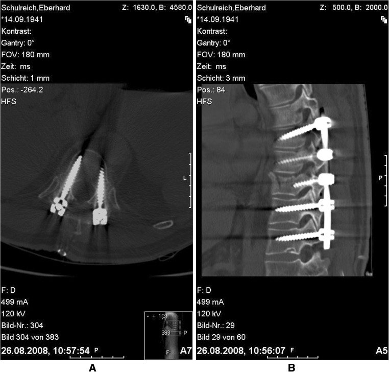
It is worthy to mention that the ideal position of the screw is achieved when the screw lies in the middle of the pedicle in both axial and sagittal reconstruction CT scans (Fig. 1).
Fig. 1.
Ideal position of the screw a axial and b sagittal CT images
Results
The overall inter-observer agreement in the free hand group was 94.2 % compared to 95.1 % in the O-arm group including both axial and sagittal reconstruction images.
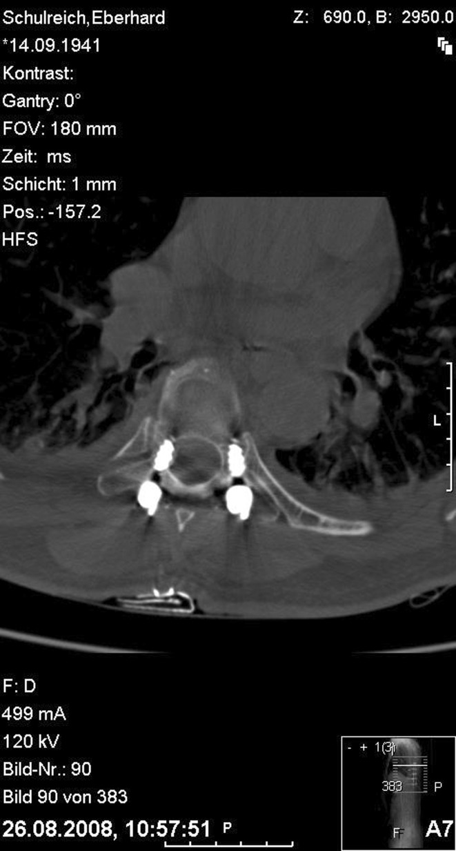
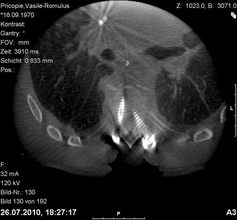
In the axial images (Table 2), 31 (28.7 %) screws showed medial (Fig. 2) and 12 (11.1 %) screws showed lateral encroachment in group I in comparison to 22 (22 %) and 3 (3 %) screws, respectively, in group II (Fig. 3). Frank penetration <3 mm was found in 7 (6.5 %) screws in group I, in comparison to 4 screws (4 %) in group II. One screw (0.9 %) in group I and another one (1 %) in group II showed medial frank penetration 3–6 mm. In group I, the malplaced screw was not associated with neurological deficits and the malplaced screw in group II was detected in Th8 in a patient with complete cord transection due to flexion rotation injury of Th5–Th6, hence there was no need to revise it. In group I, 4 screws (3.7 %) showed lateral penetration 3–6 mm. These screws did not affect the stability of fixation and they were not revised.
Table 2.
Comparison between the two groups regarding screw malplacement
| Screw malplacement | Free hand group (n = 108) | 3D group (n = 100) | P | ||
|---|---|---|---|---|---|
| No. | Percent | No. | Percent | ||
| Lateral axial | |||||
| Encroachment | 12 | 11.1 | 3 | 3.0 | 0.446 |
| FP < 3 mm | 3 | 2.7 | 0 | 0.0 | |
| FP 3–6 mm | 4 | 3.7 | 0 | 0.0 | |
| Medial axial | |||||
| Encroachment | 31 | 28.7 | 22 | 22.0 | 0.883 |
| FP < 3 mm | 4 | 3.7 | 4 | 4.0 | |
| FP 3–6 mm | 1 | 0.9 | 1 | 1.0 | |
| Anterior axial | |||||
| Encroachment | 8 | 7.4 | 3 | 3.0 | 0.836 |
| FP <3 mm | 2 | 1.9 | 1 | 1.0 | |
| Caudal sagittal | |||||
| Encroachment | 16 | 14.8 | 5 | 5.0 | 0.025* |
| FP < 3 mm | 0 | 0.0 | 2 | 2.0 | |
| Cranial sagittal | |||||
| Encroachment | 7 | 6.5 | 5 | 5.0 | 0.126 |
| FP < 3 mm | 0 | 0.0 | 2 | 2.0 | |
* Statistical significance
Fig. 2.
Axial CT image showing medial penetration 3–6 mm (right screw) and lateral penetration 3–6 mm (left screw) in the free hand group
Fig. 3.
Axial 3D image showing medial penetration 3–6 mm in the 3D group
In axial images, anterior encroachment and screw penetration <3 mm was observed in 10 screws (9.3 %) in group I in comparison to 4 screws (4 %) in group II.
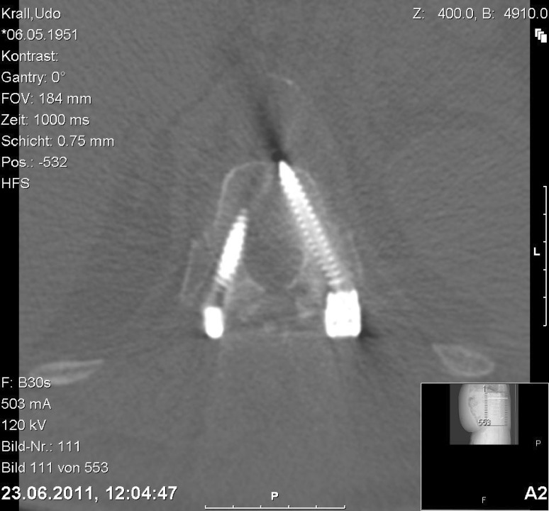
In the sagittal images (Table 2), 16 (14.8 %) screws showed caudal and seven screws (6.5 %) showed cranial encroachment in group I in comparison to 5 (5 %) and 5 (5 %) screws, respectively, in group II (Fig. 4). The difference was proved to be statistically significant. Frank penetration <3 mm was found in four (4 %) screws in group II, in comparison to group I which showed no frank penetration either caudal or cranial.
Fig. 4.
Axial CT image showing anterior penetration <3 mm in the free hand group
In group I, 6 (5.6 %) screws were revised intraoperatively after making the C-arm control. After screw revision, the C-arm control showed good position of the screws.
In our study, we considered encroachment and frank penetration <3 mm are still in the safe zone for implanting the pedicular screws because they do not endanger the efficacy of spine stabilisation as well as the neurovascular structures. Accordingly, the screw penetration more than 3 mm in any direction was considered as a screw malposition.
The accuracy rate in our work was 89.8 % in the free hand group compared to 99 % in the 3D navigated group (Table 3). No residual neurological deficits were reported in both groups.
Table 3.
Comparison between the two groups regarding accuracy of screw positioning
| Malplacement | Free hand technique (108 screws) |
3D (100 screws) |
|---|---|---|
| Encroachment | 75 | 38 |
| Minor penetration (<3 mm) | 9 | 9 |
| Moderate penetration (3–6 mm) | 5 | 1 |
| Severe penetration | 0 | 0 |
| Intraoperative screw revision | 6 | 0 |
| Accuracy | 89.8 % | 99 % |
Discussion
Insertion of pedicle screws in the thoracic spine is a demanding technique and carries the potential risks of neurological structures injury. It was shown that image-guided spinal instrumentation procedures in the cervical, thoracic, and lumbar spine have lower rates of screw misplacement than do those performed without image guidance [5, 8].
Our accuracy rate in the free hand technique was 89.8 %. Five screws showed frank penetration 3–6 mm (four lateral and one medial) and six screws were revised intraoperatively after checking their position using the C-arm. None of the malplaced screws resulted in neurological deficits.
In comparison to the free hand technique, the 3D-based navigation technique showed an accuracy of 99 %. Only one out of 100 screws showed frank medial penetration (3–6 mm). This was reported in Th8 vertebra in a patient suffering from fracture dislocation Th4–Th5 with complete paraplegia and double sphincteric incontinence. Although we have a long experience with the free hand technique and our accuracy rate approaches the results of 2D fluoroscopy-based navigation technique, the 3D-based navigation technique showed a high better accuracy rate.
The 3D-based navigation technique showed a higher accuracy rate compared to the CT and 2D fluoroscopy-based navigation techniques and a comparable accuracy rate to the 3D fluoroscopy-based navigation technique. Kosmopoulos and Schizas [8] revealed a median accuracy of 94.3 % using of neuronavigation in thoracic spine. Zausinger [27] in his work regarding the use of intraoperative computed tomography with integrated navigation system in spinal stabilizations reported a rate of 5.6 % screw malposition in the thoracic spine. Our rate of 99 % accuracy with 3D based navigation technique is still superior to the results of intraoperative CT navigation techniques.
To our knowledge, this is the first in the literature that compares the accuracy of screw placement (Table 3) between free hand and 3D navigated technique in the thoracic spine. There is no question that the 3D navigation-based technique has the best accuracy of pedicle screw placement. On the other hand, it is known that the CT navigation-based techniques prolongs the operative time [10–12] as well as the hazards of radiation compared to the C-arm control after the free hand technique.
As we started using this generic 3D system (January 2009), the time needed for positioning, performing the 3D scans as well as the navigation procedure was about 1 h. 3 months later and after using this system nearly daily, this time is reduced to 15 min. Regarding the radiation hazards, each 3D scan is equal to 60 % of an ordinary CT scan according to the radiation measurement carried by the manufactur company (Medtronic USA). For the operative team, the exposure in nearly zero because the whole surgical team leaves the theatre during the 3D scan.
We think that implanting the screws using the free hand technique in the thoracic spine is safe in the hands of the experienced surgeons. Hence, 3D-based navigation technique is more accurate and accordingly safer.
Conclusions
The 3D-based navigation technique provides high accuracy of pedicle screw placement and thus safe for the patients undergoing thoracic spine stabilization. It allows immediate detection of screw misplacement and accordingly no reoperation for malposition. In comparison to lumbar spine, placement of transpedicular screws in the thoracic spine using 3D-based navigation technique is superior to the free hand technique.
Conflict of interest
None.
References
- 1.Choi WW, Green BA, Levi AD. Computer assisted fluoroscopic targeting system for pedicle screw insertion. Neurosurgery. 2000;47:872–878. doi: 10.1097/00006123-200010000-00017. [DOI] [PubMed] [Google Scholar]
- 2.Cinotti G, Gumina S, Ripani M, Postacchini F. Pedicle instrumentation in the thoracic spine. A morphometric and cadaveric study for placement of screws. Spine. 1999;24:114–119. doi: 10.1097/00007632-199901150-00003. [DOI] [PubMed] [Google Scholar]
- 3.Foley KT, Simon DA, Rampersaud YR. Virtual fluoroscopy: computer-assisted fluoroscopic navigation. Spine. 2001;26:347–351. doi: 10.1097/00007632-200102150-00009. [DOI] [PubMed] [Google Scholar]
- 4.Fu TS, Wong CB, Tsai TT, Liang YC, Chen LH, Chen WJ. Pedicle screw insertion: computed tomography versus fluoroscopic image guidance. Int Orthop. 2008;32:517–521. doi: 10.1007/s00264-007-0358-1. [DOI] [PMC free article] [PubMed] [Google Scholar]
- 5.Holly LT, Foley KT. Intraoperative spinal navigation. Spine. 2003;28:S54–S61. doi: 10.1097/01.BRS.0000076899.78522.D9. [DOI] [PubMed] [Google Scholar]
- 6.Kim YJ, Lenke LG. Thoracic pedicle screw placement: free-hand technique. Neurol India. 2005;53:512–519. doi: 10.4103/0028-3886.22622. [DOI] [PubMed] [Google Scholar]
- 7.Kim YJ, Lenke LG, Bridwell KH, Cho YS, Riew KD. Free hand pedicle screw placement in the thoracic spine: is it safe? Spine. 2004;29:333–342. doi: 10.1097/01.BRS.0000109983.12113.9B. [DOI] [PubMed] [Google Scholar]
- 8.Kosmopoulos V, Schizas C. Pedicle screw placement accuracy: a metaanalysis. Spine. 2007;32:E111–E120. doi: 10.1097/01.brs.0000254048.79024.8b. [DOI] [PubMed] [Google Scholar]
- 9.Krag MH, Weaver DL, Beynnon BD, Haugh LD. Morphometry of the thoracic and lumbar spine related to transpedicular screw placement for surgical spinal fixation. Spine. 1998;13:27–32. doi: 10.1097/00007632-198801000-00007. [DOI] [PubMed] [Google Scholar]
- 10.Laine T, Lund T, Ylikoski M, Lohikoski J, Schlenzka D. Accuracy of pedicle screw insertion with and without computer assistance: a randomised controlled clinical study in 100 consecutive patients. Eur Spine J. 2000;9:235–240. doi: 10.1007/s005860000146. [DOI] [PMC free article] [PubMed] [Google Scholar]
- 11.Laine T, Makitalo K, Schlenzka D, Tallroth K, Poussa M, Alho A. Accuracy of pedicle screw insertion: a prospective CT study in 30 low back patients. Eur Spine J. 1997;6:402–405. doi: 10.1007/BF01834068. [DOI] [PMC free article] [PubMed] [Google Scholar]
- 12.Laine T, Schlenzka D, Makitalo K, Tallroth K, Nolte LP, Visarius H. Improved accuracy of pedicle screw insertion with computer assisted surgery. A prospective clinical trial of 30 patients. Spine. 1997;22:1254–1258. doi: 10.1097/00007632-199706010-00018. [DOI] [PubMed] [Google Scholar]
- 13.Learch TJ, Massie JB, Pathria MN, Ahlgren BA, Garfin SR. Assessment of pedicle screw placement utilizing conventional radiography and computed tomography: a proposed systematic approach to improve accuracy of interpretation. Spine. 2004;29:767–773. doi: 10.1097/01.BRS.0000112071.69448.A1. [DOI] [PubMed] [Google Scholar]
- 14.Mirza SK, Wiggins GC, Kuntz C, 4th, York JE, Bellabarba C, Knonodi MA, Chapman JR, Shaffrey CI. Accuracy of thoracic vertebral body screw placement using standard fluoroscopy, fluoroscopic image guidance, and computed tomographic image guidance: a cadaver study. Spine. 2003;28:402–413. doi: 10.1097/01.BRS.0000048461.51308.CD. [DOI] [PubMed] [Google Scholar]
- 15.Nottmeier EW, Seemer W, Young PM. Placement of thoracolumbar pedicle screwsusing three-dimensional image guidance: experience in a large patient cohort. J Neurosurg Spine. 2009;10:33–39. doi: 10.3171/2008.10.SPI08383. [DOI] [PubMed] [Google Scholar]
- 16.Panjabi MM, Takata K, Goel V, Federico D, Oxland T, Duranceau J, Krag M. Thoracic human vertebrae. Quantitative three-dimensional anatomy. Spine. 1991;16:888–901. doi: 10.1097/00007632-199108000-00006. [DOI] [PubMed] [Google Scholar]
- 17.Roy-Camille R. Osteosynthesis du rachis dorsal, lombaire et lombo-sacre par plaques metalliques vissees dans les pedicules vertebraux et les apophyses articulaires. Presse Med. 1970;578:1447. [PubMed] [Google Scholar]
- 18.Roy-Camille R, Saillant G, Berteaux D, Salgado V. Osteosynthesis of thorako-lumbar spine fractures with metal plates screwed through the vertebral pedicles. Reconstr Surg Traumatol. 1976;15:2–16. [PubMed] [Google Scholar]
- 19.Roy-Camille R, Saillant G, Mazel C. Internal fixation of the lumbar spine with pedicle screw plating. Clin Orthop Relat Res. 1986;203:7–17. [PubMed] [Google Scholar]
- 20.Silbermann J, Riese F, Allam Y, Reichert T, Koeppert H, Gutberlet M. Computer tomography assessment of pedicle screw placement in lumbar and sacral spine: comparison between free-hand and O-arm based navigation techniques. Eur Spine J. 2011;20:875–881. doi: 10.1007/s00586-010-1683-4. [DOI] [PMC free article] [PubMed] [Google Scholar]
- 21.Steinmann JC, Herkowitz HN, el-Kommos H, Wesolowski DP. Spinal pedicle fixation. Confirmation of an image-based technique for screw placement. Spine. 1993;18:1856–1861. doi: 10.1097/00007632-199310000-00022. [DOI] [PubMed] [Google Scholar]
- 22.Vaccaro AR, Rizzolo SJ, Allardyce TJ, Ramsey M, Salvo J, Balderston RA, Cotler JM. Placement of pedicle screws in the thoracic spine. Part I: morphometric analysis of the thoracic vertebrae. J Bone Joint Surg [Am] 1995;77:1193–1199. doi: 10.2106/00004623-199508000-00008. [DOI] [PubMed] [Google Scholar]
- 23.Vaccaro AR, Rizzolo SJ, Balderston RA, Allardyce TJ, Garfin SR, Dolinskas C, An HS. Placement of pedicle screws in the thoracic spine. Part II: an anatomical and radiographic assessment. J Bone Joint Surg [Am] 1995;77:1200–1206. doi: 10.2106/00004623-199508000-00009. [DOI] [PubMed] [Google Scholar]
- 24.Weinstein JN, Spratt KF, Spengler D, Brick C, Reid S. Spinal pedicle fixation: reliability and validity of roentgenogram-based assessment and surgical factors on successful screw placement. Spine. 1988;13:1012–1018. doi: 10.1097/00007632-198809000-00008. [DOI] [PubMed] [Google Scholar]
- 25.Wendl K, von recum J, Wentzensen A, Gruetzner PA. Iso-C (3 D-assisted) navigated implantation of pedicle screws in thoracic and lumbar vertebrae. Unfallchirug. 2003;106:907–913. doi: 10.1007/s00113-003-0683-9. [DOI] [PubMed] [Google Scholar]
- 26.Wiesner L, Kothe R, Ruther W. Anatomic evaluation of two different techniques for the percutaneous insertion of pedicle screws in the lumbar spine. Spine. 1999;24:1599–1603. doi: 10.1097/00007632-199908010-00015. [DOI] [PubMed] [Google Scholar]
- 27.Zausinger S, Scheder B, Uhl E, Heigl T, Morhard D, Tonn JC. Intraoperative computed tomography with integrated navigation system in spinal stabilizations. Spine. 2009;34:2919–2926. doi: 10.1097/BRS.0b013e3181b77b19. [DOI] [PubMed] [Google Scholar]


