Abstract
Purpose
Single center evaluation of the placement accuracy of thoracolumbar pedicle screws implanted either with fluoroscopy or under CT-navigation using 3D-reconstruction and intraoperative computed tomography control of the screw position. There is in fact a huge variation in the reported placement accuracy of pedicle screws, especially concerning the screw placement under conventional fluoroscopy most notably due to the lack of the definition of screw misplacement, combined with a potpourri of postinstrumentation evaluation methods.
Methods
The operation data of 1,006 patients operated on in our clinic between 1995 and 2005 is analyzed retrospectively. There were 2,422 screws placed with the help of CT-navigation compared to 2,002 screws placed under fluoroscopy. The postoperative computed tomography images were reviewed by a radiologist and an independent spine surgeon.
Results
In the lumbar spine, the placement accuracy was 96.4 % for CT-navigated screws and 93.9 % for pedicle screws placed under fluoroscopy, respectively. This difference in accuracy was statistically significant (Fishers Exact Test, p = 0.001). The difference in accuracy became more impressing in the thoracic spine, with a placement accuracy of 95.5 % in the CT-navigation group, compared to 79.0 % accuracy in the fluoroscopy group (p < 0.001).
Conclusion
This study underlines the relevance of CT-navigation-guided pedicle screw placement, especially when instrumentation of the middle and upper thoracic spine is carried out.
Keywords: Pedicle screws, Navigation, Placement accuracy, Computed tomography, Fluoroscopy
Introduction
Transpedicular screw fixation is widely used in spinal surgery and there are numerous studies concerning optimal and safe placement of pedicle screws [1–3].
Due to the lack of a generally accepted definition of “screw misplacement”, a potpourri of post-instrumentation evaluation methods is reported and placement accuracies vary extremely [4–6]. In this context, we present a single center evaluation of two different instrumentation methods for the thoracolumbar spine after placement of 4,500 pedicle screws either with conventional fluoroscopy or under CT-navigation.
Materials and methods
The present work is a retrospective analysis of 1,006 patients, operated in our clinic between 1995 and 2005, comprising 386 women and 620 men. All patients who underwent pedicle screw instrumentation in this period were included in the study. The surgical indications are specified in Table 1. There were 505 interventions carried out by means of CT-navigation (2,422 screws) in contrast to 501 cases operated under fluoroscopy (2,002 screws). At the end of the 1990s, due to a paradigm shift—that is changing from sole intraoperative fluoroscopy toward CT-navigation—comparable data with both implantation methods were available in the majority of cases.
Table 1.
Surgical indications
| Indication | Number of patients operated with fluoroscopy | Number of patients operated with CT-navigation | ||
|---|---|---|---|---|
| Tumors | 48 | 9.6 % | 46 | 9.1 % |
| Fractures | 276 | 55.1 % | 248 | 49.1 % |
| Infectious diseases | 28 | 5.6 % | 10 | 2 % |
| Degenerative disease | 149 | 29.7 % | 201 | 39.8 % |
| Total | 501 | 505 | ||
Surgery with the help of intraoperative computer tomography and three-dimensional neuronavigation and intraoperative control of pedicle screw positioning
For CT-neuronavigation-guided spinal instrumentation we used the mobile Tomoscan M-EG (Philips Medical Systems, Eindhoven, The Netherlands). This system contains a mobile CT gantry, a mobile CT examination table, which is fixed to the gantry during surgery, and a mobile workstation. All intraoperative imaging was performed with the patient being in prone position on the CT table throughout the whole surgical procedure without the need of repositioning. After skin incision and dorsal muscle preparation, multiple (n > 5) small titanium screws were temporarily implanted into the spinous processes and the vertebral laminae to serve as fiducial markers. An intraoperative CT volume image data set was obtained (120 kV, 30 mA, 0° gantry tilt, 2 mm increment, 2 mm slices, pitch = 1) under apnoea to obtain the utmost degree of picture quality. The whole volume data set was axially reconstructed by the CT computer and online transferred onto the neuronavigation system StealthStation® TREON Plus (Medtronic Navigation, Louisville, KY, USA). This was followed by a three-dimensional reconstruction on the neuronavigation workstation within the operating room. A passive open spine clamp with four reflecting signal points was tightly screwed onto one spinous process in the more caudal part of the surgical field. The fixed camera-spine system now enabled the image-to-patient registration of the implanted small titanium fiducials and the ongoing computer-guided three-dimensional neuronavigation during the whole neurosurgical procedure. The insertion of pedicle screws was performed by using this spinal navigation in all three dimensions.
An additional intraoperative CT scan (120 kV, 30 mA, gantry tilt parallel to the pedicle screws, 2 mm increment, 2 mm slices, pitch = 1) at the end of screw placement was obtained to confirm the accurate position of the implanted pedicle screws. The raw data were used to reconstruct transverse 2-mm-thick CT sections every 2 mm with a field of view adequate for visualization of the spine.
Surgery under fluoroscopy:
Insertion of pedicle screws was carried out with the help of biplanar fluoroscopy. Pedicles were cannulated using the “owl’s eye technique” [7] with the a.p.-radiation beam centred on the pedicle coaxial with its sagittal and transverse angle. A lateral beam served for control after having inserted the screw. Fluoroscopy times were not routinely recorded.
Postoperative control of screw positioning
If not done during surgery, all screws were evaluated by postoperative computer tomography with parameters similar to the intraoperative scan (120 kV, 30 mA, gantry tilt parallel to the pedicle screws, 2 mm increment, 2 mm slices, pitch = 1).
Evaluation of screw position
The intra- and perioperative CT-scans were reviewed by a spine surgeon and a radiologist, independently. According to the classification system by Learch et al. [4], screw placements were classified as follows:
Correctly placed screw completely inside the pedicle with no breach or perforation of the pedicle wall.
Minor perforation perforation of the pedicle wall less than 2 mm to either side.
Moderate displacement perforation of 2 mm to less than 4 mm to either side.
Severe displacement perforation of more than 4 mm to either side of the pedicle.
Measurements of the isthmus width of each instrumentated pedicle were made on the base of the computed tomography images acquired for the CT-navigation. In the fluoroscopy group, the postoperative images were used to determine the pedicular diameter, in case of pedicle wall breach the diameter was estimated. The minimal diameter was measured in a plane perpendicular to the pedicle axis.
Statistical analysis
The screw perforation rate between the CT-navigation and the fluoroscopy group was compared by using Fisher’s exact probability test with a significance level set at p = 0.05.
Results
Computer tomographic controls of every screw were done in 98% of cases, either during the operation or in the early postoperative period. In the remaining cases, early postoperative scans followed during the routine outpatient follow-up.
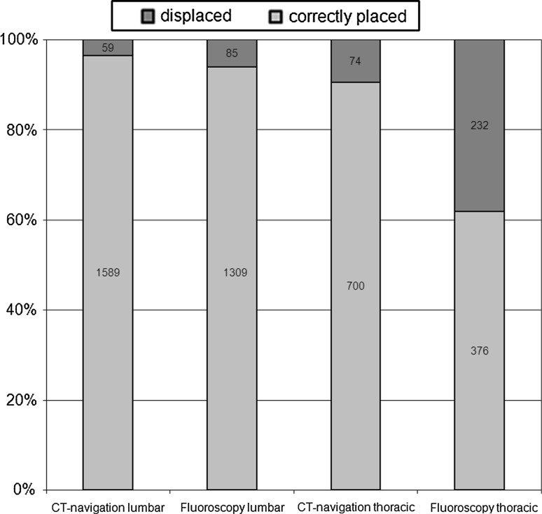
The number of all screw positions for both implantation methods is shown in Tables 2 and 3. In the CT-navigation group, a total number of 2,422 screws were placed, 774 in the thoracic and 1,648 in the lumbar spine, respectively. 74 out of the 774 thoracic and 59 out of 1,648 lumbar screws were misplaced (Table 2; Fig. 1). Correspondingly, 9.6 % of the thoracic screws and 3.6 % of the lumbar screws were misplaced when using CT-navigation.
Table 2.
Distribution of screw positions in the CT-navigation group
| Level | Total screws | Correctly placed | Minor perforation <2 mm | Displaced | |||
|---|---|---|---|---|---|---|---|
| 2–<4 mm lateral | 2–<4 mm medial | ≥4 mm lateral | ≥4 mm medial | ||||
| Th1 | 8 | 2 | 4 | 2 | 0 | 0 | 0 |
| Th2 | 11 | 8 | 3 | 0 | 0 | 0 | 0 |
| Th3 | 17 | 12 | 3 | 0 | 0 | 2 | 0 |
| Th4 | 38 | 19 | 10 | 5 | 2 | 1 | 1 |
| Th5 | 42 | 31 | 1 | 5 | 3 | 1 | 1 |
| Th6 | 51 | 30 | 13 | 6 | 2 | 0 | 0 |
| Th7 | 66 | 44 | 12 | 8 | 0 | 1 | 1 |
| Th8 | 55 | 36 | 6 | 10 | 2 | 0 | 1 |
| Th9 | 54 | 42 | 10 | 2 | 0 | 0 | 0 |
| Th10 | 56 | 48 | 4 | 2 | 0 | 2 | 0 |
| Th11 | 158 | 138 | 14 | 6 | 0 | 0 | 0 |
| Th12 | 218 | 177 | 33 | 4 | 1 | 3 | 0 |
| Total thoracic screws | 774 | 587 | 113 | 50 | 10 | 10 | 4 |
| 700 | 74 | ||||||
| L1 | 207 | 174 | 23 | 6 | 4 | 0 | 0 |
| L2 | 280 | 230 | 41 | 4 | 1 | 2 | 2 |
| L3 | 259 | 238 | 12 | 8 | 1 | 0 | 0 |
| L4 | 363 | 312 | 36 | 10 | 1 | 4 | 0 |
| L5 | 424 | 370 | 41 | 8 | 2 | 3 | 0 |
| S1 | 115 | 106 | 6 | 0 | 3 | 0 | 0 |
| Total lumbosacral screws | 1,648 | 1,430 | 159 | 36 | 12 | 9 | 2 |
| 1,589 | 59 | ||||||
| Total screws | 2,422 | 2,289 | 133 | ||||
Table 3.
Distribution of screw positions in the fluoroscopy group
| Level | Total screws | Correctly placed | Minor perforation <2 mm | Displaced | |||
|---|---|---|---|---|---|---|---|
| 2–<4 mm lateral | 2–<4 mm medial | ≥4 mm lateral | ≥4 mm medial | ||||
| Th1 | 4 | 2 | 0 | 1 | 0 | 1 | 0 |
| Th2 | 6 | 1 | 2 | 1 | 2 | 0 | 0 |
| Th3 | 11 | 0 | 4 | 4 | 2 | 1 | 0 |
| Th4 | 14 | 4 | 0 | 5 | 4 | 0 | 1 |
| Th5 | 60 | 16 | 12 | 18 | 12 | 2 | 0 |
| Th6 | 46 | 14 | 8 | 12 | 10 | 1 | 1 |
| Th7 | 84 | 34 | 16 | 14 | 16 | 3 | 1 |
| Th8 | 71 | 22 | 7 | 22 | 14 | 4 | 2 |
| Th9 | 85 | 24 | 11 | 23 | 21 | 5 | 1 |
| Th10 | 72 | 35 | 19 | 12 | 3 | 2 | 1 |
| Th11 | 75 | 60 | 11 | 2 | 1 | 1 | 0 |
| Th12 | 80 | 65 | 9 | 1 | 2 | 2 | 1 |
| Total thoracic screws | 608 | 277 | 99 | 115 | 87 | 22 | 8 |
| 376 | 232 | ||||||
| L1 | 199 | 170 | 25 | 2 | 1 | 1 | 0 |
| L2 | 208 | 151 | 52 | 1 | 2 | 2 | 0 |
| L3 | 240 | 198 | 33 | 3 | 2 | 2 | 2 |
| L4 | 268 | 204 | 52 | 4 | 6 | 1 | 1 |
| L5 | 287 | 162 | 89 | 12 | 13 | 8 | 3 |
| S1 | 192 | 98 | 75 | 2 | 12 | 3 | 2 |
| Total lumbosacral screws | 1,394 | 983 | 326 | 24 | 36 | 17 | 8 |
| 1,309 | 85 | ||||||
| Total screws | 2,002 | 1,685 | 317 | ||||
Fig. 1.
Distribution of screw positions related to either method of implantation and anatomical region
In the fluoroscopy group, 2,002 screws were placed altogether with 608 thoracic and 1,394 lumbar screws, respectively. 232 out of 608 thoracic screws (38.2 %) were misplaced, whereas 85 out of 1,394 lumbar screws (6.1 %) showed a moderate or severe displacement (Table 3; Fig. 1). The difference between both implantation methods was statistically significant in the thoracic (p < 0.001) as well as in the lumbar group (p = 0.001).
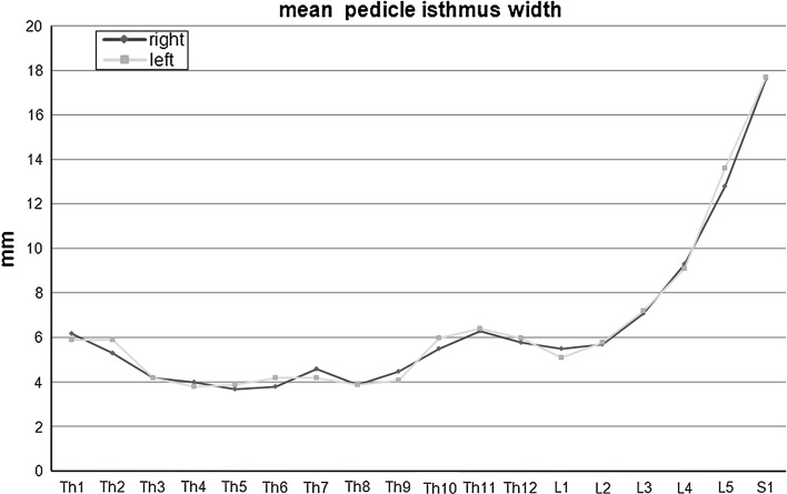
In cases of instrumentation of pedicles with an isthmus of less than 5 mm (predominantly in the thoracic spine) the in–out–in-technique is an approved tool to achieve a sufficient anchoring of the screw with an acceptable lateral breach (≤3 mm). Looking at our results, especially the rather high displacement rate in the upper thoracic levels is most notably due to the rigid definition of misplacement. In the fluoroscopy group, there were 115 lateral breaches of less than 4 mm among all thoracic screws (Table 3). Out of these, there were 103 pedicles with an isthmus width of 5 mm or less, resulting in 103 wrongly categorized screws in the misplacement group. Considering this, the misplacement rate of all thoracic screws placed with fluoroscopy decreases to still 21 %. In contrast, 39 out of the 50 thoracic screws with a lateral breach of less than 4 mm in the CT-navigation group would have been categorized as “correctly placed” leading to a misplacement rate of 4.5 %. The mean pedicle diameters measured at the isthmus perpendicular to the axis are shown in Fig. 2.
Fig. 2.
The isthmus width of each pedicle was determined perpendicular to the axis at the most narrow site
Revisions of malpositioned screws were done either based on intraoperative judgement by the surgeon or less frequently because of the compromise of neural structures by the screw. Considering the retrospective character of our study, the reproduction of circumstances and specific reasons for screw revisions was not always easy. Traceable revisions of malpositioned screws have been carried out in 22 fluoroscopy cases (4.4 %) and in 6 navigation cases (1.2 %), respectively.
There were nine nerve root lesions with paresis of the dependent muscles due to screw perforation (four cases L5, two cases L4 and S1, respectively, one case L3) in the fluoroscopy group. Furthermore, we saw two cases of incomplete myelopathy due to medial perforation in the thoracic region, which partly resolved after revision.
In the navigation group two nerve root lesions (L5 and L4) appeared.
Discussion
Accuracy of placement: lumbar spine
For the lumbar region the accuracy rate of screw placement in our series is 96.4 % when using CT-navigation and 93.9 % when using fluoroscopy, respectively, with a statistically significant difference (p = 0.001) between both implantation methods. These rates seem to be acceptable, when comparing them with accuracy rates reported in literature [8–12]. In a meta-analysis of a total of 7,533 pedicle screws, Tian and Xu [8] reported a mean accuracy rate of 90.21 % out of 6,063 in vivo placed thoracolumbar pedicle screws, independent of the method of implantation. Other reports communicate misplacement rates of up to 30 % in the lumbar spine when using conventional implantation methods for pedicle screw placement [13, 14].
In a recently published meta-analysis, Kosmopoulos and Schizas [2] report a median placement accuracy for in vivo placed navigated screws of 93.7 % (2,778 thoracolumbar screws) and 86.6 % (10,107 thoracolumbar screws) for unnavigated screws, respectively. Looking specifically onto the lumbar level, the reported median accuracy of navigated screws in this study is 96.1 %; however, on the base of only 864 screws. In comparison, without the use of navigation techniques, the median accuracy decreases to only 79.0 % (1,674 screws).
Considering our data, we can point out that the correct screw placement is safer when using CT-navigation rather than fluoroscopy, with a marginal but statistical significant difference. With both methods, we achieved an acceptable safety concerning the correct screw placement within the pedicle. However, a prolonged operating time and the requirement of technical adjuncts with a corresponding high demand on handling skills have to be kept in mind.
It seems justifiable that either method—CT-navigation and fluoroscopy-guided implantation of pedicle screws—is applied in one institution provided that the handling of both methods is routinely ensured.
Accuracy of placement: thoracic spine
Compared to the lumbar region, the placement of thoracic pedicle screws remains a challenge, despite of modern technology and computer assistance especially in the upper thoracic spine, where misplacement rates of up to 40 % have been reported [9, 15]. In a study of Rampersaud et al. [9], pedicle breaches occurred in 31.6 % (25 out of 79 screws) of all 2D navigated thoracic screws with 72 % of pedicle breaches being lateral. Using intraoperative multiplanar imaging, Bledsoe et al. [10] found 93.3 % out of 150 thoracic screws to be contained solely in the desired pedicle and all 10 pedicle violations were Grade 1. Other studies report on a comparable number of misplaced screws in the thoracic spine [16–18].
When dealing with conventional, namely fluoroscopic, support for pedicle screw insertion into the thoracic spine, the reported misplacement rates are considerably higher in comparison to screws placed under navigation, especially 3D-navigation techniques like O-arm [19–21]. Only a few publications are available regarding placement accuracy of non-navigated pedicle screws in upper thoracic levels. In a study of Guzey et al. [16], the rate of misplacement was 20.3 % for 113 upper and middle thoracic screws placed under uniplanar or biplanar fluoroscopy. An in vivo study of non-deformed spines reported on 27 and 109 screws placed at T1–T2 and between T3 and T9, respectively, from a total of 209 screws placed between T1 and T12 [22]. With fluoroscopy support, the placement accuracy was 88 % for the T1–T2 screws (15 screws solely contained in the pedicle and 9 lateral perforations less than 4 mm), and 66 % for the T3–T9 screws (15 screws completely intrapedicular, 55 lateral perforations less than 4 mm).
In a randomized controlled trial comparing navigated and non-navigated pedicle screw insertion into thoracic spine, there was a rate of 23 % of pedicle breaches in the non-navigation group compared to only 2 % in the navigation group [23].
Our results impressively underline the significance of navigation support mainly in the middle and upper thoracic area. A misplacement rate of more than 20 % (129 misplaced screws out of 608) seems to be unacceptable compared to only 4.5 % misplacements when using CT-navigation leading to the conclusion that pedicle screw instrumentation in the middle and upper thoracic area should be carried out with the help of navigation only.
The availability of an intraoperative CT seems to be of particular importance. An accurate assessment of screw positions becomes hereby possible without any significant time delay and with utmost accuracy. In terms of the assessment of pedicle screw position the accuracy of computed tomography significantly surpasses any other imaging modality [24, 25]. If necessary, a prompt revision of misplaced screws is possible as well [11]. The revision rates of lumbar as well as thoracic pedicle screws are markedly lower when using CT-navigation compared with fluoroscopy-assisted implantation.
Conclusion
Reviewing the literature, our study is the largest single center series comparing the placement accuracy of thoracolumbar pedicle screws placed either with CT-navigation or fluoroscopy. Although retrospective in nature, this work underlines the significance of CT-navigation, especially when instrumentation of the middle and upper thoracic spine is carried out. As an alternative to other modern 3D navigation techniques, the computed tomography based navigation is an indispensable tool in these cases. In the lumbar and lower thoracic spine, both methods seem comparable.
A post-instrumentation CT scan seems to be of particular importance, allowing the surgeon to evaluate the accuracy of instrumentation before wound closure and to replace it when necessary.
Conflict of interest
None.
References
- 1.Boachie-Adjei O, Girardi FP, Bansal M, Rawlins BA. Safety and efficacy of pedicle screw placement for adult spinal deformity with a pedicle-probing conventional anatomic technique. J Spinal Disord. 2000;13:496–500. doi: 10.1097/00002517-200012000-00005. [DOI] [PubMed] [Google Scholar]
- 2.Kosmopoulos V, Schizas C. Pedicle screw placement accuracy: a meta-analysis. Spine. 2007;32:E111–E120. doi: 10.1097/01.brs.0000254048.79024.8b. [DOI] [PubMed] [Google Scholar]
- 3.Laine T, Lund T, Ylikoski M, Lohikoski J, Schlenzka D. Accuracy of pedicle screw insertion with and without computer assistance: a randomised controlled clinical study in 100 consecutive patients. Eur Spine J. 2000;9:235–240. doi: 10.1007/s005860000146. [DOI] [PMC free article] [PubMed] [Google Scholar]
- 4.Learch TJ, Massie JB, Pathria MN, Ahlgren BA, Garfin SR. Assessment of pedicle screw placement utilizing conventional radiography and computed tomography: a proposed systematic approach to improve accuracy of interpretation. Spine. 2004;29:767–773. doi: 10.1097/01.BRS.0000112071.69448.A1. [DOI] [PubMed] [Google Scholar]
- 5.Beck M, Mittlmeier T, Gierer P, Harms C, Gradl G. Benefit and accuracy of intraoperative 3D-imaging after pedicle screw placement: a prospective study in stabilizing thoracolumbar fractures. Eur Spine J. 2009;18:1469–1477. doi: 10.1007/s00586-009-1050-5. [DOI] [PMC free article] [PubMed] [Google Scholar]
- 6.Pechlivanis I, Kiriyanthan G, Engelhardt M, Scholz M, Lucke S, Harders A, Schmieder K. Percutaneous placement of pedicle screws in the lumbar spine using a bone mounted miniature robotic system: first experiences and accuracy of screw placement. Spine. 2009;34:392–398. doi: 10.1097/BRS.0b013e318191ed32. [DOI] [PubMed] [Google Scholar]
- 7.Idler C, Rolfe KW, Gorek JE. Accuracy of percutaneous lumbar pedicle screw placement using the oblique or “owl’s-eye” view and novel guidance technology. J Neurosurg Spine. 2010;13:509–515. doi: 10.3171/2010.4.SPINE09580. [DOI] [PubMed] [Google Scholar]
- 8.Tian NF, Xu HZ. Image-guided pedicle screw insertion accuracy: a meta-analysis. Int Orthop. 2009;33:895–903. doi: 10.1007/s00264-009-0792-3. [DOI] [PMC free article] [PubMed] [Google Scholar]
- 9.Rampersaud YR, Pik JH, Salonen D, Farooq S. Clinical accuracy of fluoroscopic computer-assisted pedicle screw fixation: a CT analysis. Spine. 2005;30:E183–E190. doi: 10.1097/01.brs.0000157490.65706.38. [DOI] [PubMed] [Google Scholar]
- 10.Bledsoe JM, Fenton D, Fogelson JL, Nottmeier EW. Accuracy of upper thoracic pedicle screw placement using three-dimensional image guidance. Spine J. 2009;9:817–821. doi: 10.1016/j.spinee.2009.06.014. [DOI] [PubMed] [Google Scholar]
- 11.Zausinger S, Scheder B, Uhl E, Heigl T, Morhard D, Tonn JC. Intraoperative computed tomography with integrated navigation system in spinal stabilizations. Spine. 2009;34:2919–2926. doi: 10.1097/BRS.0b013e3181b77b19. [DOI] [PubMed] [Google Scholar]
- 12.Amato V, Giannachi L, Irace C, Corona C. Accuracy of pedicle screw placement in the lumbosacral spine using conventional technique: computed tomography postoperative assessment in 102 consecutive patients. J Neurosurg Spine. 2010;12:306–313. doi: 10.3171/2009.9.SPINE09261. [DOI] [PubMed] [Google Scholar]
- 13.Gertzbein SD, Robbins SE. Accuracy of pedicular screw placement in vivo. Spine. 1990;15:11–14. doi: 10.1097/00007632-199001000-00004. [DOI] [PubMed] [Google Scholar]
- 14.Laine T, Makitalo K, Schlenzka D, Tallroth K, Poussa M, Alho A. Accuracy of pedicle screw insertion: a prospective CT study in 30 low back patients. Eur Spine J. 1997;6:402–405. doi: 10.1007/BF01834068. [DOI] [PMC free article] [PubMed] [Google Scholar]
- 15.Liljenqvist UR, Halm HF, Link TM. Pedicle screw instrumentation of the thoracic spine in idiopathic scoliosis. Spine. 1997;22:2239–2245. doi: 10.1097/00007632-199710010-00008. [DOI] [PubMed] [Google Scholar]
- 16.Guzey FK, Emel E, Hakan Seyithanoglu M, Serdar Bas N, Ozkan N, Sel B, Aycan A, Alatas I. Accuracy of pedicle screw placement for upper and middle thoracic pathologies without coronal plane spinal deformity using conventional methods. J Spinal Disord Tech. 2006;19:436–441. doi: 10.1097/00024720-200608000-00011. [DOI] [PubMed] [Google Scholar]
- 17.Belmont PJ, Jr, Klemme WR, Dhawan A, Polly DW., Jr In vivo accuracy of thoracic pedicle screws. Spine. 2001;26:2340–2346. doi: 10.1097/00007632-200111010-00010. [DOI] [PubMed] [Google Scholar]
- 18.Carbone JJ, Tortolani PJ, Quartararo LG. Fluoroscopically assisted pedicle screw fixation for thoracic and thoracolumbar injuries: technique and short-term complications. Spine. 2003;28:91–97. doi: 10.1097/00007632-200301010-00021. [DOI] [PubMed] [Google Scholar]
- 19.Schnake KJ, Konig B, Berth U, Schroeder RJ, Kandziora F, Stockle U, Raschke M, Haas NP. Accuracy of CT-based navigation of pedicle screws in the thoracic spine compared with conventional technique. Unfallchirurg. 2004;107:104–112. doi: 10.1007/s00113-003-0720-8. [DOI] [PubMed] [Google Scholar]
- 20.Schizas C, Theumann N, Kosmopoulos V. Inserting pedicle screws in the upper thoracic spine without the use of fluoroscopy or image guidance. Is it safe? Eur Spine J. 2007;16:625–629. doi: 10.1007/s00586-006-0262-1. [DOI] [PMC free article] [PubMed] [Google Scholar]
- 21.Odgers CJ, Vaccaro AR, Pollack ME, Cotler JM. Accuracy of pedicle screw placement with the assistance of lateral plain radiography. J Spinal Disord. 1996;9:334–338. doi: 10.1097/00002517-199608000-00011. [DOI] [PubMed] [Google Scholar]
- 22.Kuntz C, Maher PC, Levine NB, Kurokawa R. Prospective evaluation of thoracic pedicle screw placement using fluoroscopic imaging. J Spinal Disord Tech. 2004;17:206–214. doi: 10.1097/00024720-200406000-00007. [DOI] [PubMed] [Google Scholar]
- 23.Rajasekaran S, Vidyadhara S, Ramesh P, Shetty AP. Randomized clinical study to compare the accuracy of navigated and non-navigated thoracic pedicle screws in deformity correction surgeries. Spine. 2007;32:E56–E64. doi: 10.1097/01.brs.0000252094.64857.ab. [DOI] [PubMed] [Google Scholar]
- 24.Abul-Kasim K, Strombeck A, Ohlin A, Maly P, Sundgren PC. Reliability of low-radiation dose CT in the assessment of screw placement after posterior scoliosis surgery, evaluated with a new grading system. Spine. 2009;34:941–948. doi: 10.1097/BRS.0b013e31819b22a4. [DOI] [PubMed] [Google Scholar]
- 25.Rao G, Brodke DS, Rondina M, Bacchus K, Dailey AT. Inter- and intraobserver reliability of computed tomography in assessment of thoracic pedicle screw placement. Spine. 2003;28:2527–2530. doi: 10.1097/01.BRS.0000092341.56793.F1. [DOI] [PubMed] [Google Scholar]


