Fig. 2.
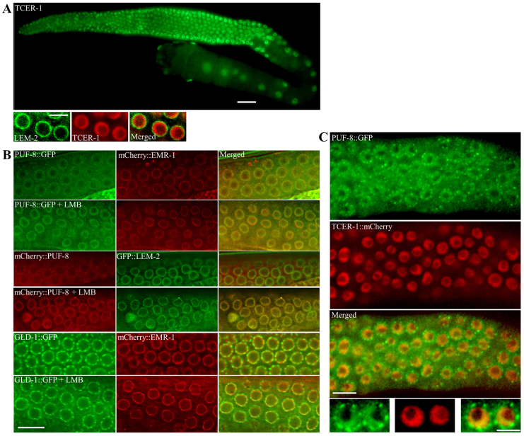
PUF-8 and TCER-1 colocalise at the inner nuclear periphery. (A) Expression pattern of tcer-1::gfp transgene in a wild-type background (top) and of gfp::lem-2, a nuclear envelope marker, and tcer-1::mCherry transgenes (bottom). Only a few germ cells from the distal part of the gonad are shown at higher magnification. Left, distal; right, proximal. (B) Row 1 (from top) shows expression patterns of PUF-8::GFP and mCherry::EMR-1, another nuclear envelope marker, in worms carrying both transgenes. Row 3, PUF-8 is fused to mCherry and the nuclear envelope is visualised using the GFP::LEM-2 marker. Row 5, expression patterns of GLD-1::GFP (shown here as a control) and mCherry::EMR-1. Rows 2, 4 and 6 are as rows 1, 3 and 5, respectively, but after treatment with leptomycin B (LMB). (C) Expression patterns of PUF-8::GFP and TCER-1::mCherry in worms carrying both transgenes. Beneath, two nuclei from each panel shown at a higher magnification. Except for A (top), images have been deconvolved using the Iterative Deconvolution module of Axiovision software to enhance the axial resolution of the fluorescence signal. (B,C) Only a part of the distal gonad, revealing a few germ cell nuclei, is shown. Scale bars: 25 μm in A (top); 10 μm in B and C (top); 5 μm in A (bottom) and C (bottom).

