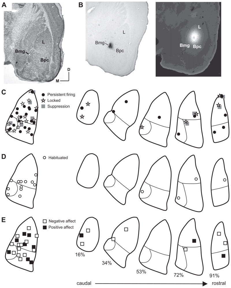
Fig 11.
Auditory responses were observed throughout the lateral and basal nuclei of the amygdala. (A) Nissl-stained, coronal section through amygdala. This section is used in outlines at left in (C–E). Abbreviations: Bpc, parvicellular division of basal nucleus, Bmg, magnocellular division of the basal nucleus; D, dorsal; L, lateral nucleus; M, medial. (B) Recording sites marked by biotinylated dextran amine deposit in Bmg (left) and by Fluoro-Gold deposit in L (right). (C–E) Anatomical distribution of different categories of auditory responses, compressed onto a single section (left) and displayed throughout a rostro-caudal series of amygdalar sections matched to site location (right). Only responses localized by tracer deposits are plotted. (C) Distribution of neurons based on temporal discharge pattern. (D) Distribution of habituating responses. (E) Distribution based on affect of call as described previously (Clement et al., 2006). Numbers below sections indicate percent location within caudal-to-rostral series.

